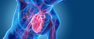
Heart block is an abnormal heart rhythm in which the heart beats too slowly. Why is this happening and what to do? Let's find out together with the experts. Each heartbeat occurs in the heart's upper right chamber (atrium) in the sinus node, a bundle of specialized cells that act as the heart's natural pacemaker. When it beats, an electrical signal from the upper chambers goes to the lower chambers, which causes the heart to contract and pump blood. Heart block disrupts this normal rhythm.
Degrees of heart block in adults
Heart block can be first, second, or third degree, depending on the electrical signal abnormality.
First degree heart block. In this case, the electrical impulse still reaches the ventricles, but travels through the AV node more slowly than usual.
Second degree heart block. It is divided into two categories:
- Type I, also called Mobitz block I or Wenckebach AV block, is a less severe form of second-degree heart block in which the electrical signal on its way to the ventricles becomes slower and slower;
- type II, also known as Mobitz block II - in this case, some electrical signals do not reach the ventricles at all, and the heartbeat may be slower than usual.
Third degree heart block. In this embodiment, the electrical signal from the atria to the ventricles is completely blocked. To compensate, part of the ventricles acts as a pacemaker, creating electrical signals. These signals cause the heart to pump blood, but much more slowly than normal.
Classification
There are several types of heart block. They are divided based on the location of the area where the conduction function of the heart is difficult or completely blocked. Due to the development of the disease in different parts of the heart, each blockade has its own characteristic features.
Photo: psodaz / freepik.com
Sinoatrial blockade
Sinoatrial block or sick sinus syndrome (SSNS) develops as a result of complete or partial dysfunction of the sinus node. This can occur both as a result of physiological disorders and due to other factors not related to the state of the body (extracardiac factors).
The main cause of SSSS can be considered fibrosis (development of connective tissue and its scarring) of the sinus node tissue. This pathology can be caused by inflammatory processes, increased tone of the vagal nerve, or taking medications.
SSSU can manifest itself:
- shortness of breath;
- general weakness of the body;
- pain in the heart;
- dizziness and loss of consciousness;
- low blood pressure;
- arrhythmia.
Interatrial block
Interatrial block develops when the propagation of electrical impulses is disrupted, both within and between the atria. The propagation of excitation occurs along special conduction pathways, which are circular and longitudinal muscle bundles, usually not described as part of the specialized conduction system of the heart.
One of the mechanisms of changes in the electrophysiological properties of the atrial myocardium, leading to a slowdown in conduction, is the development of fibrosis. There is a violation of the integrity of myocardial cells with the deposition of collagen in the intercellular space, which prevents the movement of impulses throughout the heart.
Accompanied by general weakness, pain in the chest and abnormal heart rate.
Atrioventricular block
Atrioventricular (AV) block is a partial or complete interruption of the transmission of impulses from the atria to the ventricles, leading to disturbances in heart rate and blood flow rate.
There are 3 types of atrioventricular block:
- 1st degree - due to a slow transition of impulses from the atria to the ventricles;
- 2nd degree - characterized by blocking of some impulses;
- 3rd degree - (complete atrioventricular block), associated with blocking of all impulses coming from the atria to the ventricles.
It occurs due to damage to the most sensitive area of the conduction system of the heart - the atrioventricular junction.
Intraventricular block
Intraventricular block occurs when the His bundle, the conducting region of the heart located in the region of the ventricles, is disrupted. The bundle of His consists of 3 branches: the left and right anterior branches and the posterior branch. The posterior branch is also divided into right and left.
Figure 1. Differences in ECG readings with normal rhythm and with blockades.
Intraventricular block is classified based on damage to 1 of 3 branches:
- Blocking of the anterior right branch - the innervation of the right ventricle is disrupted;
- Blocking of the anterior left branch - in case of disruption of the innervation of the left ventricle;
- Blocking of the posterior branch - the work of the His bundle is disrupted to the greatest extent, since along this branch impulses go from the atria to the ventricles, and are subsequently divided into right and left;
- Three-bundle block is associated with disruption of the conduction of the entire His bundle.
Causes of heart block in adults
The most common cause of heart block is a heart attack.
Other causes include heart disease, problems with the structure of the heart (due to defects) and rheumatism. The blockage can also be caused by damage during open heart surgery, as a side effect of certain medications or exposure to toxins. Heart block can be congenital, but it is a rare pathology. The risk of heart block increases with age and the presence of heart disease.
First-degree heart block is common in well-trained athletes, teenagers, young adults, and people with a highly active vagus nerve.
Therapy and surgical treatment
AV blockade 1st degree
It is difficult to determine the level of blockade using a regular “electrocardiogram”. PQ interval is greater than 0.20s. Each P wave corresponds to a QRS complex.
Causes:
observed in healthy individuals, athletes, with increased parasympathetic tone, taking certain medications (cardiac glycosides, quinidine, procainamide, propranolol, verapamil), rheumatic attack, myocarditis, congenital heart defects (atrial septal defect, patent ductus arteriosus). With narrow QRS complexes, the most likely level of AV block is a node. If the QRS complexes are wide, conduction disturbances are possible both in the AV node and in the His bundle.
Treatment.
If the PQ interval does not exceed 400 ms and there are no clinical manifestations, no treatment is required.
AV blockade 2nd degree
Increasing prolongation of the PQ interval until the loss of the QRS complex.
Causes:
observed in healthy individuals, athletes, when taking certain medications (cardiac glycosides, beta-blockers, calcium antagonists, clonidine, methyldopa, flecainide, encainide, propafenone, lithium), with myocardial infarction (especially lower), rheumatic attack, myocarditis. With narrow QRS complexes, the most likely level of AV node blockade. If the QRS complexes are wide, disruption of impulse conduction is possible both in the AV node and in the His bundle.
Mobitz I type.
With Holter monitoring, it is detected in almost 6% of healthy individuals, especially often in athletes. Typically, AV blockade appears during sleep, when parasympathetic tone is increased. In lower myocardial infarction, AV blockade type Mobitz I often serves as a precursor to complete AV blockade, which in such cases is accompanied by a stable AV nodal escape rhythm, is well tolerated and does not require a pacemaker.
There are no clinical manifestations:
no treatment required.
In case of hemodynamic disorders:
atropine, 0.5-2.0 mg, IV, then “electrocardiostimulation” (ECS). If AV blockade is caused by myocardial ischemia, then the level of adenosine in the tissues is increased; in such cases, an adenosine antagonist, aminophylline, is prescribed.
Type Mobitz II.
The level of blockade is distal to the AV node (in the bundle of His or its legs). High risk of progression to complete AV block with a slow idioventricular rhythm, which entails a sharp decrease in cardiac output.
Treatment.
Regardless of the clinical manifestations, temporary, then permanent “electrocardiostimulation” (ECS) is indicated.
Diagnostics
First, the cardiologist will review the patient's medical history and ask questions about your general health, diet and activity level, and family medical history.
In addition, he will ask about the medications the patient is taking, as well as about bad habits. The doctor will then listen to your heart and check your pulse for signs of heart failure, such as fluid retention in your legs and feet.
In addition, to diagnose heart block, use:
- ECG – this will show the heart rate and rhythm, as well as the timing of electrical signals as they pass through the heart;
- A Holter monitor is a portable ECG that is attached to the patient’s body and records heart activity from 24 to 48 hours;
- An event monitor is another type of portable ECG that is worn over an extended period, where the device records heart activity at specific times rather than continuously;
- Electrophysiology test - This test uses thin flexible wires (catheters) that are placed on the surface of the heart to record its electrical activity.
Pacemakers
Currently, there are many options for permanent pacing. They have one thing in common: under the skin of the anterior chest wall (usually near the collarbone) an electric pacemaker is implanted - a small metal box, the wires from which (electrodes) go through the veins to the right side of the heart. These wires sense the heart's own electrical activity (to synchronize their work with it) and transmit impulses to the heart. Modern pacemakers almost always stimulate both the atria and the ventricles. In addition, they are tuned so that the heart rate increases during exercise and decreases at rest. There are essentially no contraindications for cardiac pacing: pacemaker implantation is a simple and harmless procedure that can be performed at any age.
A separate type of cardiac pacing is the so-called biventricular stimulation. It is performed not for conduction disorders themselves, but to achieve synchronous contraction of all the walls of the left ventricle. Therefore, the installation of a biventricular stimulator is also called resynchronization therapy. Electrodes from the stimulator go to the right ventricle and to the coronary sinus (which is directly adjacent to the left ventricle). This type of treatment greatly helps some heart failure patients.
Some pacemakers also have a defibrillator function: they detect life-threatening arrhythmias and automatically deliver a shock to stop them.
Patients with pacemakers lead normal lives. They just need to avoid the influence of a strong magnetic field. Thus, magnetic resonance imaging (MRI) is contraindicated in patients with implanted pacemakers.
From time to time, patients with implanted pacemakers need to be seen by specialists: to check the serviceability of the stimulator (in particular, its battery capacity), and to adjust the stimulation parameters.
Share:
Popular questions and answers
Cardiologist Stanislav Snikhovsky answered our questions about heart block.
What complications can occur with heart block?
Clinically, all heart blocks are manifested by slowing of the pulse.
When the pulse rate decreases, fainting conditions associated with a lack of blood circulation to the brain may develop. Patients may complain of irregular heartbeat, headache and shortness of breath. With complete heart block (pulse about 40 beats per minute or less), Morgagni-Edams-Stokes syndrome develops, which is manifested by convulsions and loss of consciousness. Complete heart block causes rapid development of heart failure and can be fatal.
When to call a doctor at home if you have a heart block?
If there is sudden general weakness or fainting, a rare (less than 50 beats per minute) or very weak pulse, a feeling of irregular heartbeat, sudden shortness of breath and a feeling of lack of air, you should immediately call an ambulance.
Complete AV block.
The atria and ventricles are excited independently of each other. The frequency of contractions of the atria exceeds the frequency of contractions of the ventricles. Same PP intervals and same RR intervals, PQ intervals vary. Causes: complete AV block can be congenital. An acquired form of complete AV block occurs with myocardial infarction, isolated disease of the cardiac conduction system (Lenegra's disease), aortic defects, taking certain medications (cardiac glycosides, quinidine, procainamide), endocarditis, Lyme disease, hyperkalemia, infiltrative diseases (amyloidosis, sarcoidosis ), collagenosis, trauma, rheumatic attack. Impulse conduction blockade is possible at the level of the AV node (for example, with congenital complete AV block with narrow QRS complexes), the His bundle, or the distal fibers of the His-Purkinje system.
Treatment
— permanent “electrocardiostimulation” (ECS). If the causes of blockade are reversible (for example, hyperkalemia), if blockade occurs in the early postoperative period or with lower myocardial infarction, temporary pacemaker is limited.
Congenital complete
AV blockade is accompanied by a stable replacement AV nodal rhythm, usually does not cause hemodynamic disturbances, is well tolerated and does not require pacemaker. Tactics while awaiting pacemaker implantation depend on the type of replacement rhythm and its stability.
For hemodynamically significant bradyarrhythmias
- constant ECS. With dual-chamber and atrial pacemaker, the risk of stroke is significantly lower than with ventricular pacemaker
to the top of the page
Treatment
Treatment of SA (SA, sinoatrial, sinoauricular) blockade of 1, 2, 3 degrees and treatment of AV (AV, atrioventricular) block of 1, 2, 3 degrees is the same - implantation of an electrical pacemaker (pacemaker). A pacemaker is a minicomputer that regulates the heart and prevents it from stopping.
Do not wait for fatal complications, if you have symptoms of bradycardia, consult a doctor today, tomorrow may be too late. Take care of your health and the health of your loved ones.
Online consultations (Skype, Viber, Whats App)
Sign up for a consultation


