Sinusitis is the most common ENT disease among children and adults. Characterized by inflammatory lesions of the paranasal sinuses. It can occur in acute or chronic form, and is accompanied by a vivid clinical picture. The pathology progresses rapidly and therefore requires timely and competent treatment.
Causes
The main causes of the disease are:
- injuries of the maxillary sinuses that violate the integrity of the mucous membrane;
- mechanical damage to the nasal septum;
- not fully cured runny nose, ARVI, flu;
- congenital anomalies of the structure of the nasopharynx organs;
- penetration of pathogenic bacteria, viruses, fungi;
- constantly dry air in the room;
- chemical burns of the mucous membranes of the sinuses;
- polyps, adenoids;
- malignant neoplasms;
- allergy;
- various systemic diseases (tuberculosis, HIV and a number of others);
- radiation therapy.
How does purulent sinusitis occur?
Purulent sinusitis (in the medical literature you can find another name for the disease - maxillary sinusitis) is a purulent inflammation of the maxillary (maxillary) sinuses.
This is a paired organ. The sinuses are located to the right and left of the nose. They are connected to the nasal cavity by a small opening - the anastomosis. In a healthy adult, the sinuses are well ventilated, and the mucus secreted by their mucous membrane easily leaves the sinus cavity and comes out.
When, due to certain factors, the nasal cavity becomes inflamed and swollen, the already small anastomosis swells. As a result, the mucous masses cannot come out and remain blocked. What happens next? Pathogenic microorganisms begin to multiply quickly in it. The body uses all its strength to fight the enemy. White blood cells try to cope with bacteria. Dead leukocytes and pathogens form pus. The purulent masses stagnate, maintaining severe inflammation. At this stage, you cannot do without full treatment.
If sinusitis is not treated in a timely manner, the only way to quickly evacuate purulent accumulations and improve the patient’s condition is to pierce the sinuses. It will be impossible to manage only with conservative methods (without puncture).
Symptoms of sinusitis
The main clinical signs of the disease are:
- nasal congestion;
- the appearance of mucopurulent discharge;
- pain, heaviness, tension, pressure in the nose;
- decreased sense of smell;
- headache;
- swelling of the zygomatic part, cheeks on the side of inflammation.
If the sinus is filled with fluid, then when the head is tilted forward, the pain intensifies. In the acute course of the disease, symptoms of intoxication of the body are observed (fever, chills, general weakness). Children with sinusitis usually experience difficulty in nasal breathing, light, transparent discharge, and headaches. Also, the clinical picture depends on the type of disease. For example, with odontogenic sinusitis, nasal discharge has a foul odor.
Complications
Inflammation of the maxillary sinuses is a dangerous condition that in most cases leads to serious consequences.
If treatment is started, the pathology quickly moves from acute to chronic. Damage to the respiratory and hearing organs is often observed. These are otitis media, bronchial asthma, pneumonia, laryngotracheitis. All existing diseases in the anamnesis are aggravated.
In the most severe cases, infectious and inflammatory diseases of internal organs are recorded, most often the heart, kidneys, brain, joints and eyes. The most life-threatening are meningitis, sepsis, pericarditis, and myocarditis.
Forms of the disease
Based on the type of pathogen, the following types of sinusitis are distinguished:
- viral;
- fungal;
- allergic;
- bacterial;
- traumatic.
According to the nature of the inflammatory process:
- acute sinusitis;
- chronic sinusitis.
Based on the nature of the clinical picture, the following forms of sinusitis are distinguished:
- catarrhal;
- polyposis;
- purulent;
- atrophic;
- hyperplastic;
- mixed (several forms of the disease are combined at the same time).
Treatment methods for purulent sinusitis in adults
Sinusitis is a disease with extremely unpleasant symptoms that greatly disturb the usual rhythm of life, so it must be treated quickly and without fail. How to quickly cure purulent sinusitis?
A feature of the modern method of therapy is the combination of drug therapy with lavage of the maxillary sinuses and physiotherapeutic procedures.
It is this method of treatment, supported by the professionalism and experience of an ENT doctor, that allows you to do without a puncture.
Before telling you how to cure sinusitis, I would like to focus your attention on what treatment methods should never be used! The main mistakes patients make in choosing a therapy method, which often lead to punctures, are the following:
- warming up the sinuses with pus - boiled eggs, bags of salt, hot sand, etc. are used. This thermal method only increases the formation of pus and aggravates inflammation;
- an attempt to treat the disease on your own and randomly prescribe antibiotics to yourself. Only an ENT doctor can choose the right drug and prescribe the correct dosage.
If you choose the wrong method of therapy, precious time is lost, during which dangerous complications can arise, including meningitis.
Treatment at home
What to do with sinusitis? What first aid should be given to the patient? How to rinse the maxillary sinuses? Treatment of the disease includes drug therapy, as well as home and folk remedies that are used as auxiliary methods.
- Drug therapy. How to treat sinusitis at home without antibiotics? Therapy consists of regularly rinsing the nasal cavity with salt solutions and taking vasoconstrictor drugs (in children, often in combination with antiallergic drugs). In addition, to relieve swelling and open the natural canal that connects the sinuses to the nasal cavity, the use of local glucocorticosteroids is indicated. Antibiotics are prescribed only if the bacterial nature of sinusitis is confirmed by laboratory testing.
- Home and folk remedies. How to get rid of sinusitis without a puncture at home? This can be done using folk remedies. They are quite popular among the population and can be used as part of complex therapy for the disease (with the permission of the attending physician). Folk remedies are tinctures and decoctions of medicinal plants, pharmaceutical solutions of ointments, bee products, homeopathic and other remedies.
There are several options for using home remedies:
- external - for this purpose, use oils and ointments based on golden mustache, cyclamen root, Kalanchoe and a number of other plants;
- inhalations - use chamomile decoction, essential oils, bay leaf, soda solution, potato decoction;
- instillation into the nose - for these purposes, use golden mustache, juice of garlic, radish, onion, Kalanchoe, cyclamen root, sea buckthorn oil;
- washing the nasal cavity with a solution of sea or table salt, chamomile decoction, hydrogen peroxide.
Treatment of chronic sinusitis at home in a child or adult should be carried out under the supervision of a specialist.
Attention! Treatment of sinusitis in adults at home with medications should be prescribed by an ENT specialist. Self-medication is unacceptable!
Guide to the treatment of chronic sinusitis
What are the diagnostic signs of acute and chronic sinusitis? Are antibiotics indicated, and if so, in what case? Which patients should be referred to specialists?
Reviews concerning the diagnosis and treatment of diseases of the paranasal sinuses (PSN) most often raise many new questions, since an accurate diagnosis is complicated by the non-specificity of non-invasive examination methods. Empirical treatment, especially with antibiotics, is generally considered successful, although many cases will resolve spontaneously without any treatment.
The purpose of this review is to highlight current understanding of the nature of inflammation of PPN and to provide logical and factual justification for medical or surgical treatment.
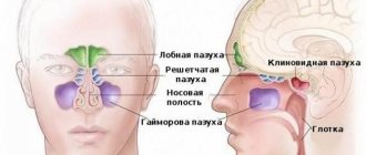
Anatomy and physiology. The nasal cavity and PPN are endowed with important physiological functions. Inhaled and exhaled air passes primarily through the nasal cavity, so the nose must have protective mechanisms that can protect the airways from inhaled pathogens and foreign bodies.
| Figure 1. Mucus flows back into the nasopharynx due to the movement of cilia. |
The glands of the nasal ciliated epithelium and the PPN produce the superficial mucous layer. It traps particles of substances, and the cilia, which are in constant motion, push them back into the nasopharynx (see Fig. 1).
Both the maxillary and frontal sinuses are ventilated through ducts that in turn pass through the anterior ethmoidal region. It is very important that these passages remain patent, since normal mucus flow is necessary to maintain air flow in the sinuses.
The important role of the anterior cells of the ethmoidal labyrinth and the middle meatus in the physiology of the PPN is confirmed by the fact that this region is called the “osteomeatal complex” (Fig. 2). It is believed that mild, localized inflammation in this area may lead to secondary infection of the maxillary and frontal sinuses. This is largely true, although the pathogenesis of sinusitis is more complex.
| Figure 2. Normal middle meatus - region of the “ostiomeatal complex” |
Microbiology. The nasal cavity and PPN are populated by normal bacterial flora; Normally, the same microorganisms are found there as in infected sinuses. Many infectious processes in the sinuses are viral in nature; bacteria are attached a second time.
In acute sinusitis, Streptococculs pneulmoniae, Heamophiluls influlenzae and Moraxella catarrhalis are most often isolated.
In chronic sinusitis, the same microorganisms are usually present, as well as anaerobes, such as strains of Fulsobacteriulm, Staphylococculs aulreuls, and occasionally gram-negative bacteria, such as strains of Pseuldomonas. In recent years, cases of diagnosis of sinusitis caused by fungi have become more frequent, usually in immunocompromised patients. Aspergilluls strains are most often detected, and the severity of clinical manifestations depends on the patient’s immune status.
| Figure 3. Pus in the middle meatus in acute sinusitis |
Allergic sinusitis is increasingly being diagnosed, often associated with nasal polyps.
Clinic. From the standpoint of otorhinolaryngological surgery, the concepts of the anatomy, physiology and pathology of PPN have radically changed with the advent of rigid endoscopy of the nasal cavity and the possibility of computer scanning (CT) of the sinuses.
However, none of these diagnostic methods are available to the general practitioner, who often has to diagnose and treat sinusitis based on clinical symptoms.
Often the complaints of patients with acute and chronic sinusitis are the same, so a timely approach suggests that when trying to distinguish between these conditions, the doctor relies on pathophysiology rather than on considerations of the duration of the disease.
| Figure 4. Computer scan of sinuses |
Sinusitis is considered acute when the infection resolves with drug therapy without leaving significant damage to the mucous membranes. Acute episodes may be recurrent in nature; Chronic sinusitis is a permanent disease that cannot be treated with medication alone. When distinguishing between these conditions, the problem is that there are always indications for surgical treatment, although in reality, long-term drug therapy is sufficient for many patients. In addition, surgery is not 100% successful.
In many patients with a history of acute sinusitis, the onset of the disease is preceded by a cold. Symptoms suggesting the development of acute sinusitis:
- purulent nasal discharge;
- nasal congestion;
- pain and tenderness during examination;
- fever and chills.
In some cases, there are local symptoms that suggest involvement of various sinuses. When diagnosing, the most reliable symptom is a complaint of purulent nasal discharge or its detection during examination (Fig. 3).
If a patient suffers from headaches or facial pain in the absence of purulent discharge, it is most likely not sinusitis.
When sinusitis is untreated, the infection sometimes spreads beyond the sinuses, leading to serious complications. More often this happens when the frontal and ethmoid sinuses are infected; Children are most susceptible to complications.
As the infection spreads forward from the frontal sinus, the soft tissues of the forehead become swollen and painful. Initially, cellulitis develops, then a subperiosteal abscess. Spread through the posterior wall of the frontal sinus leads to intracranial complications such as meningitis, subdural empyema, or anterior lobe abscess.
When the ethmoid sinus becomes inflamed, the infection spreads through the thin bone of the lamina paper, leading to damage to the orbit, accompanied by cellulitis and orbital abscess. Untreated eye socket infections almost always lead to blindness.
| Figure 5. CT scan of the sinuses demonstrating unilateral chronic sinusitis. |
If complicated sinusitis is suspected, especially if there is swelling of the soft tissues of the orbit in a child, an urgent consultation with an otolaryngologist and clarification of the diagnosis using a computer scan is necessary.
The clinical picture of chronic sinusitis is varied. As with an acute infection, nasal congestion and purulent discharge are constant symptoms. The temperature does not rise or rises moderately, and complaints of general malaise, headache and facial pain are typical. Additionally, many patients complain of a decreased sense of smell, and they feel a disgusting smell of pus in the nose.
A simple clinical examination of the nasal cavity using an otoscope can detect large polyps; small polyps are visible only during nasal endoscopy.
| Rinsing the maxillary sinus under local anesthesia is losing its former popularity, as it rarely brings long-term relief |
Over the past decade, cases of diagnosis of acute and chronic sinusitis in children have increased, especially in North America. Diagnosis and treatment of childhood sinusitis is complicated by many factors.
Recurrent symptoms of upper respiratory tract lesions in children appear quite often and, as a rule, indicate the presence of disease of the tonsils and adenoids, and not primary sinusitis. Computed tomography scans of children with upper respiratory tract symptoms often reveal abnormalities of the PPN, especially the maxillary one.
Clinical experience shows that the symptoms of sinusitis in children often go away on their own with age, but it has not yet been established whether “snotty” children grow into “snotty” adults.
There is no doubt that chronic sinusitis also occurs in children, especially if there is dysfunction of the ciliated epithelium. However, most British ENT surgeons believe that, as far as possible, conservative treatment methods for children should be followed.
Examination. In general practice, the diagnosis of sinusitis is usually made on the basis of clinical data.
| Figure 6. A “spur” of the nasal septum cutting into the middle turbinate is a possible cause of “contact pain” |
Plane radiography of the sinuses is extremely nonspecific and is not very informative for identifying pathological changes. Abnormalities on such x-rays are found in half the population. Thus, an x-ray may reveal thickening of the mucous membrane of the maxillary sinus, which does not coincide with the results of direct endoscopy. Despite this, planar films are used quite often, especially for chronic symptoms.
Guidance issued by the Royal College of Radiologists states that planar radiography is not a mandatory routine examination for PPN disease].
Review of planar films suggests that it is appropriate to prescribe a full course of topical steroids without radiography of the PPN in patients with chronic nonspecific sinusitis; If such treatment is ineffective or neoplasia is suspected, the patient should be referred for treatment to a specialist.
The most specific method for assessing the anatomy and pathology of the sinuses is computed tomography, usually in the projection of the coronal suture (Fig. 4).
Computer scanning of the sinuses provides accurate information about the patient's anatomy and the presence of pathological changes (Fig. 5). However, this study should be carried out only after a specialized examination, including nasal endoscopy.
- Treatment
Acute sinusitis. In acute sinusitis, there is no consensus on the choice of antibiotic and the duration of treatment. On the one hand, according to the recommendation of North American rhinologists, antibiotics should be taken for at least 14 days or another 7 days after the symptoms disappear. Some studies suggest that antibiotics have no benefit over placebo when it comes to treating sinusitis-like symptoms in general practice.
The presence of such opposing points of view often only confuses the general practitioner who is faced with acute sinusitis. The danger of prescribing a long course of antibiotics is the development of antibiotic resistance; in addition, patients often refuse long-term treatment. Inadequate treatment hides the risk of residual infection, and there is always a risk of complications, albeit small.
| Figure 7: Intranasal steroid therapy should be tried before referral |
Many patients presenting with sinusitis symptoms recover spontaneously without antibiotics; The doctor’s task is to determine in a timely manner whether such a recovery is possible.
It is assumed that CT scanning can help resolve this issue successfully. Patients with fluid levels or complete opacification of the maxillary sinuses require antibiotics, while patients whose scans show no abnormalities or only mucosal thickening are likely to recover spontaneously.
English GPs do not have immediate access to CT scans and are unlikely to be provided with one for the diagnosis of acute sinusitis, as it exposes the patient to significant radiation and is also quite expensive.
From a purely symptomatic standpoint, purulent nasal discharge and nasal congestion are more reliable signs of sinus infection than other symptoms such as headaches and facial pain. For patients with the first group of symptoms, the prescription of antibiotics is justified.
When choosing an antibiotic, it is necessary to take into account the possibility of the presence of penicillin-resistant strains.
First-line drugs are amoxiclav, erythromycin and cephalosporins, such as cefixime. The same antibiotics can be prescribed for chronic infections; in this case, quinolone derivatives such as ciprofloxacin are also useful.
Often, in acute sinusitis, decongestants, both local and systemic, are used as additional agents. Topical decongestants, such as xylometazoline, reduce mucosal swelling and improve air conductivity, which theoretically speeds recovery.
Steam inhalations, often with aromatic additives, such as menthol, bring relief to the patient, increasing the sensation of air flow in the nasal cavity, but do not objectively contribute to recovery.
Chronic sinusitis. The presence of a chronic PPN infection implies either a disease of the mucous membrane itself, or an anatomical obstacle to the aeration of the sinuses. In any case, chronic sinusitis does not respond to antibiotic therapy alone.
The cornerstone of treatment in this case is steroid therapy, usually via the nasal route. The point of prescribing steroids is to reduce inflammatory swelling and improve sinus ventilation.
Topical steroids are prescribed in drops or spray form. Topical betamethasone drops are often effective and should be administered with the correct position (head tilted down) (Fig. 7) and used for no more than six weeks to avoid systemic side effects. The advantage of new steroid sprays (triamcinolone, budesonide) is their single use during the day, which is more convenient for the patient.
Patients should be referred for specialist consultation if adequate medical treatment fails or if more serious diseases such as neoplasia or Wegener's granulomatosis are suspected. Often, a course of intranasal steroids alleviates the condition of patients with recurrent acute and chronic sinusitis. Such a course should be carried out before referral to an otolaryngologist.
There are a number of symptoms that raise suspicion of neoplasia and require early referral: unilateral nasal discharge, facial numbness, diplopia, deafness due to middle ear effusion, and identification of an intranasal mass on examination.
Surgical treatment is indicated for some patients, with endoscopic ethmoidectomy generally preferred by surgeons. Puncture of the maxillary sinus under local anesthesia is losing its former popularity, as it rarely brings long-term relief and is extremely disliked by patients.
New surgical and anesthetic techniques allow most centers to perform sinus surgery on a day hospital basis and avoid routine postoperative nasal packing.
Treatment of facial pain. A significant portion of a rhinologist’s working time is spent diagnosing patients with facial pain and headaches. With the advent of sinus surgery, impressive results have been achieved in the treatment of diseases accompanied by these symptoms.
Often the symptoms inherent in sinusitis and the complaints typical of migraines and cluster headaches are largely the same.
If a patient with facial pain does not have nasal congestion or purulent discharge, and the endoscopy and CT scan results are normal, then the problem is most likely not in the nose and sinuses, and sinus surgery is not effective, although the possibility of a placebo effect should not be discounted .
Recently there has been interest in so-called contact pain. In this condition, it is assumed that the nasal septum is in abnormal contact with the lateral wall of the nose. This usually occurs when a sharp spur extends from the septum and rests on the middle turbinate (Fig. 6). Typically, patients complain of pain around the central part of the face, extending to the forehead and eye sockets.
Note!
- Many PPN infections are caused by viruses; bacterial agents are secondary. Typically, Streptococculs pneulmoniae, Heamophiluls influlenzae and Moraxella catarrhalis are found in acute sinusitis
- In many patients with a history of acute sinusitis, the onset of the disease is preceded by a cold. Signs suggesting the development of acute sinusitis: purulent nasal discharge, nasal congestion, pain and tenderness during examination, fever and chills
- The most reliable symptom is a complaint of purulent nasal discharge or its detection during examination. If the patient suffers from headaches or facial pain in the absence of purulent discharge, it is most likely not sinusitis
- Plane radiography of PPN is extremely nonspecific and has little information for identifying pathological changes. Abnormalities on such x-rays are found in half the population
- Many general practitioners presenting with sinusitis symptoms recover spontaneously without antibiotics; The doctor’s task is to determine in a timely manner whether such a recovery is possible
- First-line drugs include amoxicillin/clavulanate, erythromycin, and cephalosporins such as cefixime. The same antibiotics can be prescribed for chronic sinusitis; in this case, quinolone derivatives such as ciprofloxacin are also useful
- Patients should be referred for consultation with an otolaryngologist if adequate medical treatment has failed or if more serious diseases such as neoplasia or Wegener's granulomatosis are suspected. Often, a course of intranasal steroids alleviates the condition of patients with recurrent acute and chronic sinusitis. This course should be completed before referring the patient to a specialist.
Prohibited tricks
It is worth noting that not all traditional medicine is approved by otolaryngologists and can be used in treatment.
Absolutely forbidden:
- Use radish juice, onion juice, celandine for instillation into the nasal passages.
- Lubricate the nasal mucosa with ichthyol ointment and birch tar.
- Treat the nasal passages with garlic pulp.
- Rub your nose with cloves.
- Warm up your nose. Warming is prohibited for any form of sinusitis, as it can cause complications.
- Use honey in its pure form. Even if there is no allergy to bee products, an unexpected reaction may occur in the nasopharynx.
These methods are not related to the treatment of inflammation and will only aggravate the situation, causing swelling and an acute allergic reaction.
Important to know and remember! Before using the chosen traditional treatment method, you should definitely consult with your doctor. You cannot self-medicate! This can lead to terrible, sometimes the most severe and irreparable consequences!
Diagnostics
When making a diagnosis, anamnesis, visual examination, laboratory and instrumental studies are carried out. During rhinoscopy, the lumens of the nasal passages, the condition of the mucous membrane, the nature of the discharge, and the presence or absence of polyps are assessed.
The most informative are instrumental diagnostic methods. X-rays are performed in several projections, sometimes using a contrast agent. To clarify the condition of the sinus walls, computed tomography is used. MRI makes it possible to evaluate changes in soft tissues. In some cases, endoscopy, sinus puncture followed by bacterial culture of the exudate may be required.
Nasal rinsing
To date, this method is the most effective. The main purpose of lavage is to remove the accumulated pus from the maxillary sinuses. The most common solutions are the following, which have properties for killing microbes and pathogenic microflora:
- Salt solution. Salt has antiseptic properties. It is enough to dilute the water with one tablespoon of salt and rinse your nose 5-6 times daily. It is important not to overdo it with salt, and not to use too hot water, so as not to cause a burn to the mucous membrane.
- A solution of soda and salt. The recipe is similar to the previous one - mix 200 ml of water with 1 tsp. salt and 1 tsp. soda Baking soda is a natural antiseptic. It kills germs, heals micro-wounds and thins mucus.
- Washing with propolis. Add 1 tsp to one glass of water. alcohol tincture of propolis. Can be used up to 5 times daily.
It is worth following the washing technique. You cannot inhale water through your nose on your own. The solution should flow into the nasal passage on its own. First you need to clean your nose. It is better to use a kettle or container with a long spout to prevent water from pouring past your nostrils. You need to lean over the sink and turn your head to one side. The solution is poured into the passage that is located above. If the manipulation is performed correctly, the water will flow out through the second nostril or throat.
Inhalation via nebulizer
Inhaling vapors of medicinal plants helps to destroy pathogenic microflora in the upper respiratory tract, as well as dilute pus. The following recipes are popular:
- A teaspoon of propolis tincture is diluted with 1000 ml of water. This ingredient has an antimicrobial effect.
- A combination of soda and Vietnamese “Star”. Add 1 tsp per 1000 ml of water. a spoonful of soda and one drop of star.
- Inhalation of fir oil. Add 10 drops of essential oil to one glass of boiling water.
Diagnosis of sinusitis using modern techniques
To confirm the diagnosis of sinusitis, the following types of examination are used:
- Video endoscopy of the nasal cavity and nasopharynx to identify features of the anatomical structure and determine predisposing factors for the development of sinusitis;
- radiography of the paranasal sinuses;
- Ultrasound examination of the paranasal sinuses is a safe method with no contraindications, used to diagnose sinusitis and monitor the treatment process;
- CT, MRI - according to indications;
- laboratory diagnostics according to indications in full.
X-ray characteristics of the maxillary sinuses
X-rays of the maxillary sinuses should be interpreted not by a radiologist, but by the attending otolaryngologist. Accumulations of pus in the nasal sinuses and their swelling are often equally visualized on radiographs. If you mistake the swelling for purulent accumulations, then you can make a puncture in vain, which will not give any positive results. Often, the liquid contents of the maxillary sinus with sinusitis are characterized by a clear visual level, which allows us to talk about its purulent nature.
With normal darkening of the sinuses, the results of radiography must be supported by other types of diagnosis . It is unacceptable to practice when a diagnostic puncture is performed for the sole purpose of determining the presence of pus in the sinus. An MRI or CT scan often allows one to clarify the results of an x-ray examination and differentiate between normal swelling and the presence of pus. The same applies to ultrasound scanning of the sinuses.