Therapy with Retinalamin
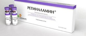
Retinalamin is available in the form of a white powder (lyophilisate), which, when diluted, produces an injection solution.
The main active ingredient of the drug is polypeptide fractions isolated from the biomaterial of the eyes of cattle and pigs. Thanks to their complex effects, photoreceptors and retinal pigment epithelium are stimulated at the cellular level, and protein synthesis processes are activated. Interaction reactions normalize the permeability of vascular walls, accelerate recovery and repair processes, help restore the light sensitivity of the retina and increase visual acuity.
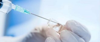
The drug is prescribed in the form of injections, which are performed intramuscularly or parabulbarly - through the skin of the lower eyelid under the eyeball. According to indications, injections are performed daily in a dosage of 5-10 mg of the substance once. The course of treatment lasts from five to ten days and can be re-prescribed after three months.
In pediatric ophthalmology, Retinalamin solution is used primarily in the form of injections performed intramuscularly.
Experience in using Retinalamin for various ophthalmological diseases
Dystrophic eye diseases such as glaucoma, AMD, diabetic retinopathy, myopia and its complications are common causes of low vision and blindness throughout the world. In addition, there is a tendency towards an increase in the frequency of these diseases, including at a young age [1–4]. The last decade has been marked by the accumulation of new data on the role and place of neuroprotective therapy in the treatment of dystrophic eye diseases. Neuroprotection can be defined as a set of therapeutic measures aimed at preventing, reducing, and in some cases reversing the processes of death of neuronal cells. One of the significant groups of neuroprotectors are biogenic peptides. The first studies on the use of this group of drugs in ophthalmology were conducted in the early 1980s. at the Department of Ophthalmology of the Military Medical Academy, i.e., the experience of their use is more than 30 years. Retinalamin, which is a representative of this group of drugs, is a complex of water-soluble polypeptide fractions. The mechanism of action of the drug is determined by its metabolic activity. Retinalamine improves metabolism in eye tissues and normalizes the functions of cell membranes, intracellular protein synthesis, regulates lipid peroxidation processes, and helps optimize energy processes. In vitro studies of the effect of Retinalamin on the survival of nerve cells and the state of cultured retinal cells under conditions of oxidative stress were carried out in 2006–2007. on the basis of the Institute of Molecular Genetics of the Russian Academy of Sciences. The effect of Retinalamin was expressed in high cytoprotective activity, increased proliferation and stimulation of the transformation of stem cells into neurons, which ensured their connections with the nervous structures of the brain and restoration of visual function. Moreover, the protective effect on cells was observed both before and after the creation of conditions of oxidative stress. Thus, the drug has indications for use for both preventive and therapeutic purposes. Thus, Retinalamin has a stimulating effect on photoreceptors and cellular elements of the retina, helps improve the functional interaction of the pigment epithelium and outer segments of photoreceptors during dystrophic changes, and accelerates the restoration of light sensitivity of the retina. Against this background, vascular permeability is normalized, reparative processes are activated in diseases and dystrophic lesions of retinal and optic nerve cells [5–10]. In 2016, an observational clinical study of Retinalamin was conducted in daily clinical practice in patients with various ophthalmological pathologies within the framework of registered indications for use. This study was approved by an independent multidisciplinary clinical research ethics review committee. Purpose of the study:
assessment of the effectiveness and tolerability of Retinalamin in patients with various ophthalmological diseases during 1 or 2 consecutive courses of treatment with an interval of 3 months.
Material and methods
The study included patients over the age of 18 years with “dry” or “wet” AMD, POAG with compensated IOP, diabetic retinopathy and myopic disease. Each patient signed an informed consent to participate in the study. Exclusion criteria included age <18 years, the presence of contraindications to the use of Retinalamin, a single eye, VA in the worse eye <0.1 with correction, any other serious diseases or conditions that, in the opinion of the study physician, could distort the results of the observational program and limit patient participation in the study, as well as pregnancy and breastfeeding. The total number of patients was 4172 people (64% women and 36% men). The average age of the patients was 59±17 years. The distribution by nosology in the study groups was as follows: “dry” or “wet” form of AMD – 894 people, average age – 51.3±9.2 years; POAG with compensated IOP – 1269 people, average age – 62.1±10.7 years; diabetic retinopathy – 943 people, average age – 42.8±7.9 years; myopic disease – 1066 people, average age – 34.5±13.6 years. Follow-up visits were scheduled at 1, 3, 4, and 6 months to assess the effectiveness and tolerability of retinoprotective therapy. from the start of therapy if 2 courses of Retinalamin are prescribed. During the 1st course of treatment with Retinalamin, patients were examined after 1, 3 and 6 months. from the start of therapy. Efficiency assessment was carried out based on changes in health and health status (expansion of health zone boundaries in total, reduction in the number of scotomas and changes in MD1, PSD2 indicators). To assess the quality of life of the study participants, a “Patient Questionnaire” was used, in which there were 7 situations for assessment, each of which contained 5 categories (1 – I have no difficulties; 2 – I have minimal difficulties; 3 – I have moderate difficulties; 4 – I have enough severe difficulties; 5 – experiencing extreme difficulty). Tolerability of Retinalamin was assessed by the frequency of adverse events (AEs).
results
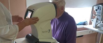
The dynamics of VA during therapy are shown in Fig. 1. There was a trend towards an increase in VA, most pronounced in most cases after 3 months. therapy. The results of the PZ study were divided into 2 groups depending on the perimetry method used. Kinetic perimetry was used in 37% of observations; its use was associated both with the equipment of diagnostic centers and with limitations for standard automatic perimetry (low VA, complexity of the technique). The dynamics of the peripheral boundaries of the PZ were assessed by the sum of 8 meridians (Σ, in degrees) and the number of scotomas (Fig. 2, 3). Changes in static perimetry results were assessed by changes in the main photosensitivity indices: MD - average decrease and PSD - pattern deviation (Fig. 4, 5).
All changes in perimetry results were statistically insignificant (p>0.05). There was a trend towards improvement in visual function during observation. Pronounced positive dynamics of VA with correction, perimetric indices, an increase in the peripheral boundaries of the VA, and a decrease in the number of scotomas were observed at 1 and 3 months. from the start of therapy. Subsequently, stabilization was observed in most of the assessed indicators. The difference in responses to the “Patient Questionnaire” at different follow-up periods was not significant, however, there was a slight decrease in the total score during Retinalamine therapy, which indicates a tendency to improve the quality of life of patients. The difference in responses between nosological groups is most likely due to the characteristics of the clinical picture of the diseases. Tolerability of Retinalamin in patients with various ophthalmological diseases during 2 consecutive courses with an interval of 3 months. was good, no adverse events were identified during the study.
Discussion
In recent years, a number of large domestic studies have been conducted on the effectiveness of Retinalamin in patients with POAG of various stages, AMD, diabetic retinopathy, and myopic disease. The use of Retinalamin in a pathology such as diabetic retinopathy contributed to the improvement of basic visual functions, electrophysiological and hemodynamic parameters (decrease in linear blood flow velocity parameters and decrease in the resistance index) [11]. With progressive myopia in children, Retinalamin had a positive effect on VA and contributed to the expansion of peripheral VA [12]. The clinical effectiveness of treatment with Retinalamin in the “dry” form of AMD has been confirmed both with subconjunctival and parabulbar administration of the drug. The effectiveness of treatment was assessed after 3 and 6 months. [13]. At the Department of Ophthalmology, Faculty of Medicine, Russian National Research Medical University, the effect of prescribing 2 courses of Retinalamin with a break of 3 months was studied. Positive dynamics of the studied indicators were revealed, and the effect increased gradually, and after 1 month. after completion of therapy, the main indicators exceeded the indicators identified immediately after the end of the course of treatment. After the 2nd course of therapy, an increase in the effect of the drug was noted [14]. In 2007, a study was conducted involving 120 patients with POAG. Starting from 3 months after treatment with Retinalamin, positive dynamics of the studied parameters were observed, including contrast sensitivity [15]. In a 2013 study, clinically significant results after using Retinalamin were noted after 3, 6, 12 months. There was an expansion of the boundaries of the PV, an increase in VA, the average thickness of retinal nerve fibers, and stabilization of the glaucomatous process according to ophthalmoscopy. In the control group, by the end of the observation period, most patients showed progression of POAG [16]. In the period from November 2013 to May 2014, a nationwide screening study was conducted on the effectiveness of Retinalamin in patients with compensated POAG stages I–III. The study included 453 patients (453 eyes) aged from 28 to 89 years. Retinalamine was administered to all patients at a dose of 5 mg IM for 10 days. The total observation period was 3 months. During this time, the protocol provided for 4 control examinations of patients: before treatment, after 10 days, 1 and 3 months. after it started. Standard ophthalmological examination, ophthalmoscopy and perimetry were performed. It was found that improvement in indicators (VA, PV, IOP) after a course of Retinalamin occurs within 3 months. [17]. Thus, the results of the clinical use of Retinalamine in various diseases do not contradict the data of previously conducted studies and confirm its effectiveness for the prevention and relief of changes in retinal tissue and improvement of its functions.
Conclusion
When prescribing Retinalamin in patients with AMD, glaucoma, diabetic retinopathy, myopic disease in the form of 2 consecutive courses with an interval of 3 months.
Positive dynamics of the average VA and a tendency to increase the photosensitivity of the retina were observed (expansion of the borders of the retina, a decrease in the number of scotomas and changes in MD and PSD indicators). Retinalamin can be recommended as a universal neuroprotector for various ophthalmological diseases associated with degenerative lesions of retinal tissue. 1 MD (mean deviation), the average deviation from the age norm, shows general depression or the presence of areas in the visual field with normal light sensitivity and defects. 2 PSD (pattern standard deviation), partial standard deviation, represents the degree of deviation of the shape of the patient’s hill of vision from the age norm.
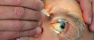
Parabulbar injections
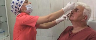
When prescribing a course of parabulbar injections with Retinolamine, they are performed by a specialist with sufficient experience in such procedures in a specially equipped clinic room or in a sterile operating room. Injections of the drug do not require special preparations.
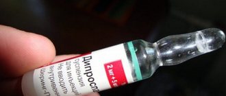
To obtain an injection solution, the leophilisate of the drug is diluted using an isotonic solution or procaine (0.5%) and water for injection. Administration is carried out with a disposable syringe with a slightly blunted needle. During the procedure, the needle enters percutaneously into the lower eyelid (outer edge) and advances approximately 1 cm in depth. The required dose of the drug is injected into the space at the back wall of the eyeball.
Retinolamine solution cannot be stored, therefore, after the injection, its remains are disposed of.
Experimental studies of the effectiveness of Retinalamin
A.A. Antonov, A.S. Makarova, V.S. Reshchikov Federal State Budgetary Institution "NIIGB"
Retinalamine is a complex of polypeptides isolated from the retina of animals. The drug regulates metabolic processes in the retina, stimulates the functions of the cellular elements of the retina, helps improve the functional interaction of the pigment epithelium and the outer segments of photoreceptors in various pathologies of the retina, enhances the activity of retinal macrophages, has a normalizing effect on blood coagulation and has a pronounced protective effect on the vascular endothelium. Retinalamin, having a pronounced protective effect on the vascular endothelium and collagen fibers of perivascular connective tissue, helps restore damaged structures of the vascular wall.
Active study of Retinalamin began in 1985, when it was first isolated. A.I. Dneprovskaya and S.V. Kharintseva (1988) conducted a series of studies on the effect of Retinalamin on the course of experimental retinopathy. The authors studied its effect at various concentrations in vitro on immunity parameters, platelet aggregation properties and fibrinolysis. Retinalamine helped stimulate the fibrinolytic activity of the blood, which may be due to the presence of a low molecular weight plasminogen activator in its composition. As experiments on fibrin plates have shown, Retinalamin contains a low-molecular-weight inhibitor of plasmin and trypsin. The data obtained indicated a normalizing effect of the drug on the state of the hemocoagulation system. In addition, the immunomodulatory effect of Retinalamin has been shown. Under its influence, the expression of receptors on T- and B-lymphocytes significantly increased, and the phagocytic activity of neutrophils increased.
Based on the concept of bioregulatory therapy, which assumes that polypeptides from various organs increase the resistance of these organs to the action of pathological agents, a number of experiments were carried out. Based on the created experimental models of retinal dystrophy, the effect of Retinalamin on the course of the pathological process was studied. The drug had a pronounced therapeutic effect in toxic retinal dystrophy caused by the administration of a 3% solution of potassium iodide. During ophthalmoscopy in experimental rabbits receiving Retinalamin, a decrease in the size of dystrophic foci and retinal edema was observed. Clinical data were confirmed by histological examination.
Much experimental work has been carried out to study the effect of Retinalamin on the regeneration processes of the neuroreceptor apparatus of the eye. Experiments were carried out on Campbell rats suffering from genetically determined retinal pigmentary degeneration. The data obtained indicated the ability of Retinalamin to inhibit the development of the degenerative process of the retina.
I.B. Maksimov (1996) studied the effectiveness of the use of peptide bioregulators (including Retinalamin) in combination with microsurgical treatment for injuries of the cornea, retina and their consequences. The author created experimental models to study reparative processes in the cornea and retina. Under the influence of the drugs, there was a decrease in the time (1.5-2 times) for restoration of the integrity of the damaged epithelial, stromal and endothelial layers of the cornea due to the membrane-stabilizing effect on damaged cellular structures (according to keratoimpedansometry data). In case of penetrating wounds of the cornea, in addition to accelerating reparative regeneration, peptide bioregulators increased the strength of the developing corneal scar by 1.8 times, enhanced the function of local immunity (according to the lysosomal cation test by 2.2-2.5 times) and contributed to the formation of insignificant in intensity corneal opacities.
The use of Retinalamin in experimental laser damage to the retina, as well as in toxic dystrophy caused by the action of monoiodacetic acid, allowed 78.4% of experimental animals to obtain a therapeutic effect, manifested in an acceleration of 2-2.5 times (compared to the control) coverage of the retinal defect pigment epithelial cells, preventing further development of the pathological process, as well as reducing (according to electroretinogram data) the degree of inhibition of the functional state of the retina. Retinalamine had a normalizing effect on the course of experimental retinal vein thrombosis caused by the administration of thrombin. Histological examination of the eyes of rabbits in the control group revealed gross pathological changes: extensive plasma and hemorrhages, fragmentation of associative neurons was noted in the internal granular layer, extensive foci of necrosis involving all layers of the retina, retinal detachment over a large area. When using Retinalamin, the histological picture was more favorable: retinal edema was observed only in the outer layers, hemorrhages were practically absent, massive foci of necrosis were not recorded, only single neurons were destroyed.
When performing a number of studies on cell cultures, the tissue specificity of the action of peptide bioregulators was established. It is possible that a set of peptides obtained from a certain tissue or organ contains those peptides that mediate a phenomenon such as autoinduction, that is, the maintenance of tissue specificity during the functioning of adult tissues. This assumption was tested by S.V. Trofimova while studying the inductive effect of Retinalamin on pluripotent embryonic tissue.
When studying the induction of Retinalamin on cells of the pluripotent tissue of the early gastrula ectoderm of Xenopus laevis, the author revealed that Retinalamin has neural inductive activity. After exposure of the drug to the ectoderm of the early gastrula, neural differentiation is triggered, including the brain, retina, and pigment epithelium. Depending on the concentration of Retinalamin, different tissues developed. It should be noted that when exposed to Retinalamine at a concentration of 50 ng/ml, in some cases not only the brain developed, but, perhaps, the pineal gland also appeared as part of it. When exposed to Retinalamin at a concentration of 10 ng/ml, the retina developed in two of the eighty cultures taken in the study and the pigment epithelium developed in one. It was established that for Retinalamin there is an indirect dependence of the inductive effect on the concentration of the inducing factor, since it had an effect even at a concentration of 2 ng/ml, which is not typical for other proteins and peptides.
The author also studied the proliferative activity of Retinalamin on cell cultures of the retina and pigment epithelium of Wistar rats. Analysis of the results of exposure showed a statistically significant dependence of the mitogenic activity of cells on the concentration of Retinalamin. It has been established that pigment epithelial cells proliferate somewhat more actively than retinal cells. The results obtained after the first week of cultivation showed that the greatest mitogenic activity is observed when retinal and pigment epithelial cells are exposed to Retinalamine at a concentration of 200 ng/ml. The mitogenic activity of pigment epithelial and retinal cells when exposed to Retinalamin over the next three weeks was maximum at concentrations of 50 and 100 ng/ml.
The data obtained indicate the significant ability of Retinalamin in a certain tissue-specific concentration to induce active proliferation of cultured cells of the retina and pigment epithelium. It is necessary to point out the multifunctional ability of peptide drugs to control the proliferative activity of cells of the corresponding tissue and trigger the process of differentiation in pluripotent tissue.
When exposed to differentiated cells of an adult organism in a short-term culture, proliferative activity of the cells is observed, but their tissue specificity is also maintained. It must be emphasized that the proliferation process does not suppress the process of maintaining tissue-specific differentiation, i.e. the two mechanisms work synchronously, and the cells do not lose the signs of differentiated tissue during the cultivation process. Peptide regulators are capable of influencing both differentiated cells of an adult organism, triggering cell proliferation and maintaining tissue specificity, and pluripotent embryonic cells, inducing tissue-specific differentiation.
The objective of the study was to evaluate the effect of Retinalamin on the organotypic tissue culture of young and old animals. For cultivation, we used retinal tissue from embryonic chickens and newborn rats, that is, young organisms with a high potential for growth and differentiation. In addition, it is of great interest to study the effect of peptide drugs on the tissues of old organisms, in which growth and adaptation abilities are less pronounced. At 1 day During cultivation, in all samples, the spreading of tissue explants on the collagen substrate and the beginning of the migration of proliferating cells along the periphery of the explant were observed. After 3 days, if the growth zone development was stimulated in the experiment, the area index of the experimental explants increased compared to the area index of the control explants. To identify effective concentrations, peptides were added to the chicken embryo tissue culture in the concentration range from 0.5 to 100.0 ng/ml. The effective concentration for Retinalamin was 20.0 ng/ml. Retinalamine significantly stimulated the growth of retinal explants by 2.49 ± 0.3 a.u.
Further increases in dose had no additional effect on the growth of tissue explants. Under the influence of Retinalamin, the tissue culture area index increased by 25%.
The effective concentration found for each peptide was used in subsequent series of experiments - on young and old Wistar rats. Retinalamine significantly stimulated the growth of retinal explants in young rats by 2.67±0.12 a.u., and in old rats by 1.68±0.12 a.u. compared to control. The effect of peptide bioregulators turned out to be different when culturing the tissue of young and old rats. The growth zone was larger when culturing the retinas of young animals than of old ones.
An experimental study of the morphological changes occurring in the posterior segment of the eyeball during glaucoma following the administration of Retinalamin was carried out in 2008 on a model of adrenaline-induced glaucoma in 38 rabbits according to the scheme of E.M. Lipovetskaya (1966). This model is an experimental analogue of POAG and is characterized by the formation of a glaucomatous symptom complex, including the development of trabeculo- and neuropathy in the eye. A morphometric analysis of retinal ganglion cells and optic nerve neurons was carried out; against the background of the effect of Retinalamine, the most pronounced protective effect was established on small ganglion cells, to a lesser extent on medium ones, and lastly on large cells.
The preservation of the total number of cells compared to the comparison group was increased by 43%. The effect of the drug was expressed in a decrease in the degree of damage to the optic nerve and the number of dying retinal ganglion cells, a decrease in atrophic processes in the layer of their axons, and an increase in the activity of Müller cells. An increase in the activity of Müller cells has been proven due to an increase in the expression of glutamine synthetase and inducible nitric oxide synthase, which reflects the influence of glia on the correction of metabolism.
In 2012, the Ufa Research Institute of Eye Diseases studied the reparative activity of the retinal pigment epithelium of rabbits using peptide bioregulators (Retinalamine).
The condition of pigment epithelial cells in two groups was assessed at different times. In the control group, flattened pigment epithelial cells with a small amount of melanin in the cytoplasm were noted even 1 month after injury. In the experimental group, many cells had a cubic shape, the cytoplasm of the cells contained melanin granules of varying intensity, and on the 30th day of treatment of the animals, a gradual restoration of the pigment epithelium of the retina of the eyeball was noted. It has been established that the use of peptide regulators in the postoperative period helps to accelerate the reparative regeneration of the pigment epithelium due to the migration of flattened pigment cells to the site of damage, intracellular accumulation of melanin granules, restoration of cell shape and reduction of the inflammatory reaction of the choroid itself.
Modern experimental studies of Retinalamin are associated with the search for alternative routes of its administration, which can facilitate targeted delivery of the drug and create its depot at the optic nerve head. The drug is used subconjunctivally, in the sub-Tenon's space, under the skin of the temples, by the endolymphatic route, and also by endonasal electrophoresis. Each of these methods has both its advantages and disadvantages, such as low concentration in the area of the pathological focus due to systemic absorption of the drug, duration of treatment, parabulbar hematomas, risk of perforation of the eyeball, etc. In this regard, on the basis of the Siberian State Medical University conducted experimental studies on the use of Retinalamin by epiretinal administration. A series of experiments was carried out on 20 Wistar rats, divided into 2 groups. Morphological examination of the retina of rats 1, 3, 7, 14 and 21 days after epiretinal injection in both the main group and the comparison group revealed no pathological changes. The retinal layers maintained their normal structure throughout the experiment.
Thus, experimental studies have proven the effect of Retinalamin on the proliferation and differentiation of various cells in the tissues of the organ of vision. The results obtained indicate its possible effectiveness in such pathologies as glaucoma, age-related macular degeneration, and traumatic injuries. The question of increasing the effectiveness of treatment with Retinalamin through its targeted delivery to target tissues remains relevant.
Side effects, complications and contraindications
Parabulbar injections of Retinalamin are considered one of the most effective ways to deliver medication to the affected area, but they are very painful and can be accompanied by bruising and itchy swelling under the eyes, as well as allergic reactions.
Among the possible negative effects of the procedure, experts also mention:
- The appearance of a veil before the eyes, which reduces visual acuity.
- Hemorrhages under the conjunctiva.
- Severance of the ligaments of zinn (rare).
- Iris prolapse (very rare).
Retinalamin injections are not prescribed if you are hypersensitive to the components of the product. Limitations also include age under 18 years (children with central retinal dystrophy are excluded) and pregnancy. Women who are breastfeeding are advised to temporarily stop breastfeeding when prescribed Retinalamin injections.