About the disease
When grown in a petri dish containing blood agar, Staphylococcus aureus colonies have a characteristic golden yellow color, hence the name. This microorganism is extremely common, colonizing about a quarter of the world's population.
It usually spreads in the nasal cavity, groin area, armpits and other parts of the skin, but in most cases it is a normal part of human skin flora and does not cause any problems.
However, as the number of bacteria on the skin increases, they begin to penetrate into the deeper layers of the skin through microcracks and can lead to skin infections and abscesses.
Pathological processes can affect not only various internal organs, such as the liver, kidneys, spleen, brain, but also muscles, bones and joints, which leads to extremely disastrous consequences, therefore, if you suspect an infection, you should immediately consult a doctor.
Antibiotics against Staphylococcus aureus
Various types of antibiotics are used for Staphylococcus aureus. It can be:
- "Azithromycin";
- "Amoxicillin";
- "Clarithromycin";
- "Roxithromycin";
- "Norfloxacin";
- "Furazolidone" and others.
Methicillin-resistant bacteria show resistance to Ciprofloxacin, but are sensitive to Levoflaxacin and Roxithromycin.
To combat Staphylococcus aureus, fatty acids containing up to 16 carbon atoms are used. The antimicrobial activity of the substance is due to the acidic environment in which microorganisms do not survive.
Types of staphylococcal infections
There are several most common types of staphylococci:
● Hemolytic staphylococcus.
Most often, this infection affects the upper respiratory tract, causing purulent sore throat, pharyngitis, tonsillitis, bronchitis and other inflammatory diseases. These bacteria are very resistant and difficult to treat.
● Golden.
This microorganism is extremely resistant to almost all types of penicillin antibiotics, antiseptics, high temperatures, and active direct sunlight. It causes various skin lesions, such as eczema, abscess, boils, lesions of the gastrointestinal tract, upper respiratory tract, mucous membranes, and in the worst case leads to toxic shock.
● Epidermal.
This microorganism lives on the surface of the skin and mucous membranes of any healthy person and does not cause any harm. But, if this bacterium enters the blood of a person with a weakened immune system, which most often occurs during surgical operations, the use of improperly processed instruments, catheters, blood poisoning occurs, which leads to inflammation of the inner lining of the heart.
● Saprophytic.
Despite the fact that this species is the least dangerous, when infected it leads to general intoxication of the body due to the release of dangerous toxins and enzymes during its life processes. These microorganisms often cause inflammation of the urethra and bladder. This is mainly typical for women due to the anatomical features of the structure of their genitourinary system. If left untreated, cystitis leads to kidney inflammation and problems conceiving a child.
Introduction
Two different types of microorganisms are present on the skin and appendages.
One of them consists of resident microorganisms or commensals, which, in most cases, are harmless and non-pathogenic, and sometimes beneficial to the host. The second group consists of transient microorganisms that can have harmful and pathogenic effects and colonize the skin after injury, which will lead to inflammation and the development of skin infection [1–3]. With the availability of bioinformatics technologies leading to the discovery of new phylogenetic approaches, many discoveries have been made in recent years. Studies have been conducted to study the diversity and topology of skin microbiota, to identify various commensal microorganisms present on the skin. This has made it possible to estimate the relative abundance of each population, and to understand their beneficial role or contribution to dermatological conditions such as acne[4–6].
Metagenomic analysis and 16S RNA ribosomal gene sequencing are the predominant bacteriological methods for analyzing the bacterial composition of microbial communities and for choosing the most effective study design, which is critical to obtaining meaningful analysis results [7].
Thus, the physiological characteristics of different skin sites have been associated with different levels of bacterial diversity, including Actinobacteria such as Propionibacteria, most recently renamed Cutibacteria (including Cutibacterium acnes, as well as Cutibacterium granulosum and Cutibacterium avidum), Proteobacteria, Bacteroides and Firmicutes; Staphylococcus epidermidis belongs to the latter group [5, 6, 8, 9].
Of these, C. acnes and S. epidermidis are the two main commensal skin bacteria [3, 10–12].
Until recently, research in the field of acne has mainly focused on the role of C. acnes, while the role of S. epidermidis has been discussed for several years but remains to be elucidated [10, 13].
The purpose of this article is to review the published data on the role of S. epidermidis in the physiopathology of acne and present future treatment prospects based on the available data and evidence.
Methods
A review of literature published between early 2000 and 2022 was performed. and available in PubMed using the following keywords: acne, acne vulgaris + microbiota, skin + microbiota, acne + microbiota + Propionibacteria acnes + Staphylococcus epidermidis, bacterial resistance, skin commensals + Propionibacterium acnes + Staphylococcus epidermidis.
results
Interaction between host skin and skin microbiota
The interaction of the host skin and microbiota in the skin immune system allows the differentiation of harmless commensal microorganisms, such as Corynebacterium sp., Staphylococcus sp. , excluding S. aureus, Cutibacterium sp., Malassezia furfur, and transient harmful pathogens such as S. aureus, Streptococcus pyogenes and Enterobacteriaceae. Even though recognition involves the human immune system, this mechanism is not fully understood. Toll-like receptors (TLRs) have been reported to be desensitized by long-term exposure to commensal microorganisms, or by decreased cell surface expression through activation of TLR kinase 3 pathway inhibitors [14, 15].
Moreover, specificity can be achieved by co-recognition of pathogen-associated molecules through their recognition receptors [6].
Thus, both such recognized commensals C. acnes and S. epidermidis interact with the host to help protect healthy skin from colonization by pathogens [9, 16].
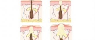
Rice. 1. Healthy skin and skin with acne. A. Healthy skin. In healthy skin, S. epidermidis controls the proliferation of C. acnes. b Formation of microcomedone after excessive colonization of the skin by C. acnes, leading to dysbiosis. Excessive colonization with C. acnes during puberty leads to dysbiosis and acne. AMP, antimicrobial peptides; TLR, Toll-like receptors; PAR, protease-activated receptors.
However, the composition of the skin microbiota is constantly evolving and changing over time. For example, imbalance or dysbiosis of previously healthy skin, caused by external factors such as trauma, stress or pollution, or endogenous factors (hormonal changes, pH changes), can cause inflammatory skin diseases such as acne, atopic dermatitis, rosacea and psoriasis [ 13, 17–21].
In particular, during puberty, sebum production increases and its qualitative change occurs due to hormonal modifications, which can lead to the development of an unbalanced skin microbiota through increased colonization of the pilosebaceous complex and the skin surface with C. acnes [6].
This over-reproduction may be related to nutrient availability [22].
Role of C. acnes
In healthy skin, C. acnes plays a beneficial role in the microbiota of the pilosebaceous complex. It limits the growth of S. aureus, methicillin-resistant strains of S. aureus, and S. pyogenes while maintaining an acidic pH in the pilosebaceous complex by hydrolyzing sebum triglycerides and secreting propionic acid [6, 23, 24].
However, during puberty, excessive colonization of the pilosebaceous complex by C. acnes can lead to loss of diversity and dysbiosis, leading to the development of acne [6, 25–27].
A recent clinical study, using a single-locus sequence-based typing method, examined subgroups of C. acnes on the face and back of patients with severe acne and healthy controls [28].
Almost 75% of acne patients had identical C. acnes phylotypes on the face and back, whereas this was the case in only 45% of healthy controls. In the healthy group, phylotypes IA1 (39%) and II (43%) were the main phylotypes, whereas in the acne group, phylotype IA1 (84%), especially on the back (96%), was the main one. This may support the hypothesis that acne severity may be associated with loss of C. acnes phylotype diversity and selection of the major IA1 phylotype among acne patients [28–30].
Therefore, different inflammatory profiles depend on the phylotype (i.e., phylotype IA1, which is mainly observed on the face and back of acne patients), which activates innate immunity through the expression of protease-activated receptors (PARs), tumor necrosis factor-α and interferon-γ, and interleukins (IL-8) [28, 31–37].
In addition, C. acnes activates the release of lipases, matrix metalloproteinases and hyaluronidases, leading to hyperkeratinization of the pilosebaceous unit and, finally, comedones, papules and pustules [31–34, 38].
In Fig. Figure 1 shows the difference between a healthy microbiome and a microbiome with dysbiosis.
Role of S. epidermidis
S. epidermidis is the most commonly isolated commensal species from the epithelium [25, 39].
It colonizes predominantly the axillae, head, and nasal passages and is usually nonpathogenic to humans[11, 39].
S. epidermidis belongs to the group of coagulase-negative staphylococci, which differ from coagulase-positive staphylococci such as S. aureus in the absence of the coagulase enzyme. According to multilocus sequence typing, there is a large diversity of S. epidermidis, with more than 400 identified types (STs), compared to 155 for C. acnes [40, 41].
Most clinical isolates are CC2, from which ST2 is often isolated. It is possible that the successful spread of ST2 may be due to the fact that all ST2 isolates contain the IS256 sequence and ica genes, two factors that correlate with the invasiveness of S. epidermidis [42, 43].
However, there is still controversy as to whether icaA is a useful S. epidermidis biofilm marker [44].
In the past, S. epidermidis was considered a harmless commensal microorganism on human skin. It is currently considered an important opportunistic pathogen. It is the most common cause of hospital-acquired infections, as is its more virulent cousin, S. aureus. In particular, S. epidermidis represents the most common source of infection on stationary medical devices [45].
Treatment is complicated by specific antibiotic-resistant genes and the formation of biofilms, multicellular agglomerations that have intrinsic resistance and tolerance to antibiotics and host defense mechanisms [46].
In addition, studies have identified specific molecular determinants that allow S. epidermidis immune invasion and the ability to cause chronic diseases [47].
It is hypothesized that S. epidermidis has its own, original functions for a non-infectious lifestyle, highlighting the random nature of S. epidermidis infections, allowing the distinction between carriage, contamination and infection. A better understanding of the physiology of S. epidermidis is important for evaluating therapeutic strategies against S. epidermidis infections [39].
Some strains of S. epidermidis can modulate the host innate immune response, especially TLR 2, and thus enable the host to fight pathogens [6, 28].
Phenosoluble modulins produced by S. epidermidis have been shown to selectively inhibit skin pathogens such as S. aureus and group A Streptococcus, and together with host antimicrobial peptides (AMPs) enhance their clearance [48–50].
In addition, S. epidermidis activates keratinocyte AMP expression through a TLR2-dependent mechanism [51].
This confirms that this commensal interacts closely with the host innate immunity [48, 49].
This has also been shown through microdissection of epidermis, dermis, adipose tissue followed by 16S ribosomal RNA sequencing, demonstrating that S. epidermidis colonization regulates T cell trafficking and function through IL-1 [52, 53].
Another recent research paper reported that S. epidermidis may also protect against skin neoplasms by producing 6-N-hydroxyaminopurine, a molecule that inhibits DNA polymerase activity [54].
In addition, butyric acid secreted by S. epidermidis allows adipose tissue stem cells to differentiate into adipocytes and accumulate lipids in the cytoplasm, leading to an increase in the dermal layer [55].
Interaction between S. epidermidis and C. acnes in acne
S. epidermidis and C. acnes use glycerol as a common carbon source to produce various short-chain fatty acids (SCFAs) used as antimicrobial agents to compete with each other. These two microorganisms play a role in the pathogenesis and development of acne. [56, 57].
There is no evidence yet that S. epidermidis plays an active role in the pathogenesis of acne, but predominantly C. acnes is currently associated with acne [9].
Some isolates of S. epidermidis have been shown to have antimicrobial activity against C. acnes using antagonism assays [27, 56].
Among S. epidermidis strains with increased antimicrobial activity, differences in the diameter of the inhibitory zones were observed, indicating that the antimicrobial substances were of a different nature. Most S. epidermidis strains had small zones of inhibition (2–4 mm) against C. acnes, and some strains produced light-opaque zones of inhibition. One strain, FS1, produced very large inhibitory zones (>10 mm) but was not active against all C. acnes strains tested. Another strain, 14.1.R1, inhibited all strains of C. acnes but produced only small zones of inhibition (2–5 mm). There were no differences in the antimicrobial activity of S. epidermidis strains isolated from normal and acne skin, respectively. Similarly, the origin of C. acnes strains did not determine their susceptibility to the antimicrobial activity of S. epidermidis, since strains isolated from acne lesions and healthy skin did not differ significantly in their susceptibility patterns to S. epidermidis. On the contrary, the study showed higher antimicrobial activity among C. acnes phylogroup I-2, against S. epidermidis. The authors hypothesized the possible presence and secretion of a bacteriocin or bacteriocin-like substance specific to phylogroup I-2 of C. acnes [13].
The dependence of the severity of acne (mild, moderate or severe) on antimicrobial activity varied from 29 to 39%. Therefore, C. acnes type IA1 CC18 strains, mainly involved in acne, did not exhibit higher antimicrobial activity compared with healthy strains.
Various antagonism studies have shown that in vivo S. epidermidis controls C. acnes proliferation through the release of succinic acid, a fatty acid fermentation product that inhibits keratinocyte surface TLRs, tumor necrosis factor, and C. acnes-mediated IL-6 [27, 56, 58 ].
Since both C. acnes and S. epidermidis are present on the skin, suppression of inflammation caused by C. acnes may depend on the activation of the LTA miR-143 pathway on keratinocytes by staphylococci, which is necessary to reduce the inflammatory response. Studies have shown that the mechanism of LTA-miR-143-mediated suppression of TLR2 signaling is through miR-143 targeting the 3'UTR of TLR2. This reduces the amount of TLR2 protein and reduces inflammation caused by C. acnes [58–60].
Thus, it helps regulate skin homeostasis and suppress inflammation caused by C. acnes [58, 61].
Accordingly, an unbalanced balance between C. acnes and S. epidermidis in the pilosebacial units of acne patients in favor of the IA1 CC18 phylotype of C. acnes (75–80 cases) may prevent S. epidermidis from fully playing its role as a regulator of natural skin homeostasis and limiting the growth of C. acnes. Figure 2 visually shows the interaction between C. acnes and S. epidermidis.
Figure 2. Bacterial interactions on the skin. AMP, antimicrobial peptides.
S. epidermidis and its implications for current and future acne treatments
Current topical acne treatments include various retinoids, such as adapalene and tretinoin, which reduce inflammation by altering the innate immunity activated by C. acnes [62–65].
In addition to benzoyl peroxide, the use of antibacterial agents such as erythromycin and clindamycin in monotherapy resulted in the development of antibiotic-resistant strains of not only C. acnes but also S. epidermidis within 4–6 weeks [30, 57, 66].
Systemic antibiotics have been reported to induce only minor resistance but may lead to adverse effects in the gut microbiota [30, 33, 67].
Oral isotretinoin normalizes the innate immune system response to C. acnes by inhibiting its proliferation [68].
Thus, eliminating C. acnes alone may promote the proliferation of S. aureus, causing skin inflammation and proliferation of S. epidermidis, leading to other unbalanced skin homeostasis and the risk of nosocomial infections.
AMPs are effector molecules of the skin's innate immune system. They are amphipathic (both lipophilic and lipophobic) and destroy the lipid membrane of the bacterium, leading to lysis and cell death, interacting preferentially with negatively charged bacterial membranes rather than with neutrally charged membranes of mammalian cells. The importance of the contribution of AMPs to human immunity is undeniable, since changes in AMP expression are associated with various pathological processes. However, data on the role of AMPs in acne vulgaris are limited. A recently published study reports that the AMPs hBD-1 and cathelicidins play an important role in the pathogenesis of acne [69].
AMPs have shown activity against a wide range of Gram-positive and Gram-negative bacteria, as well as some fungi, parasites and enveloped viruses [70].
They are produced as a response of keratinocytes and sebocytes. However, they also contribute to the development of additional inflammatory reactions. AMP production includes β-defensins, RNase 7, psoriasin S100 protein, and cathelicidins and is mediated by the MyD88 pathway and IL-1 signaling. Additionally, it has been demonstrated that more cells express TLR-2 as acne severity increases[33].
This may be one explanation why agents targeting TLR-2, such as topical retinoids, are more effective in patients with more severe acne [71].
Cytokines are also produced as a result of the interaction between C. acnes and TLR-2, defensins and matrix metalloproteinase through activation of PAR-2R [72].
Another treatment approach may be regular oral or topical supplementation of the skin microbiota with beneficial microorganisms (probiotics) for acne patients to balance the skin microbiota [73, 74].
In 2010, Arck et al. [75] suggested that there is not only a gut-brain or skin-brain axis, but also a gut-skin axis. The authors showed that oral supplementation of a Lactobacillus strain in mice attenuated stress-induced neurogenic skin inflammation and inhibited hair loss. Their concept suggested that modulation of the microbiota through the use of probiotics reduces neurogenic skin inflammation. These observations provide hope that the right type of bacteria may have beneficial effects on skin homeostasis, inflammation, and the response of peripheral tissues to perceived stress. Other human studies have confirmed this hypothesis [76–78].
Thus, probiotics may be effective in acne and other inflammatory skin diseases such as atopic dermatitis and possibly psoriasis [79–83].
From this point of view, studies show that S. epidermidis can control dysbiosis caused by C. acnes and reduce the severity of acne [27, 56].
However, it remains unclear which SCFA in the glycerol fermentation products of S. epidermidis primarily contributes to anti-C. acnes effect. It is also not determined whether SCFAs act together with other antimicrobial molecules in fermentation products to provide anti-C. acnes effect. Anti-S. acnes effect of the fermented medium persisted after boiling the said medium, suggesting that antimicrobial proteins/peptides may not be a major contributor to anti-C. acnes effect of fermented media [56].
Prodrugs such as SCFA pivaloyl methylbutyrate (AN-9) have been developed to achieve pharmacological concentrations of such SCFAs in vivo [84].
Moreover, live S. epidermidis can potentially be used as an active component in probiotics for acne[27, 56].
Discussion and conclusion
Acne is a chronic and multifactorial inflammatory disease of the skin and pilosebaceous unit. Dysbiosis in acne patients is associated with decreased numbers of S. epidermidis and overcolonization of certain C. acnes phylotypes in the pilosebaceous unit, resulting in varying levels of innate immune activation and varying degrees of acne severity. Recent studies appear to support a beneficial role of S. epidermidis in acne physiopathology by limiting C. acnes skin colonization and inflammation [9, 33].
However, an imbalance in favor of S. epidermidis may also lead to other health consequences, such as hospital-acquired infections. Therefore, balanced skin homeostasis should be the ultimate goal of any acne treatment.
From this perspective, other treatment options can be considered, such as S. epidermidis-derived probiotics to restore a natural, balanced microbiota, and regulation of host AMP mediators without increasing the local epidermal S. epidermidis population.
Symptoms and forms of staphylococcus in adults and children
The symptoms experienced by the patient vary greatly depending on the method and location of infection and the state of the immune system. But a number of the most characteristic features can be identified:
● elevated temperature;
● suppuration of cuts, swelling, purulent rashes on the skin and mucous membranes, itching and redness of the eyes;
● lack of sense of smell and nasal breathing;
● sore throat, pain when swallowing, dry cough;
● nausea, vomiting, general weakened state;
● severe headaches, epileptic attacks, severe shortness of breath;
● painful sensations in the joints.
One of the most common forms of staphylococcus in adults is its asymptomatic carriage. A healthy person does not feel any signs of the disease until a provoking factor appears that gives impetus to the development of the disease.
For women, the most dangerous form is the saprophytic form, which can lead to cystitis. This is due to the fact that the women’s urethra is located very close to the vagina, which is why there is a high probability of pathogenic microflora, including staphylococcus bacilli, entering the urethral canal, and then into the bladder, causing its inflammation.
Since young children do not have a strong immune system, the risk of developing an infection in a child is quite high. Moreover, even those types of staphylococcus that cause practically no problems in adults can lead to serious complications in children. Staphylococcus is especially dangerous in children under one year of age, as it can easily spread throughout the body in the shortest possible time and lead to serious consequences, including death.
What is cutaneous staphylococcus?
The content of the article
Staphylococcus epidermidis, a close relative of MRSA bacteria. It is so common that it is found on almost every person's skin. Cutaneous staphylococcus is part of the natural bacterial flora and is not dangerous when you are healthy - it is a deadly microorganism that is found throughout the world and remains dormant in most cases.
This type of staph is often ignored in clinical trials, and due to its ubiquity, it can evolve and genetically mutate between bacteria very quickly.
The problem with cutaneous staphylococcus occurs when immunity is reduced - then Staphylococcus epidermidis infection can cause a number of complications and become a risk factor for many diseases. Cutaneous staphylococcus is especially dangerous for people who have undergone invasive procedures.
Causes
Most often, the bacterium enters the human body through wounds and microcracks in the skin. The infection, having penetrated through the wound, begins to multiply in the blood, spreading throughout the body and affecting the lungs, heart, brain, liver, kidneys, and joints.
With staphylococcus, diseases can be very different, such as pneumonia, meningitis, osteomyelitis, endocarditis, sepsis and many others.
Staphylococcus infection can occur in the following ways:
● Through contact and household use when using the patient’s personal items;
● Airborne droplets during close contact with an infected person;
● Fecal-oral on dirty fruits, vegetables and other food products, dirty dishes and hands;
● Vertical when a child passes through the birth canal of an infected mother during childbirth.
● Infection often occurs during surgery through medical instruments and during various manipulations.
How is staphylococcus transmitted?
You can become a carrier by accident: in transport, at work, at school, in nature, by getting damaged skin or by “capturing” the microorganism along with food or water. Signs of the disease appear after the following contacts:
- household
- airborne
- fecal-oral
- vertical (from mother to child)
The microorganism is resistant to temperature and physical influences, so it can linger on surfaces for up to six months.
Diagnostics
Only a doctor can diagnose the presence of infection based on the results of a staphylococcus test.
It is worth remembering that testing is recommended only if symptoms of the disease are present. The presence of bacteria in biological material may mean that a person is its carrier, which in itself is the norm.
For analysis, material is taken from the area where the infection is believed to be developing. To detect a pathological process, several tests are carried out to track the dynamics of bacterial growth. If their number increases rapidly, the presence of a staphylococcal infection can be diagnosed. Also, additional analysis will determine the specific type of infection so that the doctor can choose a personal treatment regimen.
Treatment
Staphylococcus can be treated surgically if there is a purulent lesion in the area of infection. It may also be necessary to remove an infected implant, catheter, etc.
As a conservative treatment for staphylococcus, antibiotics and antibacterial agents are used in combination with immunomodulatory therapy. The drugs are selected taking into account the resistance of staphylococcus to a wide range of medications. The use of brilliant green solution for treating infected wounds has shown greater effectiveness.
What tests need to be taken
First of all, to determine the presence of infection, a staphylococcus test is taken.
For diseases of the respiratory tract, a swab is taken from the nasopharynx and oropharynx, for cystitis - urine, for gastrointestinal disorders - feces, for skin lesions - a scraping from the skin, and if there is a suspicion of extensive infection, they donate blood.
Also, before starting treatment with antibiotics, it is required to be tested for resistance to these medications.
Which doctor should I contact?
Advanced cases of the disease are dealt with by an infectious disease specialist.
But if you just have suspicions and want to get an accurate diagnosis, first you should consult a therapist, or with children, a pediatrician. The doctor will conduct a diagnosis and, based on the results obtained, will write you a referral to the necessary doctor.
Symptoms
Bacterial gastroenteritis is a fairly aggressive disease characterized by acute symptoms. Violations of intestinal functions make themselves felt already on the first day of the development of the disease. The following symptoms may appear:
- Nausea and profuse vomiting.
- Prolonged diarrhea.
- Cramping abdominal pain and abdominal muscle cramps.
- Presence of blood in the stool.
- Fever.
- Dizziness and impaired consciousness.
- Weakness and loss of appetite.
- Headache.
- Reduced blood pressure.
The appearance of fever and abnormal blood pressure may require urgent hospitalization. The most dangerous symptoms of bacterial gastroenteritis are associated with dehydration, which can cause death in the patient. In addition, you should consult a doctor if symptoms do not go away within five days.