Rectum
The rectum is a tube with muscular walls consisting of longitudinal and transverse layers, it is fixed in the pelvis by ligaments and has a length of about 15 cm. The rectum then passes through the rectosigmoid junction into the sigmoid colon. The rectosigmoid junction has the appearance of a bend of 70-120 degrees. For 10-15 cm from the anus, the rectum has practically no painful endings and does not contact the small intestine located in the abdomen. Due to these features, when performing sigmoidoscopy, discomfort occurs only when it is necessary to examine the rectosigmoid section and the final segment of the sigmoid colon and is caused by passage through the rectosigmoid junction and contact through the wall of the sigmoid colon with the small intestine, on the surface of which there are many pain receptors, while diagnosis of the rectum itself is practically painless.
Which doctor should you contact if symptoms occur?
If you or your loved ones have the described complaints, you should contact a coloproctologist as soon as possible, who, after consultation and examination, will establish a preliminary diagnosis and determine further tactics and urgency of treatment, or, if in doubt about the diagnosis, a list of necessary additional studies.
If surgery is required, you will be offered hospitalization in a hospital, where, after assessing the results of the studies, which, as a rule, do not take much time and are easy to perform, the operation will be performed.
Anal canal
The anal canal is the terminal part of the rectum, it ends with the anus and has a pronounced muscular wall - the anal canal sphincter complex. This complex consists of external and internal sphincters, which are responsible for voluntary and involuntary retention of feces and gases. In the anal canal, the rectal mucosa transitions into the epithelium (anodermis) of the anal canal, which then passes into the skin of the perianal area. In the skin part of the anal canal there is a very large number of pain receptors, and on the mucous membrane they are practically absent. When an anal canal fissure develops, it is located on the skin part, its bottom is the subcutaneous/superficial portion of the sphincter (constituting no more than 1/10 of its total volume), and with mechanical irritation (bowel emptying), a pronounced spasm of this muscle develops, and intense pain syndrome. That is why performing a drug blockade or dosed sphincterotomy in the case of a fissure with pain that is not amenable to conservative treatment leads to its rapid healing.
Symptoms of acute paraproctitis depend on the localization of the process, the type of pathogenic microorganisms and the reactivity of the body. The disease begins acutely after a short prodromal period with malaise, weakness, and headache. Fever, headache, and increasing pain in the perineum and pelvis appear. If the inflammatory process in the perirectal tissue is not limited and proceeds like phlegmon, septic signs occur. As the abscess forms, the pain increases and becomes pulsating. This period ranges from 2 to 10 days. Subsequently, the abscess breaks into the rectum or onto the skin of the perineum. In 70% of cases, a breakthrough of the abscess is manifested by a short-term improvement in the condition. In 30-70% of cases, acute paraproctitis becomes chronic. If an acute inflammatory process occurs against the background of fistulas, then this form is called chronic recurrent paraproctitis. After opening the abscess, the internal opening remains open. The hole in the skin does not close, and bloody or purulent discharge periodically appears from it. Temporary closure of the internal opening leads to remission. The period of temporary well-being can last several months, or even years. Subcutaneous paraproctitis. This is the most common form, accounting for 50% of all types of paraproctitis. Patients complain of rapidly increasing pain in the anus, perineum, pulsating in nature, intensifying with changes in body position, coughing, and defecation. There is stool retention. When the abscess is localized in front of the rectum, dysuric phenomena may occur. Body temperature rises to 38-39°C with chills.
On examination: the skin of the perineum on the affected side is hyperemic. The radial folding at the anus is smoothed out. A protrusion of the skin appears, which takes on a spherical shape. When the abscess is located in the anus, the latter becomes deformed, becomes slit-like, and sometimes gapes. In such cases, gas incontinence, liquid feces, and mucus leakage occur. Palpation is sharply painful. In 50% of cases fluctuation is determined. A digital examination of the rectum reveals a painful infiltrate and a smoothed anal canal. Instrumental research is extremely painful and even impossible. Acute submucosal paraproctitis is a mild form of paraproctitis. Occurs in 2-6% of cases. Patients complain of mild pain in the rectum, which intensifies during defecation. Within a week, pus, as a rule, breaks into the lumen of the rectum and recovery occurs. Upon examination, submucosal paraproctitis manifests itself when the process extends below the pectineal line (swelling of the corresponding semicircle of the anus). A digital examination reveals a painful, round, tightly elastic formation under the mucous membrane above the pectineal line. Ishiorectal paraproctitis. Occurs in 35-40% of cases. Initially, patients complain of a deterioration in their general condition, fever, and poor sleep. Subsequently, vague heaviness and dull pain in the rectum appear. By the end of the 1st week, the patient's condition worsens. The temperature rises to 39-40°C. The pain becomes acute, throbbing, intensifies with defecation or sudden movements. When the abscess is localized in the area of the prostate or urethra, dysuric disorders occur.
On the 5-7th day of illness, swelling, swelling, and slight hyperemia of the skin of the perineum appear,
Anesthesia and pain relief after surgery
The vast majority of surgical interventions for rectal fistulas are performed under subarachnoid or, as it is also called, spinal anesthesia. It refers to regional anesthesia methods that block the transmission of nerve impulses in a specific part of the body. A distinctive feature of any method of regional anesthesia is the minimum number of complications, in particular from the cardiovascular system, respiratory system, and brain. At the same time, you are conscious or, at your request, are in a superficial medicated sleep (sedation).
In the case of subarachnoid anesthesia during perineal surgery, the blockade of the nerve impulse occurs at the lumbosacral level. In our clinic, anesthesiologists and resuscitators prefer to perform a “saddle” block, which provides ideal anesthesia of the perineum with virtually no motor block, i.e. with preservation of motor function. If necessary, anesthesia can be supplemented with intravenous sedation. All this ensures maximum comfort for the patient and ideal working conditions for the surgeon.
If you are constantly taking a number of medications, for example, antihypertensives, those that affect blood clotting, or others, be sure to inform your anesthesiologist-resuscitator about this a few days before surgery.
Surgical interventions on the perineum can also be performed under general anesthesia.
After the operation, you will be prescribed pain relief as planned for several days, which can be easily done at home using tablets. These include local anesthetic drugs, intravenous analgesics and, in case of severe pain, potent drugs.
What complications can there be after surgery?
Currently, with the availability of standardized technology and an integrated approach to the diagnosis and treatment of rectal fistulas, unpleasant consequences are minimized. However, it should be noted that the risk of complications always exists.
The most serious complication after surgery for rectal fistulas is the development of anal incontinence. The risk of its occurrence is especially high during repeated interventions, when the anatomy is significantly changed and the holding function may be initially compromised. It is worth noting that when operations are performed by an experienced specialist who performs this type of intervention on an ongoing basis, the risk of incontinence is practically absent.
In addition, bleeding may develop both in the early postoperative period and several days after surgery. The nature and severity of the complication are determined only after examination. Stopping bleeding is usually possible in a dressing room. In some cases, repeat surgery may be required.
In addition, when treating complex and recurrent fistulas, due to pronounced scar changes, the sutures fixing the mucomuscular flap may diverge, which leads to inflammation in the wound and requires repeated interventions.
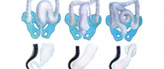
Features of the structure and function of the large intestine
The colon is the lower section of the intestine that extends into the anus.
The intestine consists of two sections: the large and small intestine. The colon is the lower section of the intestine that extends into the anus. The anatomy of the colon has its own characteristics and differences. The large intestine is shorter but wider than the small intestine.
The small intestine can reach a length of 4 meters, and the thick intestine - 1.5-2 meters. The diameter of the colon can reach 8 cm, tapering towards the anus. The large intestine is different in color from the small intestine. The colon is grayer. Visually, they are very easy to distinguish both by size and color.
The large intestine has three longitudinal muscle bands along its entire length. In other words, a thin strip of muscle stretches along the intestine. On the large intestine, or rather on the muscle bands, there are also omental processes. These are small accumulations of fat. The muscle layer of the colon is quite uneven, which creates the appearance of bulges on the body of the colon. It resembles a cluster of swellings and constrictions.
Like the small intestine, the large intestine consists of a mucous layer, submucosal, muscular and serous. The outer serous layer may be absent in some areas due to the unevenness of the surface of the intestine itself. Before digested food reaches the rectum, it can remain in the sections of the large intestine for a day.
There are many more microorganisms living in the large intestine than in the small intestine. There are at least 500 species here. The large intestine performs several important functions:
- Digestive. Digested food coming from the small intestine continues to be digested in the large intestine. Remains of food along with bile are compacted, and feces are formed.
- Excretory. Various toxic substances and other waste products unnecessary for the body are eliminated through the colon. If this process is delayed, poisoning may occur. Food moves through the large intestine more slowly than through the small intestine. Peristalsis intensifies only when the next portion of food enters the stomach. Thus, the colon prevents food from stagnating in the digestive system.
- Protective. It is no secret that the intestines are responsible for human immunity. Various bacteria live on the intestinal mucosa and perform a protective function. It is important to maintain this balance so that the person’s immunity does not suffer.
- Suction. The colon absorbs remaining water, vitamins and nutrients from leftover food.
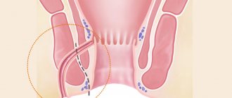
Treatment of complex fistulas should be divided into 2 stages
At the first stage, a drainage ligature is installed into the fistula tract - a thin, almost invisible suture material (Fig. 7). The main goals of this stage of treatment are: the formation of a direct fistula tract around the ligature, drainage of possible leaks, reduction of the inflammatory process, prevention of self-closure of the external opening for constant drainage of inflammatory and/or purulent discharge.
Figure 7. Drainage ligature
At the second stage, after the acute inflammation subsides (4-6 weeks), the main surgical treatment is performed - excision of the fistula.
Important! Two-stage surgical treatment provides conditions under which radical surgery becomes possible with minimal complications.
Currently, in the treatment of rectal fistulas, various biological materials based on connective tissue (Permacol), fibrin glue, and collagen paste are widely used, which are introduced into the fistula tract, tamponing it. These methods have a lesser effect on the rectal closure apparatus; their use does not lead to the formation of a massive wound defect. However, the rate of disease recurrence is significantly higher than radical surgery.
Promising modern methods for treating fistulas are endoscopic (VAAFT) and laser (FiLaC) technologies for removal of affected tissue. These technologies practically eliminate the possibility of damage to the sphincter apparatus, however, today, the effectiveness in leading clinics in the world does not exceed 50-60%.