general characteristics
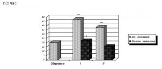
The pancreas is the only source of trypsin, so determination of its content may be more important for judging pancreatic damage than other enzymes. All pancreatic proteases, especially trypsin and chymotrypsin, bind very quickly to serum immediate and delayed inhibitors. The most accurate method for determining the total fraction of trypsin and trypsinogen in the blood is radioimmunological, based on the principle of a competitive protein-binding assay with double antibodies. The advantages of radioimmunoassay include: high sensitivity, specificity, reliability, accuracy. The level of trypsinemia depends on the form and stage of the disease. Acute attacks of pancreatitis are characterized by an increase in trypsin content, which is observed more often than an increase in the activity of amylase and lipase. The progressive, relapse-free course of chronic pancreatitis or long-term pancreatitis is accompanied by a gradual decrease in serum trypsin levels. An increase in trypsin concentration in the blood serum of children in the first few weeks after birth indicates the presence of cystic fibrosis, and therefore the determination of this indicator is considered an effective screening method. As the disease progresses and true pancreatic insufficiency develops, the concentration of trypsin in the blood serum decreases.
Micromethod for determining trypsin activity in biological fluids
Alekhin S.A. Lipatov V.A.
Kursk State Medical University, department. of surgical diseases No. 2, department. operative surgery and topographic anatomy
www.drli.h1.ru
Determination of trypsin activity is an important criterion in the diagnosis of acute pancreatitis. Until now, the main method for determining trypsin activity was the Erlanger method modified by Shaternikov. The disadvantages of this method are its high cost, the error associated with the nonlinearity of the dependence of the change in the optical density of a standard p-nitroaniline solution on its concentration used to construct a calibration graph, and the complexity of calculations.
The goal of our work was to eliminate the above-mentioned shortcomings of the existing methodology.
We have developed a micromethod for determining trypsin activity in biological fluids.
The method is based on trypsin cleavage of a synthetic colorless substrate - benzoylarginine-p-nitroanilide - with the formation of colored p-nitroaniline, the amount of which we determine on a BACKMAN-DU65 spectrophotometer with microcuvettes at a wavelength of 410 nm.
The reagents used in our method are the same as in the Erlanger method as modified by Shaternik.
Calibration is carried out according to the described method; the difference is the construction of a graph, which is carried out to automate the process and maintain a database in Microsoft Excel.
Since the calibration graph cannot have a clear linear relationship, to express the dependence of the optical density of the solution on the concentration of p-nitroaniline, it is necessary to determine the equation that describes this graph. Such an equation is a polynomial fitting equation, which can also be plotted on a calibration graph in Microsoft Excel.
Determination progress:
0.4 ml of benzoylarginine-p-nitroanilide is added to the test and control tubes and 0.025 ml of physiological solution is added. Then 0.025 ml of blood plasma is added, immediately after this 0.05 ml of 0.5 N HCl solution is added to the control tube. 0.5 ml of the contents is placed in a microcuvette and the optical density value is measured at a wavelength of 410 nm and this value is designated as EK(410). Since in some cases the solution becomes turbid during the enzymatic reaction, to apply a correction for turbidity, the optical density of the liquid must also be determined at a wavelength of 550 nm. The resulting data is labeled EK(550). Next, the test tube is placed in a thermostat at a temperature of 370C for 30 minutes. After thermostatting, the optical density of the solution is determined at wavelengths of 410 and 550 nm in a test tube and the data obtained are marked as ЕОп(410) and ЕОп(550).
Calculation of trypsin activity is carried out in three stages
1. Calculation of the difference in optical densities at different wavelengths, it will be equal to:
DE = (EOp(410) – EK(410)) – (EOp(550)- EK(550))
2. Calculation of the amount of p-nitroaniline formed using a polynomial equation, which in general will look like this:
y = b + c1*x + c2*x2 +……. +c6*x6 , where b and c are constants
An example of a polynomial equation for calculating the concentration of p-nitroaniline: y = 874.45*x1 + 84.94*x2
In this case, the constant b is equal to 0 and is therefore omitted. x is DE, and y is the amount of p-nitroaniline formed.
Each laboratory must perform a calibration and calculate a polynomial equation for the calibration data.
3. Direct calculation of trypsin activity.
Since this is a micro-variant of the method for determining trypsin, the amounts of reagents and blood plasma are proportionally reduced in relation to the classical method, and the conversion factors will be the same as in the classical method.
The amount of p-nitroaniline calculated using a polynomial equation shows how much of the substance was formed during the enzymatic reaction in 0.5 ml of substrate for 30 minutes when adding 0.025 plasma, and the calculated amount of trypsin in units of activity shows how much of the substance is formed in 0.5 ml of substrate when adding 0.1 ml of plasma in 1 minute.
Therefore, A - the amount of trypsin in units of activity will be equal to: A = (y * 4) / 30.
Thus, calculating trypsin activity using this method significantly reduces the consumption of reagents (10 times compared to the Erlanger method as modified by Shaternikov); the economic efficiency of the first of them is obvious. Due to the absence of fundamental differences between the two methods, the results of measuring trypsin activity will be similar (the difference between the results will be within the error range of the method). It is also important to reduce the amount of blood taken for testing. So, according to our proposed plasma method, only 0.05 ml is needed. and according to the classical method 0.5 ml. The use of automatic micropipettes allows you to reduce the error associated with working with microquantities of reagents and biological materials. Another positive aspect of our modification is the simplification of calculations and construction of a calibration graph and their automation.
Using the polynomial approximation equation makes it possible to reduce the error of the technique associated with the nonlinearity of the dependence of the change in the optical density of a p-nitroaniline solution on its concentration. In general, the calculation method is easier to understand than the classical one, and the use of Microsoft Excel allows not only to fully automate the calculation, but also to maintain a database.
Alekhin S.A., Lipatov V.A. Micromethod for determining trypsin activity in biological fluids. // Materials of the 66th scientific conference of students and young scientists “Current problems of medicine and pharmacy”, Kursk, 2001, -p. 57 - 59.
Nosological classification (ICD-10)
- H11.3 Conjunctival hemorrhage
- H20 Iridocyclitis
- H20.9 Iridocyclitis, unspecified
- H59 Lesions of the eye and its adnexa after medical procedures
- H60.9 Otitis externa, unspecified
- H66.0 Acute suppurative otitis media
- I80 Phlebitis and thrombophlebitis
- J01.9 Acute sinusitis, unspecified
- J32.0 Chronic maxillary sinusitis
- J86 Pyothorax
- J90 Pleural effusion, not elsewhere classified
- K05.4 Periodontal disease
- L89 Decubital ulcer
- M86.9 Osteomyelitis, unspecified
- S05.9 Injury to unspecified part of eye and orbit
- T14.1 Open wound of unspecified body area
- T30 Thermal and chemical burns of unspecified location
Links[edit]
- PDB: 1UTN; Leiros HK, Brandsdal BO, Andersen OA, Os V, Leiros I, Helland R, et al (April 2004). "Trypsin specificity revealed by LIE calculations, X-ray structures, and association constant measurements". Protein Science
.
13
(4): 1056–70. DOI: 10.1110/ps.03498604. PMC 2280040. PMID 15044735. - Jump up
↑ Rawlings ND, Barrett AJ (1994).
"Families of serine peptidases". Methods in enzymology
.
244
: 19–61. DOI: 10.1016/0076-6879 (94) 44004-2. ISBN 978-0-12-182145-6. PMC 7133253. PMID 7845208. - ↑
German physiologist Wilhelm Kühne (1837-1900) discovered trypsin in 1876.
See: Kühne W (1877). "Uber das trypsin (pancreas enzyme)". Verhandlungen des Naturhistorisch-medicinischen Vereins zu Heidelberg
.
New episode. 1
(3): 194–198 - via Google Books. - Engelking LR (2015-01-01). "Chapter 7 - Protein Digestion." Textbook of veterinary physiological chemistry
(Third ed.). Boston: Academic Press. pp. 39–44. DOI: 10.1016/B978-0-12-391909-0.50007-4. ISBN 978-0-12-391909-0. - ↑
Kühne W (6 March 1876).
"Ueber das Trypsin (Enzym des Pankreas)" [On trypsin (pancreatic enzyme)]. In Naturhistorisch-medizinischen Verein (ed.). Verhandlungen des Naturhistorisch-medizinischen Vereins zu Heidelberg
[
Negotiations of the Natural History Medical Association in Heidelberg
] (in German). Heidelberg, Germany: Universitätsbuchhandlung by Karl Winter (published 1877). pp. 194–8 - via Archive.org. - "Protein Digestion". Elective course (Clinical biochemistry)
. Ternopil National Medical University. July 14, 2015. Retrieved April 11, 2022. - Polgár L (October 2005). "Catalytic triad of serine peptidases". Cellular and Molecular Life Sciences
.
62
(19–20): 2161–72. DOI: 10.1007/s00018-005-5160's. PMID 16003488. S2CID 3343824. - Voet D, Voet JG (2011). Biochemistry
(4th ed.). Hoboken, NJ: John Wiley and Sons. ISBN 9780470570951. OCLC 690489261. - "Classification of modified trypsin" (PDF). www.promega.com. 2007-04-01. Retrieved February 8, 2009.
- Jump up
↑ Rodriguez J, Gupta N, Smith RD, Pevzner PA (January 2008).
“Does trypsin cut before proline?” (PDF). Journal of Proteomic Research
.
7
(1): 300–5. CiteSeerX 10.1.1.163.4052. DOI: 10.1021/pr0705035. PMID 18067249. - Chelulei Cheison S, Brand J, Leiba E, Kulozik U (March 2011). "Analysis of the effect of temperature changes combined with different alkaline pH on the hydrolysis pattern of β-lactoglobulin and trypsin using MALDI-TOF-MS/MS." Journal of Agricultural and Food Chemistry
.
59
(5):1572–81. DOI: 10.1021/jf1039876. PMID 21319805. - Gudmundsdóttir A, Pálsdóttir HM (2005). "Atlantic cod trypsins: from basic research to practical applications." Marine Biotechnology
.
7
(2): 77–88. DOI: 10.1007/s10126-004-0061-9. PMID 15759084. S2CID 42480996. - Noone PG, Zhou Z, Silverman LM, Jowell PS, Knowles MR, Cohn JA (December 2001). "Cystic fibrosis gene mutations and risk of pancreatitis: association with epithelial ion transport and trypsin inhibitor gene mutations." Gastroenterology
.
121
(6):1310–9. DOI: 10.1053/gast.2001.29673. PMID 11729110. - "Trypsin-EDTA (0.25%)". Stem cell technologies. Retrieved February 23, 2012.
- Debrisol. drugs.com.
- "Protease - GMO Database". GMO Compass
. European Union. 2010-07-10. Archived from the original on 2015-02-24. Retrieved January 1, 2012. - Voet D, Voet JG (1995). Biochemistry (2nd ed.). John Wiley and Sons. pp. 396–400. ISBN 978-0-471-58651-7.
- Levilliers N, Perón M, Arrio V, Pudles J (October 1970). “On the mechanism of action of proteolytic inhibitors. IV. Effect of 8 M urea on trypsin stability in trypsin-inhibitor complexes.” Archives of Biochemistry and Biophysics
.
140
(2):474–83. DOI: 10.1016/0003-9861 (70) 90091-3. PMID 5528741.
Mechanism[edit]
The enzymatic mechanism is similar to that of other serine proteases. These enzymes contain a catalytic triad consisting of histidine-57, aspartate-102 and serine-195. [7] This catalytic triad was previously called the charge relay system, implying the abstraction of protons from serine to histidine and from histidine to aspartate, but due to data , provided by NMR, the resulting alkoxide form of serine will have a much stronger proton impact than the imidazole ring of histidine, according to modern thinking, instead serine and histidine have virtually equal proton shares, forming short, low-barrier hydrogen bonds with them. [8] [ pg.
] Thus, the nucleophilicity of serine in the active site is increased, which facilitates its attack on the amide carbon during proteolysis. The enzymatic reaction catalyzed by trypsin is thermodynamically favorable, but requires significant activation energy (kinetically unfavorable). In addition, trypsin contains an "oxyanion hole" formed by the main chain amide hydrogen atoms Gly-193 and Ser-195, which through hydrogen bonding stabilize the negative charge that accumulates on the amide oxygen after nucleophilic attack on the planar surface. the amide carbon by serine oxygen causes that carbon to adopt a tetrahedral geometry. This stabilization of this tetrahedral intermediate helps to lower the energy barrier to its formation and is accompanied by a decrease in the free energy of the transition state. Preferential binding of a transition state is a key feature of enzyme chemistry.
The negative aspartate residue (Asp 189), located in the catalytic pocket (S1) of trypsin, is responsible for attracting and stabilizing positively charged lysine and/or arginine and is thus responsible for the specificity of the enzyme. This means that trypsin preferentially cleaves proteins on the carboxyl side (or "C-terminal side") of the amino acids lysine and arginine, except when either bound to the C-terminal proline, [9] although large-scale mass spectrometry data indicate cleavage occurs even with proline. [10] Trypsin is considered an endopeptidase, i.e. cleavage occurs within the polypeptide chain and not at the terminal amino acids located at the ends of the polypeptides.
Directions for use and doses
V/m. Adults - 5-10 mg 1-2 times a day; children - 2.5 mg 1 time per day. For injection, immediately before use, dilute 5 mg of crystalline trypsin in 1–2 ml of 0.9% NaCl solution or 0.5–2% procaine solution. The course of treatment is 6–15 injections. Electrophoresis with trypsin is also used: for 1 procedure, 10 mg of trypsin (dissolved in 15–20 ml of distilled water), administered from the negative pole.
Inhalation: 5–10 mg in 2–3 ml of 0.9% NaCl solution through an inhaler or through a bronchoscope. After inhalation, rinse your mouth with warm water and rinse your nose.
Eye drops: conjunctivally, 0.2–0.25% solution, which is prepared immediately before use.
Intrapleural: 1 time per day, 10–20 mg, dissolved in 20–50 ml of 0.9% NaCl solution.
Intrapleural: 50–150 mg in 5–30 ml of phosphate buffer solution; after administration, frequent changes in body position are desirable; On the 2nd day after instillation, a liquefied exudate is usually released.
Local: A three-layer cloth made of dialdehyde cellulose, soaked in a trypsin solution, is applied to the wound (after treatment) and secured with a bandage, left on the wound for no more than 24 hours. Before use, the cloth is moistened with distilled water or a furatsilin solution. The time for complete cleansing of the wound from necrotic tissue and pus is 24–72 hours. If necessary, reapply.
Isoenzymes [edit]
These human genes encode proteins with trypsin enzymatic activity:
| Alto. symbols | TRY1 |
| NCBI gene | 5644 |
| H.G.N.C. | 9475 |
| OMIM | 276000 |
| RefSeq | NM_002769 |
| UniProt | P07477 |
| Other data | |
| EU number | 3.4.21.4 |
| Locus | Chr. 7 q32-qter |
| Alto. symbols | TRYP2 |
| NCBI gene | 5645 |
| H.G.N.C. | 9483 |
| OMIM | 601564 |
| RefSeq | NM_002770 |
| UniProt | P07478 |
| Other data | |
| EU number | 3.4.21.4 |
| Locus | Chr. 7q35 _ |
| Alto. symbols | PRSS4 |
| NCBI gene | 5646 |
| H.G.N.C. | 9486 |
| OMIM | 613578 |
| RefSeq | NM_002771 |
| UniProt | P35030 |
| Other data | |
| EU number | 3.4.21.4 |
| Locus | Chr. 9p13 _ |
Other trypsin isoforms may also be found in other organisms.


