General information
Hypokalemia means a decrease in serum potassium content to below 3.5 mmol/l, the deficiency of which can be caused by both a decrease in its reserves in the body and pathological movement into cells.
Most of the potassium (120–160 mmol/L) is normally found in the intracellular space (about 90%), and the remainder is found in the blood/bone tissue. The normal level of potassium in the body is its serum concentration from 3.5 to 5.5 mmol/l. The concentration of potassium in the blood is relatively constant, which is regulated by the processes of its entry into cells/excretion by the kidneys. The process of moving potassium from the blood through cell membranes is determined by the concentration of potassium outside the cell, the state of the membranes and cellular metabolism. The binding of potassium inside the cell occurs during the formation of protein and glycogen: it has been established that for 1 g of glycogen, 1 g of protein nitrogen and 0.3 mmol of potassium ions enter the cells. Potassium enters the body with food in an average amount of 2-3 g/day and is absorbed in the gastrointestinal tract (small intestine); excreted in urine (2-5 g), to a lesser extent in feces (less than 10 mmol). The volume/rate of potassium excretion is determined by many factors: potassium concentration in the blood, osmolarity, acid-base balance, the influence of vasopressin / aldosterone , and the rate of cell turnover.
The daily potassium requirement of an adult varies between 40–100 mmol/l. Vitamin B6 increases the efficiency of potassium absorption , while alcohol negatively affects its balance in the body. It should be taken into account that potassium from food is poorly absorbed in case of magnesium deficiency. Therefore, the lack of potassium and magnesium in the body is often interrelated. Normally, potassium balance is constantly maintained by the process of its excretion and intake with food. At the same time, the potassium content in the body is inversely proportional to the sodium content, that is, the more potassium, the less sodium, and vice versa.
Potassium takes an active part in the transmission of nerve impulses, the formation of cellular potentials, the contraction of smooth/skeletal muscle fibers, and cardiomyocytes. The macroelement increases stress resistance/physical endurance, coordinates heart rate, is responsible for normalizing blood pressure, and helps eliminate allergens/toxins from the body. Lack of potassium in the body in women and lack of potassium in men is a dangerous condition, since its functions in the body are extremely wide: potassium maintains the bioelectrical activity of cells, acid-base balance/water-salt balance; Helps stabilize heart rate, blood pressure, and maintain pH levels.
There are no reliable statistics on the prevalence of hypokalemia in the general population. It is important to understand that both manifest and especially subclinical hypokalemia are very common in clinical practice, but often remain unrecognized. However, potassium deficiency is detected in 3-20% of patients in gastroenterology/cardiology departments. The reason for the difficulty in recognizing hypokalemia is the variability/dynamism and variety of its clinical manifestations, and therefore hypokalemia imitates various organ/systemic pathologies.
Associated disorders
Symptoms of the following disorders include hypokalemia. Comparisons can be useful for differential diagnosis:
- Bartter's syndrome is a metabolic disorder affecting the kidneys. Main symptoms include slow growth, weakness, excessive thirst and excessive urination. Bartter's syndrome is characterized by excessive loss of potassium through the kidneys.
- Hypokalemic periodic paralysis (HPP) is a disorder characterized by episodes of paralysis with loss of deep tendon reflexes and inability of muscles to respond to electrical stimulation. The cause of HPP is unknown. The paralysis may be limited to certain muscle groups or may affect all four limbs. Attacks usually last from 24 to 48 hours. Potassium levels are usually abnormally low (ie, hypokalemia).
- Metabolic alkalosis is a disorder characterized by elevated levels of bicarbonate in the blood. Symptoms include irritability, neuromuscular hyperexcitability, low blood potassium levels (hypokalemia), muscle weakness, gastrointestinal dysmotility, and excessive urination.
To find other disorders that include hypokalemia as a symptom, enter “Hypokalemia” as a search term in the disease database.
Pathogenesis
The pathogenesis of hypokalemia is diverse and determined by its form. The kidneys play a major role in maintaining potassium homeostasis. The level of potassium is determined by a combination of several processes, namely: potassium filtration, secretion and reabsorption. Since potassium in the blood plasma is in a free state, it is completely filtered in the renal glomeruli; potassium ions passing through the filter are almost completely reabsorbed in the distal/proximal tubules.
The process of potassium excretion by the kidneys is under humoral control, the main role in which belongs to mineralocorticoids ( aldosterone ), which both increases the penetration of potassium ions into the cells of the distal tubule and increases the permeability of the cell membrane to potassium, promoting potassium secretion. insulin also takes part in the processes of regulation of potassium excretion by the kidneys , which reduces the excretion of potassium by the kidneys. The state of acid-base balance has a significant influence on kaliuria. Thus, with alkalosis , hypokalemia develops as a result of the transition of potassium found in the blood plasma into cells. Potassium deficiency is observed with increased/accelerated breakdown of proteins in the body, which helps to force the release of potassium.
Causes and risk factors
Hypokalemia always results from excessive loss of potassium in urine, sweat, or stool. The condition is always a symptom of another disorder, rather than a disease that occurs on its own.
Excess urinary potassium excretion (kaliuresis) may result from the use of diuretics (which increases urination), magnesium deficiency in the blood, excess mineralocorticoids such as aldosterone in the blood that affect the electrolyte and fluid balance of the body (usually caused by endocrine diseases), kidney problems, or the use of high doses of penicillin.
Loss of potassium through the gastrointestinal tract is usually caused by prolonged diarrhea or vomiting, chronic laxative abuse, insufficient dietary potassium intake, intestinal obstruction, or infections such as intestinal fistulas that continuously drain intestinal fluid from the body. Additionally, excessive sweating due to hot weather or exercise can cause hypokalemia.
Causes
The causes of hypokalemia are quite varied, the main ones of which include:
- Insufficient intake of potassium from food (poor nutrition/strict diets), monotonous diet.
- Excessive consumption of sugar, coffee-containing drinks, alcohol.
- Excessive intake of sodium, cesium, thallium and rubidium into the body.
- Potassium loss due to various diseases of the gastrointestinal tract ( ulcerative colitis / pancreatitis ), various intestinal infections that manifest themselves with repeated diarrhea / vomiting .
- Primary/secondary hyperaldosteronism . Under the influence of aldosterone, the process of excretion of potassium ions by the kidneys is stimulated.
- Endocrine diseases ( thyrotoxicosis Itsenko-Cushing disease/syndrome , genetically determined dysfunction of the adrenal cortex).
- Kidney diseases ( interstitial nephritis / renal tubular acidosis ), occurring with tubular dysfunction, which leads to increased potassium excretion.
- Redistribution (changes in the ratio) of potassium between the interstitium and the cell - the transition from the extracellular space of K+ into the cells. Occurs with alkalosis (pH shift to the alkaline side).
- Neuropsychic overload, excessive stress factors.
- Use of high doses of glucocorticoids.
- Taking certain medications. The table below shows the main drugs that induce hypokalemia and the mechanisms of its development.
- Other causes are large area burns hypomagnesemia , Barter-Gittelman syndrome .
Signs and symptoms
Most often, hypokalemia is asymptomatic, without obvious signs of the disorder. However, symptoms of hypokalemia may include bouts of severe muscle weakness, eventually leading to paralysis and possibly respiratory failure.
Muscle failure can lead to intestinal paralysis, low blood pressure, painful muscle twitching (tetany), and mineral deficiencies. Severe hypokalemia can also lead to the destruction of skeletal muscle cells, especially during exercise. The normal physical response to exercise requires local release of potassium from the muscles. In potassium-depleted muscles, the lack of potassium prevents adequate dilation of blood vessels, resulting in decreased muscle blood flow, cramping, and skeletal muscle breakdown.
Hypokalemia can also impair the kidneys' ability to concentrate urine, leading to excessive urination (polyuria) and excessive thirst (polydipsia). Other symptoms may include loss of appetite, nausea and vomiting. There may also be disturbances in the functioning of the heart caused by changes in the electrocardiograph, confusion, bloating, and decreased mental activity.
Symptoms
Symptoms of hypokalemia depend on the level of potassium in the body, however, it should be borne in mind that plasma potassium concentrations do not accurately reflect the state of potassium balance. Symptoms and complaints of patients accompanying a decrease in potassium concentration in the body are nonspecific and quite varied, so in most cases we are not talking about the clinical picture, but about various clinical masks of hypokalemia.
The main signs of potassium deficiency are observed when its total amount decreases in the range of 10-30%; a decrease in potassium content below 1.5 mmol/l causes paralysis of the respiratory muscles. Symptoms of potassium deficiency in the body in women and potassium deficiency in the body in men manifest themselves with the same symptoms.
The first signs of potassium deficiency are associated with impaired neuromuscular excitability, which is caused by impaired polarization/depolarization of cell membranes and is manifested by asthenia / muscle weakness , increased fatigue , paresthesia /muscle spasms, and decreased tendon reflexes. With chronic hypokalemia, functional disorders and structural damage to both the peripheral and central nervous systems appear, which is realized by psycho-emotional disorders in the form of hypochondriacal, asthenic or anxiety-depressive syndromes.
Sensory disturbances manifest as mild paresthesia of the limbs/face or loss of tactile/pain sensitivity or severe hyperesthesia . Movement disorders correlate with the severity/duration of hypokalemia, ranging from mild muscle weakness of the upper and lower extremities, decreased tendon reflexes, to paralysis, including respiratory muscles. Hypokalemia can also manifest itself in symptoms/changes in the digestive system (intestinal paresis, flatulence , vomiting, paralytic intestinal obstruction constipation ), as well as in the genitourinary system ( polyuria , atony of the bladder , necrosis in the kidneys).
One of the most common manifestations of hypokalemia are disorders of cardiovascular activity, which are characterized by dilation of the cavities of the heart, decreased contractile function of the myocardium, the presence of systolic murmur at the apex of the heart, and decreased blood pressure .
The damaging effect of potassium ion deficiency in the body is well reflected in the ECG, which allows it to be used as a kind of indicator of latent hypokalemia. Characteristic ECG signs of hypokalemia include ventricular extrasystoles , T wave depression/inversion, QRS prolongation, pronounced U wave, and ST segment depression (Figure below).
As a rule, with potassium deficiency, there is a decrease in mental/mental activity, manifested in lethargy, apathy, and with a significant deficiency, the development of a coma is possible.
Standard Treatments
The underlying cause of hypokalemia must first be addressed. For severe hypokalemia, potassium chloride can be administered orally or intravenously. Treatment should be carefully monitored by a physician. Any associated acid-base disorders or hormonal disorders should be assessed before treatment is planned. Administration of potassium and potassium-sparing diuretics is generally not recommended in patients with kidney disease, diabetes mellitus, or autonomic nervous system dysfunction. An imbalance of external and internal potassium levels in these individuals may predispose them to life-threatening degrees of hyperkalemia (i.e., excessively elevated levels of potassium in the body).
Hypokalemia in individuals with high blood pressure taking diuretics may be improved by replacing lost potassium in the diet through certain fruits or potassium supplements. Hypokalemia can also be minimized by dietary salt restriction, as high sodium excretion rates promote urinary potassium loss. People who participate in vigorous sports or exercise in warm weather should be sure to replace the potassium that is lost through excessive sweating. This can be achieved through diet planning.
Tests and diagnostics
The diagnosis is based on laboratory and instrumental methods of examining the patient:
- Laboratory research. The blood CBS and concentrations of potassium, magnesium, and sodium are determined. A biochemical blood test determines the concentration of urea and creatinine; in urine analysis - the presence of chlorine/relative density.
- Hormone tests. The level of aldosterone/renin is determined, and the renin-aldosterone ratio is calculated. According to indications, tests are carried out for 17-OH-progesterone, cortisol , thyroid-stimulating hormone .
- Electrocardiography. The ECG looks for characteristic ECG signs of hypokalemia (QT interval prolongation, ST segment depression, presence of a U wave).
- Instrumental research. Ultrasound/CT of the adrenal glands, ultrasound of the kidneys with Dopplerography. According to indications - echocardiography.
- In addition to determining the potassium content, diagnosing the causes of hypokalemia is also important, the algorithm of which is shown in the figure below.
Diet
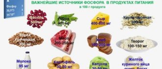
There is no special diet, but adjusting the diet to include foods rich in potassium is a critical component of correcting hypokalemia. For this purpose, it is recommended to include in the diet dairy products (milk, cottage cheese and sour cream), legumes, oatmeal/buckwheat, mushrooms, potatoes, bread with bran, tomato, sea fish (mackerel, pollock, salmon, tuna, sprat), cabbage (cauliflower/white cabbage/broccoli), carrots, cucumbers, greens (parsley, spinach), seaweed, fresh fruits (peaches, bananas, kiwi, avocados, apricots), nuts, dried fruits (prunes, dried apricots, raisins), compotes, black tea, cocoa, tomato juice, freshly squeezed juices (grape, orange, carrot), chocolate.
Unnatural sugar-containing drinks (lemonade, Coca-Cola, Fanta, Pepsi) should be limited/completely excluded from the diet. The consumption of salt is also subject to restrictions, but at the same time, a salt-free diet is not allowed. It is important to consider that most of the potassium dissolves in water during heat treatment, so you need to boil foods in a minimum amount of water.
Prevention
Preventive measures boil down to:
- A balanced diet with a sufficient amount of potassium-containing products without violating the rules of heat treatment.
- Eliminating low-calorie diets and giving up bad habits.
- Sleep/wake optimization.
- Limiting mental and nervous overload.
- Replacing (if possible) diuretics and other medications that help remove potassium from the body. If it is impossible to replace them, it is necessary to regularly undergo laboratory testing to determine the concentration of potassium in the serum.
- Limiting table salt consumption.
- Periodic use for the preventive correction of hypokalemia Panangin (1 tablet 3 times a day) for 3 weeks.
For specialists
Kozlovsky A.A., Gomel State Medical University
Minerals are necessary to ensure normal functioning of the body. They maintain a constant internal environment, acid-base balance, and water-salt metabolism. Almost all chemical elements of the periodic table D.I. Mendeleev are involved in physiological and pathological processes in humans. The human body consists of 60% water, 34% organic substances and 6% inorganic substances. The organic elements from which our organic component is formed are carbon, hydrogen, oxygen, nitrogen, phosphorus, sulfur. The inorganic component necessarily contains 22 chemical elements: Ca, P, O, Na, Mg, S, B, Cl, K, V, Mn, Fe, Co, Ni, Cu, Zn, Mo, Cr, Si, I, F ,Se; 12 components in the human body are structural, since they form 99% of its elemental composition: C, O, H, N, Ca, Mg, Na, K, S, P, F, Cl. All presented chemical elements can be divided into vital (essential), conditionally necessary and elements whose role has not yet been sufficiently studied (Fig. 1). Essential chemical elements are elements, the absence or deficiency of which disrupts the normal functioning of the body [15]. Scientists have long noticed that many diseases are associated with a deficiency of one or another chemical element in the body (hypoelementosis). Minerals and metals were used for medicinal purposes in Ancient China, India, and Mesopotamia. In the British Pharmacopoeia of the mid-19th century. thousands of medicines containing chemical elements are presented [30]. Hypoelementosis can cause not only temporary disturbances in the body, but also contribute to the development of serious diseases [13]. The development diagram of elementoses is shown in Fig. 2. In recent years, there has been increasing interest in studying the biological role of macro- and microelements.
It is no coincidence that special attention is paid to potassium and magnesium as one of the most common in the human body. Potassium and magnesium in ionized form are positive ions - cations, respectively, with one (K+) and double positive charges (Mg2+). These are some of the most common elements on Earth. There is especially a lot of potassium and magnesium in the water of the World Ocean, the electrolyte composition of which is close to the electrolyte composition of blood serum [14]. Potassium is the main intracellular cation of tissues of various organs, approximately 98% of it is concentrated inside cells. Under normal conditions, its content in the cell is 150–160 mmol/l, and in the blood serum – 3.7–5.5 mmol/l. Most of the body's potassium is intracellular, so significant loss of intracellular potassium is possible without large changes in serum potassium. The sodium-potassium pump, located in the cell membrane, is the main mechanism for maintaining the balance between intra- and extracellular potassium. Potassium ion is a very important component in maintaining homeostasis, especially in critical conditions. Potassium plays an essential role in the implementation of bioelectrical activity of cells and the maintenance of neuromuscular excitability and conductivity. Under normal conditions, potassium is supplied through the diet and absorbed through the gastrointestinal (GI) tract, followed by excretion of excess through the kidneys. Vitamin B6 contributes to the effective absorption of potassium, and alcohol, on the contrary, has a negative effect on the balance of this element [1]. A child’s daily need for potassium depends on age: 1–3 years – 400 mg, 3–7 years – 600 mg, 7–11 years – 900 mg, 11–14 years – 1500 mg, over 14 years – 2500 mg. High potassium content is observed in dried apricots, apricots, beans, prunes, peas, nuts, sorrel, potatoes, spinach, parsley, black currants, apricots and ranges from 1780 to 300 mg% (products are listed in descending order of potassium content). Hypokalemia is a persistent decrease in serum potassium concentration (less than 3.5 mmol/l). The body of an adult weighing 70 kg contains 136.85 g or 3500 mmol of potassium. Reasons for the development of hypokalemia: 1. Insufficient (less than 10 mEq/day) intake of potassium into the body from food (during fasting or limiting the intake of foods containing potassium). 2. Excessive excretion of potassium from the body as a result of: – chronic profuse diarrhea (intestinal secretions contain a large amount of potassium); – repeated vomiting (the potassium content in gastric juice is low, but the development of hypovolemia causes secondary hyperaldosteronism and an increase in the excretion of K+ ions by the kidneys); – increased excretion of potassium by the kidneys due to improper use of diuretics, primary and secondary hyperaldosteronism, renal tubular defects (Bartter’s syndrome, renal tubular acidosis), damage to renal tissue by nephrotoxic substances, including drugs (penicillins, gentamicin, amphotericin B), hypomagnesemia (promotes not only the release of potassium from cells, but also increases its excretion in the urine). 3. Redistribution of potassium ions from the blood and/or intercellular fluid into cells under conditions of: – an increase in the level of insulin in the blood; – hypercatecholaminemia (as a result of the use of drugs adrenaline, norepinephrine, dopamine, with pheochromocytoma, acute stress); – overdose of folic acid or vitamin B (these substances stimulate cell proliferation and their consumption of potassium ions). The symptoms of disturbances in potassium homeostasis depend on its content in the body (at the same time, potassium concentrations in plasma do not accurately reflect the state of potassium balance, although they have a fairly narrow range of fluctuations). The main manifestations of hypokalemia are associated with a violation of the electrical properties of the membranes of excitable tissues. Complaints and symptoms accompanying a decrease in potassium levels in the body are varied and nonspecific, which allows us to speak not about the clinical picture, but about numerous clinical masks of hypokalemia. The most common of them are neuromyopathic and psychoemotional disorders, cardiac syndrome, polyuria-polydipsia syndrome (Fig. 3). Potassium imbalance leads to disruption of polarization and depolarization of cell membranes, disruption of folinesterase function. The main result of these changes is a disorder in the process of transferring excitation from nerve to muscle, which is clinically expressed by fatigue, muscle weakness, spasms of the leg muscles, paresthesia in the extremities, and extinction of tendon reflexes [23]. Nonspecific symptoms of hypokalemia are loss of appetite, loss of concentration, apathy. Hypokalemia is also manifested by disorders of cardiovascular activity, characterized by inhibition of myocardial contractile function, the occurrence of systolic murmur at the apex of the heart and expansion of its cavities, and a decrease in blood pressure [22]. In relation to gastroenterology and urology, this means that damage to smooth muscles leads to intestinal paresis, weakening of bowel sounds, vomiting, flatulence, constipation, and bladder atony [18]. The cardiodamaging effect of potassium ion deficiency in the body is early reflected in the ECG, and therefore it can be used as an indicator of latent hypokalemia. Constant, although nonspecific, ECG signs are frequent ventricular extrasystoles, QRS prolongation, decreased ST segment, depression or inversion of the T wave, pronounced U wave. Patients taking cardiac glycosides are especially sensitive to hypokalemia [3, 17].
Chronic hypokalemia is accompanied by functional and structural damage to the central and peripheral nervous system. Dysfunction of the central nervous system is realized by psycho-emotional disorders in the form of shallow asthenic, anxiety-depressive or hypochondriacal syndromes. Polymorphic sensory disorders are represented by mild paresthesia of the face and limbs or loss of pain and tactile sensitivity or, on the contrary, severe hyperesthesia. Neuromotor symptoms usually correlate with the depth and duration of hypokalemia, ranging from weakness of limb muscles and low tendon reflexes to general paralysis, including respiratory muscles [6]. Neurovegetative disorders are predominantly sympathoadrenal in color and can be represented by tremor, hyperhidrosis, and spontaneous erythroderma. Diagnostic tests to determine hypokalemia: – serum potassium concentration less than 3.5 mmol/l – indicative test; – potassium content in urine: values of more than 20 mmol/l indicate potassium loss by the kidneys, and less than 20 mmol/l indicate extrarenal causes of potassium deficiency in plasma; – the transtubular gradient of potassium concentration, defined as the product of the ratio of urine potassium to serum potassium and the ratio of serum osmolality to urine osmolality, reveals the cause of increased potassium content in the urine; – arterial blood gases (metabolic alkalosis is more often associated with hypokalemia due to nasogastric intubation, diuretic therapy, hyperaldosteronism, and hyperkalemia also accompanies metabolic acidosis with diarrhea, renal tubular acidosis); – electrocardiography (characterized by a decrease in the ST segment, flattening of the T wave, the presence of a U wave, ventricular arrhythmias). It is not advisable to assess changes in potassium levels in the blood separately from magnesium levels, since magnesium is an important cofactor in both the absorption of potassium and ensuring its optimal intracellular level. In 1992, it was proven that simultaneous deficiency of potassium and magnesium can lead to hypokalemia, resistant to treatment if magnesium deficiency is not corrected [10]. Magnesium is not synthesized in the human body, but comes in the form of Mg2+ ion with food, water and salt. In nature, it ranks 8th in prevalence; Sea water is saturated with magnesium; it is an integral part of more than 200 minerals and is part of chlorophyll. The daily requirement for magnesium is met by 40% from foods, and 60% from ionized magnesium water. The bioavailability of magnesium from drinking water is significantly higher than from solid food [11]. Because many geographic regions have low levels of magnesium in drinking water and soil, the concentration of the element in plant foods depends on where the plants are grown. In industrialized countries, agricultural and food production technologies and lifestyle changes contribute to the loss of magnesium and an increase in the population with magnesium deficiency [14]. High magnesium content (from 540 to 102 mg%) is noted in sesame, wheat bran, sunflower seeds, nuts, soybeans, buckwheat, oatmeal, peas, dried apricots, dried prunes (products are listed in descending order of magnesium content). According to the Institute of Nutrition of the Russian Academy of Medical Sciences, the magnesium requirement of an adult is 300–400 mg/day. At a young age, as well as in people engaged in physical labor, athletes have a higher need for magnesium. Recommended average daily intakes of magnesium are given in table. 1. Ionized magnesium supplied with water and food enters the stomach, where some of the magnesium ions are split off from the magnesium salts of food and absorbed into the blood. Magnesium absorption can be increased in the presence of vitamin B and some organic acids (lactic, orotic and aspartic) [27]. The absorption of magnesium in the gastrointestinal tract decreases when there is a large amount of protein and fat in the diet, since magnesium forms insoluble or sparingly soluble compounds with them. Magnesium absorption decreases with excess calcium and phosphates. The main part of the sparingly soluble magnesium salts passes into the intestines and, after combining with fatty acids, is absorbed into the blood. These complex magnesium compounds enter the liver, where they are used for the synthesis of biologically active compounds.
In the alkaline environment of the small intestine, 30% of magnesium is absorbed, mainly passively, a small part of the ions is absorbed actively using the transport function of protein [7]. Magnesium balance is regulated by the kidneys. They can reabsorb up to 99% of the magnesium filtered through the glomerular membrane. Up to 100 mg of magnesium is excreted in the urine per day. Urinary magnesium loss increases under the influence of catecholamines and corticosteroid hormones. With a reduced intake of magnesium from food, its excretion by the kidneys decreases, and with excess, it increases [1]. Magnesium compounds entering the blood are distributed unevenly in the body: about 60% enters the bones, muscles, heart, kidneys, 39% is the intracellular fraction and only 1% is the extracellular fraction. Magnesium is a macroelement, a natural calcium antagonist and regulator of vascular tone, blood pressure and peripheral circulation. The total magnesium content in the body is 21–28 g. In cells, magnesium ions occupy the second place in content after potassium, while 80–90% of magnesium ions are combined into complexes with ATP. The concentration of ionized magnesium in the cell is maintained at a constant level, despite sharp fluctuations in its extracellular content. This occurs due to the relatively limited permeability of the plasma membrane for the cation and the presence of a magnesium transport system into the cell to maintain a high transmembrane gradient. The highest magnesium content is noted in tissues with intense metabolism (myocardium, nervous tissue) [6]. A direct sign of intracellular magnesium deficiency is a decrease in its concentration in the blood serum to a level below 0.8 mmol/l [11].
Magnesium deficiency is the most common type of mineral deficiency in the population in many countries, particularly in the United States. Thus, studies conducted in the USA in the 1990s showed that hypomagnesemia occurs in 47.1% of cases, but clinical signs of magnesium deficiency were observed in more than 72% of American adults [28]. According to the results of a study of magnesium levels in 16,000 residents of Germany in 2001, suboptimal levels (below 0.76 mmol/l) were found in 33.7% of those examined [20]. According to Russian researchers, the prevalence of magnesium deficiency in the population ranges from 16 to 42% [9]. Magnesium, participating in ensuring the most important biochemical and physiological processes in the body, affects energy, plastic, and electrolyte metabolism and is currently considered as one of the most important intracellular macroelements [2, 8]. Being a universal regulatory factor, magnesium has a normalizing effect on the functional state of almost all organs and systems in the body (Table 2). The European Food Safety Authority (EFSA) has confirmed the improvement in the health of patients due to adequate dietary intake of magnesium due to the regulation of electrolyte balance, normalization of general metabolism, regulation of cell division, preservation of bone tissue, normalization of various physiological functions, and relieving symptoms of fatigue. , increasing endurance [26].
The main reasons for the development of magnesium deficiency in children:
– insufficient intake (dietary restrictions, heat treatment of foods, excess carbohydrates, fats, phosphates and calcium in the diet, excessive consumption of coffee, sweet carbonated drinks, regions with “soft” water);
– increased need (stress, physical overexertion, physical inactivity, emotional stress, periods of growth and after viral and bacterial diseases);
– impaired absorption of magnesium in the intestine associated with gastrointestinal diseases (chronic gastroduodenitis, intestinal dysbiosis, malabsorption syndrome, etc.);
– increased excretion by the kidneys in diseases of the urinary system;
– dysregulation of magnesium metabolism;
– endocrine pathology (hyperthyroidism, hyperparathyroidism, hyperaldosteronism, diabetes);
– drug therapy (glucocorticosteroids, cytostatics, aminoglycosides, fluoroquinolones, cardiac glycosides, diuretics, anthelmintic drugs, folic acid, cyanocobalamin);
– parenteral nutrition.
Signs of hypomagnesemia can be identified in a child by the following manifestations:
– syndrome of a hyperactive, “difficult” child;
– increased fatigue, irritability, chronic fatigue, depression, apathy, memory loss, dizziness, decreased ability to concentrate and maintain attention;
– muscle spasms, muscle cramps, difficulty falling asleep or restless sleep, chilliness (in the absence of obvious signs of other diseases);
– loss of appetite, nausea, vomiting, tendency to diarrhea, constipation, weight loss;
– immunodeficiencies;
– cardialgia, arrhythmias, fluctuations in blood pressure.
The risk of developing hypomagnesemia increases sharply with:
– an insufficiently varied diet containing little unrefined cereals and especially green, yellow, red vegetables and fruits;
– low protein diet;
– frequent coffee consumption (more than 2–3 cups per day);
– drinking alcohol, including drinks such as beer and “gintonika”;
– drug use;
– smoking;
– taking diuretics.
A number of researchers have found that with significant educational loads and high responsibility of a child who attends several schools, clubs, and frequent visits to noisy discos with light and sound effects, the consumption of magnesium in the body increases, and in case of insufficient intake of the ion from food, its deficiency develops [16 ]. Athletes are also at risk for developing magnesium deficiency. The living conditions of athletes, including significant physical and psycho-emotional stress, correction of body weight towards increase or decrease over short periods of time using medications (diuretics), significant loss of magnesium ion through sweating, constant need for high-energy nutrition, cause an increased need for magnesium [ 5]. Magnesium deficiency often accompanies physical and mental stress. An increase in the content of catecholamines and steroids in the blood during acute stress causes depletion of the extracellular and intracellular pool of magnesium, and also increases its excretion in the urine, since in a stressful situation there is an increase in the level of adrenaline and norepinephrine. Magnesium deficiency has no pathognomonic clinical signs. However, the polysymptom nature of this condition makes it possible, based on the clinical picture, to suspect magnesium deficiency in the patient with a high degree of probability [7].
Initial signs of magnesium deficiency in the body appear:
– palpitations, arrhythmia, tachycardia, hypo- or hypertension;
– heart rhythm disturbances, autonomic dysfunction syndrome;
– insomnia, nightmares, difficult awakening, tearfulness or attacks of melancholy;
– a state of anxiety, anxious arousal, nervousness, fear, impaired skin sensitivity (hyperesthesia);
– fatigue, frequent headaches, difficulty concentrating; sudden dizziness, loss of balance, morning fatigue, even after a long sleep, a feeling of heaviness in the body;
– hair loss, brittle nails, dental caries;
– sensitivity to weather changes;
– reduced body temperature, cold hands and feet, tingling in the legs, spasms;
– twitching of the eyelids; fog, flickering dots before the eyes;
– enhanced start reflex, impatience, desire to do many things at the same time that a person starts and does not finish.
As hypomagnesemia progresses in patients, the following are detected:
– acute, spasmodic pain in the stomach, often accompanied by diarrhea;
– functional and chronic gastrointestinal diseases;
– bronchospasm, laryngospasm;
– muscle spasms, muscle twitching (tetany), tremors, pain when stretching or straining muscles;
– anemia;
– formation of kidney stones;
– tissue calcification, characteristic of hypercalcemia, but against the background of normal calcium levels;
– formation of thymoma (increase in the size of the thymus gland), impaired immunity.
Some researchers believe that children with attention deficit hyperactivity disorder (ADHD) may exhibit symptoms of mild hypomagnesemia: irritability, decreased attention span. In one clinical study, 95% of children with the above syndrome had hypomagnesemia. In another clinical study, children with ADHD who received magnesium showed significant improvements in behavior compared to those who received standard ADHD medication without magnesium supplementation [5]. On the ECG, magnesium deficiency is manifested by slowing of atrioventricular conduction, widening of the QRS complex, prolongation of the QT interval, nonspecific decrease in the ST interval, flattening of the T wave and the formation of a pronounced U wave. Magnesium ion deficiency is quite significant, but its diagnosis presents certain difficulties. Diagnosing magnesium deficiency is not easy either by clinical signs, which is associated with polysymptomatic manifestations, which are caused by the participation of the microelement in the regulation of many physiological processes of the human body, and by a blood test, which provides incomplete information about the content of the microelement. To assess the magnesium content in the body, various methods are used: intracellular determination of magnesium content in erythrocytes and mononuclear cells, study of magnesium levels in hair, determination of magnesium excretion in urine (stress test), assessment of magnesium concentration in blood plasma (normally 0.8–1 .2 mmol/l). The most common method is to measure plasma magnesium concentrations, but its clinical value is limited. Since Mg2+ is an intracellular ion, its concentration in serum is not very informative for assessing its total amount in the body and diagnosing the presence or absence of magnesium deficiency. With hidden (intracellular) deficiency, this indicator remains within the normal range [25]. Methods for correcting potassium and magnesium deficiency include dietary measures and pharmacotherapy. A daily intake of the required amount of potassium and magnesium can be achieved through a balanced diet. Pharmacotherapy provides three options for mineral correction – therapeutic (replacement), preventive and elimination (removal of excess in hyperelementosis) [6]. The treatment strategy is used in the presence of clear clinical or subclinical signs of hypoelementosis. The correct tactics of replacement therapy should be based not only on the clinical picture of the imbalance of micro- and macroelements, but should also take into account a preliminary quantitative assessment of the concentration of minerals in various human substrates. Therapeutic correction involves the use of more intensive and longer courses of treatment. Preventive correction is recommended for patients at risk for the development of hypoelementosis if they have minimal clinical manifestations of micro- and macronutrient deficiency. Considering the importance of potassium and magnesium ions in the development of pathology in the body, the need to correct potassium-magnesium deficiency is indisputable. In this case, the combination of potassium and magnesium ions in one preparation is preferable, since it is justified by the fact that potassium deficiency (especially in gastrointestinal diseases) is often accompanied by magnesium deficiency and requires simultaneous correction of the content of both ions in the body. When the levels of these electrolytes are simultaneously corrected, an additive effect is observed. There are quite a large number of drugs to compensate for the deficiency of these ions. However, this is very difficult due to the fact that both potassium and magnesium are, as noted above, mainly intracellular ions. That is why it is advisable to use replenishment of the deficiency of these ions with components that promote the penetration of potassium and magnesium ions into the intracellular space or “select” the optimal direction of metabolic intracellular processes [12].
Components that facilitate the penetration of the discussed ions into the cell include, in particular, aspartate. L-aspartate binds metal ions and transports them into the cell through its own transport system [21]. It is L-aspartate that enters cells and is involved in metabolic processes [24]. Aspartic acid itself takes an active part in amino acid metabolism, being the starting material for the synthesis of non-essential amino acids in the body, which should be taken into account when choosing drugs to provide nutritional support [4]. At the same time, a mixture of potassium and magnesium salts of aspartic acid significantly increases overall endurance and activates anabolic processes in muscle tissue [19]. Currently, there are several drugs containing potassium and magnesium for replacement therapy. One of the most effective drugs is panangin, which, in addition to potassium and magnesium, contains aspartate, which ensures the active transport of potassium and magnesium ions across the cell membrane, thereby affecting normal cellular metabolism, and also provides myocytes with an energy substrate for oxidative phosphorylation [31] .
Therefore, Panangin is considered the drug of choice in the complex treatment and prevention of hypokalemia and hypomagnesemia. The cardioprotective effect, positive effect on liver function, reduction in blood pressure and the risk of arrhythmia when taking Panangin significantly expands the range of its clinical use [28, 29]. For the preventive correction of hypokalemia and hypomagnesemia, it is recommended to prescribe Panangin 1 tablet 1-3 times a day after meals, depending on the patient’s age (up to 8-9 years - 1 tablet, from 10 to 14 years - 2 tablets, from 15 years - 3 tablets per day). The duration of treatment is determined individually and is usually 3–4 weeks, due to the slow saturation of tissue depots. In conclusion, it should be noted that subclinical and overt hypokalemia and hypomagnesemia are very common in clinical practice, but often go unrecognized. A doctor of any specialty must constantly remember the clinical manifestations of potassium and magnesium deficiency in the body in order to prevent them in a timely manner by prescribing adequate therapy.
L I T E R A T U R A 1. Agadzhanyan N.A., Tel L.Z., Tsirkin V.I., Chesnokova S.A. Human physiology. – St. Petersburg, 1998. – 528 p. 2. Andrianova M.Yu., Dementyeva I.I., Maltseva A.Yu. // Anesthesiology and resuscitation. – 1995. – No. 6. – P. 73–76. 3. Buldakova N.G. //Rus. honey. magazine. – 2008. – No. 29. –P. 1956–1958. 4. Gorodetsky V.V., Talibov O.B. Magnesium preparations in medical practice. Small encyclopedia of magnesium. – M., 2003. – 44 p. 5. Gromova OA Elemental status in children with various consequences of perinatal damage to the central nervous system: abstract. dis. ...Dr. med. Sci. – Ivanovo, 2001. – 42 p. 6. Gromova OA, Nikonov AA // Journal. neurology and psychiatry named after. S.S. Korsakov. – 2002. – No. 12. – P.45–49.
Consequences and complications
The most significant complication of hypokalemia is cardiac arrhythmia ( ventricular fibrillation / ventricular tachycardia ), which can be fatal in the absence of timely qualified medical care. In some cases, respiratory failure may develop due to weakness of the intercostal muscles/diaphragm. Hypokalemia increases sensitivity to cardiac glycosides, which is accompanied by the risk of glycoside intoxication. Rarer complications are intestinal obstruction and the development of chronic renal failure .
Symptoms of hypokalemia
The development of hypokalemia is accompanied by rapid fatigue, muscle weakness (especially in the lower extremities), and sometimes muscle cramps.
If the concentration of potassium ions decreases to 3.0 mmol/l and below, the following are added to the described symptoms:
- neurological and mental disorders - paresthesia, irritability, deterioration of concentration, apathy, etc.;
- functional myocardial failure, heart rhythm disturbances (people receiving cardiac glycosides have a high risk of developing life-threatening arrhythmia);
- gastrointestinal pathologies - nausea, vomiting, constipation, heaviness in the epigastric region, dynamic intestinal obstruction.
Severe acute hypokalemia leads to the development of ascending paralysis affecting the diaphragm and intercostal muscles, which requires the patient to be connected to a ventilator.
List of sources
- Nascimento L. Hyperkalemia and hypokalemia // Difficult diagnosis: In 2 volumes / Ed. R.B. Taylor, vol. 1. M., Medicine, 1992, p. 302-322.
- Manual of clinical laboratory diagnostics. Part 3: Clinical biochemistry/Ed. prof. M.A. Bazarnova, prof. V.T. Morozova. K., Vishcha school, 1986, p. 216-223.
- Sokovets T.G., Bogdanov E.I. Hypokalemic myoplegia//Kazan Medical Journal. 2013. pp.933–938.
- Molashenko N.V., Platonova N.M., Yukina M.Yu. and others. Primary aldosteronism. Collection of methodological recommendations. Ed. E.A. Troshina. Tver: Triad; 2022.
- Heitz U. Water-electrolyte and acid-base balance. Per. from English M.: Binom; 2009.