Age-related macular degeneration is one of the common causes of low vision and blindness in people over 50 years of age. This disease is associated with the development of pathological processes affecting photoreceptors, Bruch's membrane and the pigment epithelium of the retina. Let's look at the reasons why macular degeneration of the retina occurs and how it is treated.
After about 45 years, age-related changes inevitably occur in the human body. The visual system is no exception. According to statistics, about 300 people per 100 thousand population annually turn to ophthalmologists with the problem of age-related macular degeneration (AMD). This is a serious disease that, in the absence of timely professional treatment, inevitably provokes complete blindness. Most often it is observed in people of the older age group. The pathology occurs due to a slowdown in metabolism in the retinal structure, which affects cells in its central part (macula), where the light beam is focused. The disease can be localized in only one eye, but in severe cases, bilateral damage to the organs of vision is observed. It should be noted that degenerative disorders in the functioning of the macular zone of the retina lead to low vision and complete blindness. Therefore, it is important to receive qualified medical care in a timely manner.
Age-related macular degeneration (AMD) – what is it?
Age-related macular degeneration
(
AMD, macular degeneration
) is the presence of various types of degenerative changes in the macular region of the retina. Age-related macular degeneration of the retina is the most common cause of irreversible vision loss in people over 50 years of age. In clinical practice, dry and wet forms of AMD are distinguished.
- The dry form of age-related macular degeneration or age-related maculopathy (AM) can be the result of a pronounced process of normal aging of the body, has a benign course, is characterized by the presence of individual yellow spots (drusen) in the macular zone and areas of retinal hypopigmentation. As a rule, the dry form of AMD does not significantly reduce visual acuity, does not cause complaints in Patients, does not progress and does not require treatment. However, it requires dynamic observation by specialists to prevent transition to the wet form.
- The wet form of AMD develops with further progression of the process of macular degeneration. The size and number of drusen and their fusion increase, detachment of the pigment epithelium and neuroepithelium appears, retinal edema develops, foci of proliferation of newly formed vessels are formed, which leads to the formation of scar tissue, hemorrhages and ends in irreversible loss of vision.
Mechanism of development of macular degeneration
The exact pathogenesis of the disease has not yet been identified, but there are many different hypotheses.
Doctors believe that the most common mechanism for the development of macular degeneration is oxidative or oxidative stress to the retina of the eyeball.
Oxidative stress is a special condition of the human body in which an increased amount of free radicals is formed, which, in turn, damage or completely destroy cells, replacing them with scar tissue.
Reasons for development
There are several causes of AMD. Each of them or their combination can “trigger” the development of the disease:
- Age (the average age of patients with AMD is 55-80 years, but, according to observations in recent years, the number of Patients among people of pre-retirement age is steadily growing);
- Gender (women suffer from AMD 2 times more often than men);
- Heredity (if among direct relatives there are Patients with AMD, then the risk of getting the disease increases significantly);
- Cardiovascular diseases (arterial hypertension, cerebral atherosclerosis);
- Diabetes;
- Unbalanced diet (overweight, obesity, high blood cholesterol, deficiency of vitamins and antioxidants);
- Smoking (a proven risk factor for the development of AMD);
- Prolonged and intense exposure to direct rays of sunlight, as well as features of professional work (for example, working with a laser or sources of ionizing radiation);
- Bad ecology;
- Previous eye diseases or injuries.
Hereditary predisposition is one of the main causes of retinal degeneration. If any of your close relatives are affected by this disease, you should visit your eye doctor regularly to monitor the risk of developing and developing retinal dystrophy.
Treatment
The dry form of macular degeneration is an irreversible degenerative process, therefore, unfortunately, there is no treatment that can restore visual acuity. However, there are ways to slow the progression of the disease. For this, the doctor, as a rule, prescribes dietary supplements, vitamins and minerals: zinc, copper, vitamins C and E, carotenoids, omega-3 unsaturated acids.
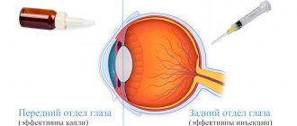
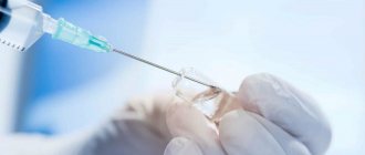
In the wet form of macular degeneration, to prevent neoangiogenesis (formation of abnormal vessels), intravitreal injections of drugs that block vascular endothelial growth factor (Eylea or Lucentis) are performed. This procedure not only stops the process of vision loss, but also helps to improve it in 30% of cases. In some cases, laser or photocoagulation of pathological vessels is used as an alternative, which also leads to a slight improvement in vision.
Patients with wet macular degeneration also need to take dietary supplements, vitamins and minerals to reduce the risk of vision loss in the other eye.
Symptoms of AMD
As a rule, the patient simply does not notice the onset of the disease due to the absence of any “acute” vision problems. But, given that retinal dystrophy affects central and color vision, the standard sequence of disease development can be described as follows.
- First, the brightness and contrast of color perception are lost and visible lines are distorted, and the patient also lacks the illumination that was sufficient before.
- At the next stage, when reading and writing texts with the usual glasses, the Patient notices the loss of individual letters or entire words.
- Then there is a loss of visual acuity at near and far distances.
- Later, as the disease progresses, a spot appears in front of the eye, at first translucent, then completely opaque, sharply worsening vision, depriving the Patient of the ability to distinguish objects and people’s faces.
Age-related macular degeneration is a disease of a paired organ, the damage extends to both eyes. Most often, retinal dystrophy manifests itself and progresses more actively in one eye. The manifestation of the disease in the fellow eye may not occur immediately, even 5-8 years after the diagnosis of AMD. If the Patient does not undergo regular check-ups with an ophthalmologist, he himself may not immediately notice any vision problems that have arisen, since the better-seeing eye takes on the additional load.
Prevention of macular degeneration
A complete cure for macular degeneration is impossible; this disease can only be prevented.
- If you smoke, you need to quit. Smoking has a detrimental effect on blood vessels, including the blood vessels in the eyes.
- Wear dark glasses when out in the sun. Don't forget about the hat.
- Eat right, eat more vegetables and fruits. Doctors especially recommend spinach and cabbage.
- Take vitamins if necessary. However, remember that excess vitamins are also bad. Hypervitaminosis may develop.
- Play sports. This strengthens the entire body, including blood vessels.
Check your vision regularly. This disease cannot yet be cured, but its development can be stopped, and it is better to do this at the initial stage.
Methods for diagnosing AMD
Today, it is possible to diagnose retinal dystrophy in a Patient in various ways. First of all, the Patient can independently suspect problems with the retina using the Amsler Grid (Amsler test), when the lines of the grid in question become uneven. Along with the traditional ones - checking visual acuity and the condition of the fundus (ophthalmoscopy), examining the visual field (perimetry), there are modern computerized methods. Such as:
- Optical coherence tomography (OCT)
- Fundus photography
- Computer perimetry
- Fluorescein angiography
- Electrophysiological studies
- Computed retinotomography
It is coherence tomography that is the gold standard for diagnosing fundus diseases and allows obtaining the highest quality images of the retina, recognizing the smallest and earliest changes that accompany age-related macular degeneration.
Self-monitoring of age-related macular degeneration
You should immediately contact an ophthalmologist if any of the following complaints occur:
- Decreased visual acuity both at distance and at close range;
- Difficulties that arise when reading and writing with the previous glasses;
- The need for more lighting;
- The appearance of a translucent fixed spot in front of the eye;
- Distortion of the contours of objects, their colors and contrast.
The diagnosis of “age-related macular degeneration” can only be established by a medical specialist. People with reduced visual function who wear glasses should be very careful about the risk of AMD and regularly have their vision checked by an ophthalmologist, since the initial symptoms of retinal dystrophy may go unnoticed due to habitually reduced visual acuity.
Development and consequences of AMD
The most common, “dry” form of retinal dystrophy develops extremely slowly. The patient does not notice any problems with lateral vision and distance vision for several years, in some cases even decades. But, at close range, and primarily during reading or writing, difficulties may appear.
In the rare but extremely dangerous “wet” form of retinal dystrophy, the disease progresses very quickly. In just a few months or weeks, distance vision deteriorates and may be completely lost.
Glasses and AMD
Glasses cannot solve the problem with the development of age-related macular degeneration of the retina. Imagine that glasses are a camera lens, and the retina is light-sensitive photographic film. And this film is damaged. No matter how “strong” the optics of your lens are, you will not get a high-quality photograph - all the shortcomings of the film will be reflected in the picture. The same is with the eyes - even with the highest quality lenses, the visual image projected onto the damaged retina will be distorted. Magnifying glasses or loupes will only help you make the most of your remaining vision. An ophthalmologist can select special correction tools for visually impaired Patients diagnosed with AMD - lenses or electronic devices that increase the size of text or images.
Diagnosis
Primary diagnosis for macular degeneration consists of taking a medical history and fundoscopy (visual examination of retinal tissue with preliminary expansion using special drops).
Additional diagnostic methods for AMD may include:
- Amsler test (can also be used for self-diagnosis);
- FAG of the retina (fluorescein angiography);
- OCT of the retina (optical coherence tomography);
- ophthalmoscopy.
Modern methods of treating age-related macular degeneration
Unfortunately, modern world medicine cannot yet offer an effective method that allows you to completely cure age-related macular degeneration of the retina and restore 100% vision. This is why early diagnosis and control of AMD is so important.
For the treatment of patients with age-related macular degeneration the following are used:
- Diet;
- Drug treatment (vitamins, minerals, antioxidants, peptide bioregulators, drugs to lower cholesterol and prevent blood “thickening”);
- Laser therapy (laser coagulation, photodynamic therapy or transpupillary therapy);
- Surgical treatment (intravitreal administration of angiogenesis inhibitors, removal of newly formed membranes or macular translocation).
All existing eye drops can only work on the surface of the eye and are not able to reach the retina, and therefore are not medications for the treatment of AMD.
Only a specialist doctor can select the most effective treatment for retinal dystrophy for you, having determined the form and stage of the disease, taking into account individual contraindications and possible concomitant diseases (such as diabetes, atherosclerosis or hypertension). Self-medication for AMD is strictly contraindicated!
Diet for AMD
Diet is an important and mandatory aspect for the prevention of the early stages of age-related macular degeneration of the retina.
- It is necessary to exclude foods with high cholesterol content from the diet.
- Fill your daily diet with vitamins, microelements and antioxidants.
- The retina needs carotenoids - lutein and zeaxanthin, which are found in egg yolks, spinach, broccoli, bell peppers, pumpkin, tomatoes, carrots, beans, cabbage, grapefruit, kiwi, etc.
- Antioxidants for the eyes contain blueberries, red currants, and red grape seeds.
- Vitamin A (retinol) is part of the visual pigment; it is found in eggs, milk, and the liver of sea fish.
- B vitamins support the nervous and immune systems, healthy growth and reproduction of cells in the body. Their sources are yeast, cottage cheese, cheese, milk, sprouted grains, and legumes.
- Vitamin C has a general strengthening effect on the body as a whole and is important for the prevention of AMD. White cabbage, kiwi, red currants, citrus fruits, green peas, spinach, etc. are rich in it.
- Vitamin E enhances the effect of antioxidants and improves blood circulation in the eye area. Its sources are vegetable oils, nuts, spinach.
- The diet for the prevention and treatment of AMD recommends a varied and balanced diet. You need to eat often and in small portions - 5-6 times a day.
Treatment of age-related macular degeneration at the Eye Clinic of Dr. Belikova.
Our ophthalmology center uses all modern techniques to stop the development of the disease and alleviate the condition of Patients.
Complex therapy for AMD
Complex drug therapy for age-related macular degeneration of the retina includes the Patient sequentially taking a tablet drug at home, and administering the drug topically, “under the eye,” as well as in the form of intramuscular/intravenous injections.
Drug therapy is combined with physiotherapeutic treatment (magnetic or laser therapy).
The period and duration of treatment is determined by the attending physician individually depending on the clinical picture of the disease. Courses of treatment can be prescribed every 4-8 months.
Treatment of the “wet” form of age-related macular degeneration
In the “wet” form of retinal dystrophy, the disease develops rapidly and requires completely different therapy. In this case, our clinic uses the following methods:
- Intravitreal (into the eye cavity) administration of steroids;
- Photodynamic therapy (PDT);
- Laser treatment (laser coagulation and transpupillary thermotherapy - thermal irradiation of the retina with a laser through the pupil);
- A combination of the above methods.
Review of retinal treatment at the Excimer Clinic
Sh. Svetlana, Moscow
Thank you for the prompt and high-quality work. Today we did a comprehensive examination and strengthened the retinas of both eyes. Many thanks to the doctor, a man of his profession (unfortunately I forgot to ask his last name) received me in room 307a. I was left with a pleasant impression;) After a while I will undergo vision correction. Thanks to all!
A. Alexander Isakovich, Moscow
I, Alexander Isakovich Alterman, am simply shocked by the attention and sensitivity of Dr. Kalashi Zurab Yusupovich and Irina Srgeevna Yusupova. I am a disabled person of the 2nd group, a labor veteran born in 1938.
Pilipchak Snezhana, Moscow
On Saturday (May 16, 2015) I had laser coagulation (strengthening the retina), done by Olga Aleksandrovna Plyukhova!! I express my deep gratitude to her for her HUMANITY, for her understanding and patience... it was a bit painful for me, but if you compare the coagulation 9 years ago , Olga Alexandrovna has golden hands!!!! Next Saturday, May 23, there will be laser correction, I'm terribly afraid)) And about the clinic. Before the consultation in Excimer, I had a consultation at the Moscow Eye Clinic (Moscow, m. Semenovskaya), earth and sky!!! At the MGK they did not find a retinal dehiscence during the examination and immediately recommended me to do a correction (and what would have happened to me???!!!!) I wonder!! Yes, Excimer is several times more expensive, but I’m not buying sausage, I’m buying good eyesight!! You can't regret it!!! Thanks everyone! I don’t understand where the bad reviews come from, everyone is so nice, the equipment is at the highest level, as is the qualifications of the nurses and doctors!
All reviews on the topic