Echinococcosis - symptoms and treatment
Echinococcosis is a long-term chronic disease that develops when humans are parasitized by the larval (cystic) stages of tapeworms of the genus Echinococcus (echinococcus). Depending on the location, they affect the liver, lungs, brain, and less often the heart and other organs. They cause slowly increasing compression of organs and tissues, disruption of their functions, and poison the body with the products of their vital activity. When an echinococcal cyst ruptures, anaphylactic shock may develop.
Etiology
Kingdom - Animals
Type - flatworms
Class - tapeworms
Order - cyclophyllidea (Cyclophyllidea)
Family - Taeniidae
Genus: Echinococcus (Echinococcus) and Alveococcus (Alveococcus)
To date, several genotypes of echinococci have been identified, which are carried by animals and have different habitats and structure.
Pathogens [1]:
- Echinococcus granulosus - causes the hydatid (cystic) form of echinococcosis (echinococcosis). Most often localized in the liver and lungs, it leads to the formation of cysts.
- Echinococcus multilocularis - causes the alveolar form of echinococcosis (alveococcosis). Usually affects the liver and can lead to death.
New pathogens have been discovered in the New World (Central and South America):
- Echinococcus oligarthrus - causes the so-called “neotropical echinococcosis”;
- Echinococcus vogeli - causes polycystic form;
- Echinococcus oligarthrus - causes an extremely rare unique monocystic form.
Echinococcus and alveococcus are types of tapeworms that, when sexually mature, parasitize the small intestines of carnivores: the first affects dogs, wolves, jackals, the second affects foxes, coyotes, and sometimes dogs.
The length of echinococcus is 2-7 mm, alveococcus is 1-4 mm. Their body consists of a head and 4-6 segments, with the first two being asexual, the third containing a hermaphroditic organ system (ovary, vitelline, testes), and the terminal segment is mature, has a spherical uterus and is approximately half the length of the body (contains on average up to 800 eggs).
The oval-shaped eggs of the parasite are released with a detached terminal segment. Passing through the intestines, they enter the environment with animal excrement, and immediately become infectious. They pollute the soil, grass, water, can stick to the animal’s fur, and remain on its face for some time when licked.
In soil, parasite eggs can remain viable for up to a year; they tolerate dry climates well, becoming dust elements. Standard disinfectants have no effect on them. Eggs die only when boiled for 20-30 seconds.
Infection of the intermediate host (including humans) occurs through ingestion of eggs. They hatch in the small intestine, then penetrate through its wall into the bloodstream and are carried by the bloodstream to various organs, subsequently undergoing specific changes: the formation, preservation and cloning of cysts - secondary echinococcosis.
Hydatid cysts consist of an inner germinal membrane and an outer cuticle. Inside they contain a colorless liquid with mature embryos floating in it - scolex. The cuticular membrane resembles the structure of insect chitin - a dense protective shell. It is impermeable to microflora and host proteins, including antibodies, but at the same time provides the parasite with access to low molecular weight nutrients.
The size of cysts can range from a millimeter (not visible on ultrasound) to 40-50 cm. They may contain daughter cysts, i.e. they are multi-chambered. Over time, a dense fibrous capsule forms around the cyst as a result of immune reactions (impeding the growth and nutrition of the cyst), and sometimes calcification occurs.
When eating raw or insufficiently heat-treated meat of infected animals (goats, sheep, etc.), the definitive host becomes infected: the parasite loses its shell, emerging from the larval stage, attaches to the mucous membrane of the small intestine and develops into an adult in 32-80 days.
Adults of E. vogeli reach a length of up to 5.6 mm, and E. Oligarthrus - up to 2.9 mm. They have no fundamental differences in structure and life cycle. Their cysts are similar to cysts in cystic echinococcosis, but they are multilocular [1][3][6][11].
Epidemiology
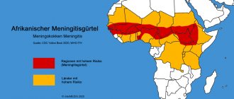
Echinococcosis is widespread everywhere (with the exception of the Arctic and Antarctic). It is more common in agricultural pastoral regions, where dogs are fed the organs of dead and slaughtered animals. The spread of the disease depends on compliance with the sanitary culture of the population.
Alveococcosis is common in the Northern Hemisphere (Central and Northern Europe, Northwestern Russia, Central Asia, Japan, Northern USA, Alaska and Canada). Elderly people get sick more often.
Echinococcosis caused by E. vogeli and E. oligarthrus occurs in Central and South America.
The source of infection for humans is sick animals.
The main hosts of echinococcus are wild and domestic canine animals (dog, wolf, jackal, coyote, fox). Intermediate hosts are wild and domestic ungulates (goats, sheep, cattle, pigs).
The main hosts of alveococcus are red foxes. Other canids (dogs, including raccoons, wolves) can also be hosts, although much less frequently. Intermediate hosts are rodents (mice, voles, lemmings, etc.).
The only known primary host of E. vogeli is the bush dog. Theoretically, domestic dogs can also be the host of this type of parasite. Intermediate hosts are paca (a large rodent) and agouti (rodents similar to guinea pigs).
The definitive host of E. Oligarthrus is wild felines (puma, jaguar, ocelot, etc.), the intermediate host is various rodents.
Mechanism of infection : fecal-oral (entry of parasites from the intestines of an infected person into the body of a healthy person through the mouth).
Routes of transmission of the parasite:
- nutritional (with food, berries, mushrooms and vegetables contaminated with parasite eggs);
- aquatic (through water contaminated with parasites);
- contact-household (as a result of cutting the skins of infected carnivores, too close contact with dogs, including mutual licking, non-compliance with hygiene rules).
A sick person is not contagious to others. It is impossible to become infected by eating raw and insufficiently cooked meat , since mature embryos of the parasite are not able to develop in the human body: for this they need a final host - a predatory mammal [1][3][7][8].
Liver echinococcosis.
This type of infection accounts for from fifty to seventy percent of all cases of echinococcosis. The parasite larvae travel through the bloodstream from the intestinal walls to the liver, where most of them settle on small capillaries. Over time, the cysts slowly but progress, thereby breaking into the internal environment of the liver.
The earliest symptoms of liver echinococcosis include:
- Heaviness and pain in the epigastric region
- Heaviness and pain in the right hypochondrium
This is due to the development of a cyst in the right lobe of the liver.
1.General information
Echinococcus is one of the most dangerous parasites, the larva of which can be transmitted to humans from infected animals (mainly canines), with water, unwashed berries, etc. At a certain stage of development, echinococci form spherical cysts with a diameter of 1-5 to 20 cm (or more) in vital organs: liver, lungs, kidneys, brain, etc. Most often, echinococcosis is registered in livestock-raising regions, where the most favorable conditions for the spread of the helminth exist, but in the era of total migrations and travel, you can become infected, essentially, anywhere.
The lungs are affected in approximately 15% of all cases of echinococcal invasion, and this is the second most common site of occurrence (after the liver) of the main focus. The severity of pulmonary echinococcosis is due to the objective difficulties of diagnosis, the abundance of complications and the obvious danger of the very localization of cysts in the respiratory system.
A must read! Help with hospitalization and treatment!
Pulmonary echinococcosis.
The development of pulmonary echinococcosis is less common than infection in the liver and accounts for twenty to thirty percent of all cases of infection. Once the infection enters the lung tissue, the disease begins to progress.
As it grows, it can cause:
- Chest pain
- Cough (from a dry cough to a wet cough with the presence of blood)
- Manifestation of shortness of breath
- In cases of the development of a particularly large cyst, deformations of the chest are noticed
Diagnostic procedures
Correct diagnosis begins with examination. In addition to complaints and medical examination, there are a number of other procedures and tests that help identify this pathology. It is more difficult to identify echinococcus in the early stages, when the disease is asymptomatic. Suspicion of the disease is based on the following facts:
- There is or is no contact with animals. In what region does the person live, what are the conditions of his home, and whether he is engaged in activities that require contact with animals.
- Does the disease manifest itself and what is its character? Elimination of signs similar to other diseases.
- Laboratory tests of urine and blood. An increase in the level of eosinophils is observed in the blood. The level of leukocytes changes, the clumping of red blood cells increases during inflammatory processes in the body.
- Blood test for antibodies.
- Casoni analysis.
- CT, MRI, radiography help to most accurately confirm the diagnosis. Thanks to instrumental methods, it is possible to accurately determine the location of the renal cyst, size and number. And also, determine the spread of the disease and identify the presence of daughter bubbles.
- Microscopic studies. The biomaterial that was taken for analysis is examined under a microscope, for example, fluid from a cyst, sputum, tissue during biopsy or probing.
The main diagnostic method is x-ray and Casoni reaction. The combination of these methods makes it possible to most accurately and quickly detect the disease. And also, evaluate the condition and type of the cyst, the image of which is clearly visible in the picture.
Manifestation of symptoms
Signs of the disease appear only after a long time from the moment of infection. After the cyst breaks into the ureter, the first symptoms can be noticed. The main indicator of helminth damage to the kidneys is acute pain in the lumbar region, as well as in the kidneys and under the ribs. A foreign body may be palpable. Palpation should be painless. Its surface is smooth, it feels soft and elastic to the touch. If fusion of the cyst is observed, its movement will be limited.
When a cyst breaks into the pelvis area, the disease occurs with renal colic. With such symptoms, fragments of chitinous tissue and bubbles can be found in the urine. This disrupts the urination process. If a single-chamber cyst has suppuration, then elements of pus appear in the urine. It is very rare that echinococcus breaks through into the intestines or abdominal cavity. Sometimes, there are cases where it breaks into the space between the layers of the pleura, or directly through the skin to the outside. Hydatid tremors are not an accurate indicator for echinococcus. It appears extremely rarely, due to friction of the bubbles in the cyst.