So-called rheumatism or acute rheumatic fever (according to new terminology) is a systemic inflammatory disease of connective tissue, in which the pathological process has a tropism towards the membranes of the heart and joints.
Even at the beginning of the 20th century, the concept of “rheumatism” meant almost any disease of the joints - doctors did not have the need or opportunity to differentiate these ailments, especially since the range of healing procedures was not diverse. Today, a rheumatologist has a wide range of diagnostic methods in his arsenal, allowing him to distinguish rheumatism from many other rheumatological diseases, each of which has its own treatment strategy.
Rheumatism is a disease predominantly of children aged 6 to 15 years, and only 1 child in 1000 suffers in this age group.
Primary rheumatism in adult patients is observed less frequently. According to statistics, women are 2-3 times more likely to suffer from this disease than men.
Rheumatism usually begins some time after a streptococcal infection of the nasopharynx, and after a few years it can turn into a chronic, incurable disease. Fortunately, only 1-3% of patients who have an infection become victims of rheumatic fever.
1
Consultation with a rheumatologist
2 Consultation with a rheumatologist
3 Consultation with a rheumatologist
Causes of rheumatism
Why do some people get rheumatism, while others under the same conditions do not? People who often suffer from ENT diseases are more likely to get rheumatism. Those at risk are those whose relatives have this disease and those who have the B-cell marker D8/17 in their blood.
So, the risk factors:
- streptococcal infection (angina, scarlet fever, pharyngitis);
- defects of the immune system, the presence of autoimmunity;
- genetic predisposition to the disease.
What happens if you run it?
After the first attack, it can take from several months to several years. Even in the absence of symptoms, the dangerous consequences of rheumatism develop.
The most harmless of them is erythema, a reddening of the skin in the form of a red circle with clear edges. It occurs in most rheumatic diseases. In a quarter of cases, inflammation of the heart tissue leads to the appearance of defects: the heart becomes larger, murmurs appear when listening, and arrhythmia appears. Damage to the mitral valve can lead to heart failure.
Disruption of the nervous system leads to “minor chorea” - involuntary movements of the limbs, muscles of the face, body, impaired coordination, handwriting, and speech defects.
Do not start the disease, consult a doctor (pediatrician or therapist) when the first symptoms appear - both streptococcal infection and signs of rheumatism. To prevent illness, follow the prevention tips.
Clinical picture of rheumatism
The trigger for the development of rheumatism is the entry of streptococcus into the body, as a result of which the immune system begins to produce antibodies to fight the infection. However, in the body itself, namely in connective tissues and heart muscle, there are molecules of the same structure. Due to the presence of this factor, the immune system begins to “fight” its cells. As a result, connective tissue is damaged, and this is fraught with heart defects and joint deformation.
Forms of rheumatism
- cardiac form (cardiac rheumatism), when all the membranes of the heart are affected (pancarditis), myocardium (myocarditis), endocardium (endocarditis);
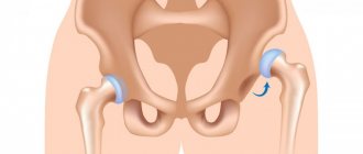
- articular form (rheumatism of the joints);
- cutaneous form;
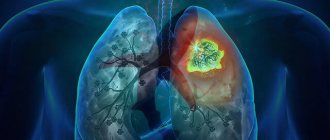
- pulmonary form (pleurisy);
- rheumatic chorea (St. Vitus' dance).
Symptoms
The manifestation is very diverse and is largely determined by the localization of the process. There may also be a hidden latent current.
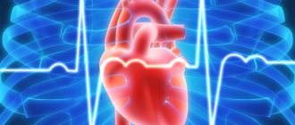
In 100% of cases with rheumatism, the heart is affected, so-called rheumatic carditis develops, which manifests itself:
- severe shortness of breath
- rapid heartbeat, irregular heartbeat
- pain in the heart
- swelling
- Expansion of the borders of the heart is detected during examination, and a heart murmur is detected during listening.
Rheumatism also affects the joints - typically an acute onset, damage to predominantly symmetrical large joints, rapid spread from one joint to another (“volatility”), pain of varying severity, up to sharp pain in the joints with limited mobility, swelling of the periarticular tissues, redness. A rapid response to adequate therapy with a decrease in symptoms is also typical.
When the central nervous system is damaged, the following manifestations of the disease may appear: movements of various involuntary muscles (facial muscles, upper and lower extremities, neck and torso). This is manifested by grimaces, antics, theatrical movements, sudden changes in handwriting, and slurred speech.
Also possible:
- increase in body temperature (in some cases up to 40 degrees)
- bouts of severe chills
- pronounced sweating
- general loss of strength
The first rheumatic attack may go unnoticed. After months or even years, a long remission may suddenly end. Repeated attacks occur with similar manifestations, which often lead to severe joint deformities and heart defects.
Types of rheumatism
There are four forms of the disease:
- cardiovascular
- polyarthritic
- nodose
- cerebral
Each form of the disease has its own distinctive signs, according to which only a cardiologist is able to assess the situation and prescribe competent treatment.
Symptoms of rheumatism
Rheumatism has a wide variety of manifestations: damage to the heart, joints, nervous and respiratory systems. 2-3 weeks after contracting a sore throat or pharyngitis, the first signs of rheumatism appear: fever, weakness, fatigue, headache. In some people, acute rheumatism begins 1-2 days after hypothermia, even without connection with infection.
Rheumatism of the heart
Already at the beginning of the disease, pain in the heart begins, increased heartbeat, shortness of breath even at rest.
Rheumatic arthritis
With articular rheumatism of the legs and arms, pain appears in the knee, elbow, wrist, and shoulder joints. The joints swell, active movements in them are limited. As a rule, after taking non-steroidal anti-inflammatory drugs, pain from rheumatism quickly disappears.
Skin rheumatism
With cutaneous rheumatism, vascular permeability increases. Therefore, skin rashes appear on the lower extremities.
Rheumatic pleurisy
A fairly rare manifestation of the disease. Main symptoms: body temperature remains above 38 degrees, severe pain in the chest, dry cough, shortness of breath, pleural noise can be heard on auscultation. More often the disease is limited to a fairly mild form of pleurisy.
1 ECG
2 Echocardiography with Doppler analysis
3 X-ray examination
Rheumatic manifestations of damage to the nervous system
Sometimes rheumatism can provoke damage to the meninges, subcortex and medulla. One of the manifestations of the disease is the dance of St. Vitus. With this complication, convulsive, involuntary contraction of the muscles of the face, torso of the arms and legs occurs. With a sharp contraction of the glottis, suffocation can occur, which is very dangerous for human life.
Abdominal syndrome
This type of complication is typical for children and adolescents. Accompanied by elevated body temperature, nausea, vomiting, and abdominal pain in the form of contractions.
Rheumatism should not be left to chance or treated by relying on the advice of friends and relatives, even those familiar with this disease. Loss of time leads to progression of the disease and dangerous complications in rheumatism. There is a threat of developing atrial fibrillation and myocardiosclerosis. Possible damage to the lungs and kidneys. And the most dangerous thing is thromboembolism (blockage of the pulmonary artery by a blood clot), which can suddenly end the patient’s life.
Rheumatism
Clinical picture:
Despite the polymorphism of clinical manifestations characteristic of rheumatism, a wide range of course options, this disease is characterized by a number of features, namely:
- connection with a previous acute streptococcal infection;
- the presence of “absolute signs of rheumatism”, according to A. A. Kisel - Kisel-Jones criteria;
- tendency to develop heart disease.
Three periods can be distinguished in the development of rheumatism. The first period lasts 2-4 weeks after streptococcal infection, is asymptomatic or with phenomena characteristic of prolonged convalescence. The second period is a clinically obvious disease with the development of polyarthritis, carditis and other clinical, morphological and immunobiochemical changes characteristic of primary rheumatism. The third period is characterized by diverse manifestations of recurrent rheumatism with progression of the severity of heart defects and the development of hemodynamic disorders. This characteristic of rheumatism reflects all stages of its development - from initial to final, accompanied by functional failure of the most affected organ - the heart.
Rheumatic polyarthritis remains one of the main clinical manifestations and diagnostic criteria, mainly primary rheumatism, less often recurrent, in which polyarthralgia predominates.
Rheumatic polyarthritis is characterized by damage predominantly to the knee, ankle, elbow, shoulder and, less commonly, wrist joints, with the migrating nature of joint damage. A rapid effect is noted after the administration of acetylsalicylic acid and other non-steroidal anti-inflammatory drugs with the disappearance of all joint manifestations within a few days, and often hours.
The severity of rheumatic arthritis varies - from unbearable pain, swelling and redness of the skin to a barely noticeable deformation, which can only be noticed due to severe pain. In the modern course of rheumatism, especially recurrent, sharp volatile polyarthralgia can essentially be considered as an equivalent to rheumatic migratory polyarthritis.
Usually, rheumatic arthritis undergoes complete reverse development, but with frequent recurrences, chronic post-rheumatic arthritis Jacques rarely develops in patients with heart disease, characterized by damage to the small joints of the hands and feet, ulnar deviation of the hands in combination with flexion of the metacarpophalangeal joints and extreme hyperextension of the distal interphalangeal joints. In recent years, it has been established that chronic seronegative Jacques arthritis is also observed in other chronic diseases with predominantly tendon-muscular and periarticular pathology, for example, chronic systemic lupus erythematosus.
Rheumatic carditis determines the nosological specificity of rheumatism and the outcome of the disease as a whole, being the most common sign of the disease and one of its main criteria.
Rheumatic carditis is characterized by the involvement of all membranes of the heart in the pathological process, while myocardial damage is an early and almost obligatory sign, against which endocarditis and pericarditis develop. Rheumatic pancarditis (formerly a common manifestation of rheumatism in children and adolescents) is now extremely rare. Early recognition of rheumatism and active anti-inflammatory therapy significantly softened the course of carditis, but did not change its essence - the outcome of heart defects. Since against the background of current myocarditis it is not always easy to recognize valvulitis, and even more so parietal or chordal endocarditis and (or) erased pericarditis, the term “rheumatic carditis” has become widespread in the clinic as a generalizing concept of heart damage due to rheumatism, which, however, obliges the doctor to use all laboratory and instrumental diagnostic methods at his disposal in order to recognize the rheumatic process in any of the membranes of the heart. Many years of clinical experience show that rheumatic carditis is characterized by sequential involvement of the myocardium, pericardium and endocardium in the pathological process, although there may be various combinations that ultimately determine the clinical and instrumental manifestations of the disease.
Three forms of rheumatic carditis - severe, moderate and weak, corresponding to the morphological definitions known in the past, such as diffuse and focal rheumatic carditis. The advantage of the clinical classification is that it reflects clinical and laboratory instrumental signs of the severity of active carditis, and its disadvantage is that these criteria are applicable mainly to primary rheumatic carditis and recurrent rheumatic carditis without heart disease. In case of recurrent rheumatic carditis against the background of a formed heart defect and hemodynamic disturbances, determining the form of rheumatic carditis is extremely difficult. But, apparently, the clinical significance of such an allocation is insignificant, since it is known that as rheumatic carditis recurs and heart disease progresses, there is a tendency for rheumatism to have a protracted and latent course, in which moderate or more often mild rheumatic carditis is usually observed.
Severe rheumatic carditis is usually found in the acute and subacute course of primary rheumatism. Its clinical picture is determined by widespread inflammation of one, two, rarely three layers of the heart (pancarditis). With severe rheumatic carditis, patients are bothered by shortness of breath and palpitations when moving, and if the pericardium is involved in the process, pain.
An objective examination reveals tachycardia that does not correspond to body temperature, but bradycardia can often be present. As a rule, patients experience moderate hypotension and a distinct increase in the borders of the heart to the left or in all directions upon percussion. According to auscultation and phonocardiographic examination, heart sounds are muffled, weakened and (or) deformed I tone, systolic (high-frequency) murmur, less often mesodiastolic murmur at the apex of the heart, pathological III and IV sounds with the appearance of protodiastolic and protodiastolic gallop rhythms. The appearance of protodiastolic aortic murmur, pericardial friction murmur, as well as radiological and echocardiographic symptoms of pericardial effusion is of diagnostic significance.
Severe carditis is also characterized, according to ECG data, by dysfunction of excitability and repolarization processes, slowing of atrioventricular conduction, prolongation of electrical systole and changes in the atrial complex.
When carrying out effective anti-inflammatory therapy (Fig. 11), dynamic clinical, radiological, electrical and phonocardiographic signs are characteristic.
Moderately severe rheumatic carditis develops with primary and recurrent rheumatism, its acute and subacute course.
Of practical importance is the recognition of moderately severe rheumatic carditis in the primary protracted course of rheumatism, which is characterized by a high incidence of heart defects due to the frequent combination of myocarditis and valvulitis in such patients. Patients often complain of persistent cardialgia and palpitations. On percussion, the left border of the heart is expanded, which is confirmed x-ray by an enlargement of the left ventricle in primary rheumatism, and in recurrent rheumatism, a decrease in the size of the heart during anti-inflammatory therapy.
On auscultation and on FCG, the first sound is weakened, the third sound is distinct, there is a systolic and transient diastolic murmur. The ECG shows disturbances in repolarization processes, intraventricular conduction, and sinus arrhythmia. There are disturbances in the contractile function of the myocardium. Noteworthy is the low dynamics of all clinical and instrumental indicators under the influence of anti-inflammatory therapy.
Mild rheumatic carditis can be observed in any variant of the course of primary and recurrent rheumatism. In the acute and subacute course of primary rheumatism, clinical laboratory signs of high activity of the process are characterized by extracardiac syndromes, and in recurrent rheumatic carditis, a mild process (carditis) against the background of heart disease can be obscured by hemodynamic disturbances.
Patients with mild primary rheumatic carditis do not complain, and objectively, only a retrospective analysis after treatment allows one to grasp the dynamics of the size of the left border of the heart. With a certain alertness of the doctor, patients can detect a tendency to tachycardia, or rather, pulse lability, a slight muffling of tones (on FCG - a mild decrease in the amplitude of the first tone), a weak systolic murmur, recorded as mid-frequency noise.
The ECG shows signs of deep and persistent disturbances of atrioventricular conduction, blockade of the atrioventricular bundle, rhythm disorders such as atrial fibrillation, extrasystole, paroxysmal tachycardia, diffuse changes in the myocardium.
Recurrent rheumatic carditis retains the features of the primary one, but the process is more severe and, as new exacerbations occur, it increasingly occurs with associated and combined heart defects, acquiring a chronic, protracted or latent course with an unstable effect of antirheumatic therapy. To a certain extent, progressive circulatory failure, which occurs not so rarely in these cases, requires the exclusion of recurrent rheumatism, although it may be associated with acute metabolic necrosis in the myocardium.
Rheumatic lung disease develops mainly in children with an acute or continuously recurrent course of rheumatism in the form of rheumatic pneumonia or pulmonary vasculitis, usually against the background of severe carditis (pancarditis).
Rheumatic pneumonia is manifested by increased shortness of breath, increased body temperature, and an abundance of different-sized sonorous moist rales from one or both sides of the lungs in the absence of dullness of pulmonary sound. X-ray reveals local strengthening, thickening and deformation of the pulmonary pattern with multiple small foci of compaction. With a bilateral root process, a typical “butterfly wings” pattern is formed. The dynamics of clinical and radiological changes under the influence of antirheumatic therapy is characteristic.
Rheumatic pulmonary vasculitis is characterized by cough, often hemoptysis, and shortness of breath. Usually, in patients in the absence of any percussion changes in the lungs, a significant number of sonorous moist rales are heard, and a diffuse increase in the pulmonary pattern is determined by x-ray. With the development of vasculitis, the effectiveness of anti-inflammatory therapy is observed. However, it is not always easy to make a differential diagnosis with congestion in the lungs, especially with recurrent rheumatic carditis against the background of heart disease.
Rheumatic pleurisy is one of the most common manifestations of rheumatic polyserositis, often occurring at the onset of the disease simultaneously with migratory polyarthritis and accompanied by pain when breathing, pleural friction noise in the area of exudate accumulation and an increase in body temperature. Pleurisy with large effusion is observed nowadays extremely rarely, mainly in children with a rapid course of rheumatism; more often, small effusion in the sinuses or adhesions (pleuroliagmyalnmr, pleuroperchka “dial”) are detected during x-ray examination. There is a rapid reversal of changes under the influence of anti-inflammatory treatment. Only with continuously relapsing pleurisy against the background of heart defects are recurrent unilateral pleurisy observed.
Kidney damage in rheumatism varies from transient toxic post-infectious nephritis to glomerulonephritis and congestive kidney in severe heart failure. The subtle symptoms of rheumatic glomerulonephritis are the main reason for the rare diagnosis in the clinic of this systemic symptom of rheumatism.
Abdominal syndrome occurs rarely, mainly in childhood, during the acute course of primary or recurrent rheumatism. Clinical symptoms are characterized by the sudden appearance of diffuse or localized abdominal pain, accompanied by nausea, less often vomiting, stool retention or increased frequency. The pain is migrating in nature, varying in severity, accompanied by fever, slight tension in the abdominal wall, and pain on palpation. The basis of the abdominal syndrome is rheumatic peritonitis, therefore the abdominal syndrome is often combined with polyarthritis and serositis of other localizations. Peritoneal symptoms disappear after a few days, and there are usually no relapses.
Rheumatic chorea is one of the main manifestations of rheumatism (“an absolute sign”, as defined by A. A. Kisel). Lesser chorea develops mainly in children and adolescents, more often girls, and pregnant women with rheumatism. The clinical symptoms of chorea are very characteristic. The child’s mental state suddenly changes: selfishness, emotional instability or, on the contrary, passivity, absent-mindedness, fatigue, and aggressiveness develop. At the same time, motor restlessness with hyperkinesis and muscle weakness with muscle hypotonia occur. Hyperkinesis is manifested by grimacing, slurred speech, dysarthria, impaired handwriting, inability to hold tableware while eating, general motor restlessness, and uncoordinated erratic movements. Sometimes muscle hypotonia becomes of primary importance, as a result of which the child cannot sit, walk, the swallowing process, physiological functions, etc. are disrupted (pseudoparalytic form of chorea). Among the individual clinical signs of chorea, they describe the symptoms of “flabby shoulders” (when lifting the patient by the armpits, the head sinks deeply into the shoulders), Cherny (retraction of the epigastric region when inhaling), “Filatov’s eyes and tongue” (the inability to simultaneously close the eyes and stick out the tongue), “ choreic hand" - flexion in the carpal ray and extension in the metacarpophalangeal and interphalangeal joints of the arm extended forward. Gordon (delay in reverse flexion of the lower leg during excitation of the knee reflex as a result of tonic tension of the quadriceps femoris muscle). Choreic hyperkinesis intensifies with excitement, less often with physical activity, and disappears during sleep. Tendon reflexes with chorea minor are somewhat increased, sometimes a mildly expressed clonus of the feet is detected, with muscle hypotonia there are no reflexes.
Among the nonspecific manifestations of damage to the nervous system during rheumatism, rheumovasculitis with one or another localization of damage to various parts of the nervous system, hypothalamic syndrome, etc. are described.
Skin lesions in rheumatism , which is based on rheumatic vasculitis, are usually manifested by annular erythema and rheumatic nodules, which are pathological signs of the disease (the main diagnostic criterion). However, in recent years, skin lesions have been observed extremely rarely.
Clinically, erythema annulare is a pale pink, barely noticeable rash in the form of a thin ring-shaped rim with a clear outer and less clear inner edge. The elements merge into bizarre shapes on the shoulders and torso, less often on the legs, neck, and face. The skin rash is not accompanied by any subjective sensations and usually goes away without a trace.
Rheumatic nodules, ranging in size from millet grains to beans, are dense, inactive, painless formations located in the fascia, aponeuroses, along the periosteum, joint capsules, and subcutaneous tissue. Favorite localization is the extensor surfaces of the elbow, knee, metacarpophalangeal joints, ankles, spinous processes of the vertebrae, etc. Rheumatic nodules appear unnoticed by patients and also quickly disappear or undergo reverse development within 1-2 months without residual effects.
Diagnosis of rheumatism
When diagnosing the disease, the following methods are used:
- ECG;
- Ultrasound of the heart;
- X-ray examination (helps to see changes in the size and shape of the heart);
- general blood test (can show ESR level, anemia, etc.);
- immunological blood test (appearance of C-reactive protein, presence of streptococcal infection).
1 General blood test
2 Immunological blood test
3 Diagnostics of rheumatism in MedicCity
Prevention Tips
- Prevention of streptococcal infections - administration of the antibiotic Bicillin-5. Bicillin prophylaxis is for those who have already suffered from rheumatism (in this case, prophylaxis is year-round), as well as those who have been in contact with patients. The doctor will select the right dose or replace the drug if you are allergic to penicillin antibiotics.
- Strengthening the immune system - there are no “magic” pills for him, but a clear daily routine, good nutrition, and regular physical activity will help him a lot.
You can go jogging, swimming, or your favorite sport. Up to 20 years of age, strength training and activities that develop flexibility and joint performance (dancing, fitness) are suitable. After 35-40 years, you need to take care of the health of your spine and choose yoga and Pilates. Moderate cardio exercise (walking, running, skating) is also suitable.
- Timely and competent treatment of upper respiratory tract diseases: when the first symptoms appear, you need to consult a doctor (pediatrician, therapist), do not refuse antibiotics if they are prescribed by a specialist, and also do not interrupt the course of treatment at the first improvement. Isolation of patients with streptococcal infection is also necessary: keeping the patient in a separate ventilated room. He must have his own hygiene items and utensils. The patient needs to buy a set of non-woven masks and change them every 3 hours.
After an infection, you need to undergo a comprehensive diagnosis: urine and blood tests, and an ultrasound of the heart. Children need to be observed for 3 months by a pediatrician and rheumatologist.
Forecast
The prognosis for this disease is made taking into account the nature of the disease and the degree of myocardial damage.
The frequency of observation by a cardiologist depends on:
- phases of the disease
- attack frequency
- severity of attacks
- associated septic condition
- circulatory failure.
An unfavorable prognosis is observed in young patients with associated septic process, when death is possible. Deaths are observed in patients due to acute coronary circulatory disorders.
Don't risk your health! Timely contact with a qualified doctor and the adoption of emergency measures, as well as competent organization of the regime in the active phase of the disease, can prevent the development of irreversible heart damage and improve the prognosis.
Effective treatment of rheumatism
Effective treatment of rheumatism should be aimed not only at suppressing streptococcal infection, but also at preventing relapse and suppressing inflammation, especially in the heart and joints. You need to know that getting rid of this serious disease must necessarily be accompanied by limiting the patient’s physical activity, which negatively affects the condition of tissues damaged by inflammation. In order to recover, it is necessary to adhere to bed rest.
If streptococcal tonsillitis is detected, the patient is prescribed internal penicillin, which is prescribed for ten days. Residual infection in a sick child is eliminated by injections of penicillin. To eliminate pain and inflammation, non-steroidal drugs, including aspirin, are prescribed.
A child who suffered from rheumatism in childhood should receive penicillin internally or intramuscularly until the age of eighteen. This is necessary in order to prevent relapse of the disease. If a person has suffered heart damage during the course of his illness, then throughout his life the patient must take an antibiotic before any operation (even tooth extraction).
Detection of the disease in the early stages: diagnosis of rheumatism
Timely diagnosis of rheumatism becomes possible by analyzing the main symptoms. In addition, the disease is determined by a blood test: in children, this study reveals a high content of leukocytes and an increased erythrocyte sedimentation rate, and antibodies to streptococci are detected.
Heart rhythm disturbances that accompany its inflammation, caused, in turn, by rheumatism, are detected using an electrocardiogram. In order to timely diagnose and begin treatment of heart valve damage, echocardiography is performed.