How does an adenoma turn into carcinoma?
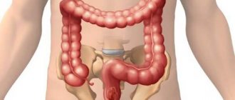
Rectal cancer.
Rectal cancer is one of the most studied malignant tumors. Typically, colorectal cancer develops from benign intestinal polyps (adenomas). This process may take more than one year. The change from polyp to cancer involves irreversible changes (mutations) in the genes of the cells lining the intestinal wall. This leads to the loss of natural mechanisms of cell division, which, in turn, causes their uncontrolled growth, that is, transformation into cancer cells. They ignore the normal boundaries of surrounding tissues and grow into nearby structures. This process is called invasive tumor growth. These cells penetrate the thickness of the intestinal wall, pushing aside and destroying the natural cell mass. Having reached the blood and lymphatic vessels, they are transferred to other parts of the human body, forming daughter tumors. These colonies are called metastases. The process of carcinoma formation from a benign intestinal polyp can take approximately 5-10 years. The risk of cancer increases with age. Most patients with colorectal cancer are over 50 years old. Gene mutations can also be hereditary, that is, transmitted by inheritance. In such cases, the process of tumor formation proceeds faster and cancer may occur at a younger age.
Risk factors predisposing to the development of rectal cancer.
An increased risk of colorectal cancer is associated with:
- bad habits (diet with a large amount of meat and fat, and a small amount of plant fiber, smoking, alcohol, excess weight and low physical activity);
- inflammatory bowel diseases (ulcerative colitis, Crohn's disease);
- benign intestinal polyps (adenomas);
- cases of colorectal cancer or polyps in previous generations of the family (hereditary predisposition);
- previous or concomitant other types of cancer (breast, ovarian, uterine cancer).
What are the symptoms of colorectal cancer?
Colon cancer does not develop or grow overnight. It takes months, even years, before the tumor grows into noticeable symptoms. This is why cancer can occur without clinical manifestations or have few symptoms.
The main symptoms of colorectal cancer include:
- changes in bowel habits (stool retention, diarrhea, “ribbon-like stools” or frequent false urge to defecate without passing stool);
- blood or mucus in the stool;
- recurring pain attacks in the abdomen;
- persistent bloating;
- general symptoms such as decreased performance or weight loss.
These symptoms can occur with a number of other diseases. This requires additional caution and urgent clarification of the reasons for these complaints.
What methods are used to prevent and early diagnose cancer?
Digital rectal examination.
Digital rectal examination is the main and simplest diagnostic method in the arsenal of a coloproctologist, which does not require the use of special equipment. During this manipulation, the specialist examines the rectum by inserting the index finger into the intestine. When performing a digital rectal examination, it is possible to determine the presence of intra- and extraintestinal formations at a height of up to 10 cm from the edge of the anus, assess the nature of the formation (benign or malignant), and the involvement of neighboring organs and tissues in the tumor process. In the presence of intraluminal formations, patients must undergo a biopsy (a piece is taken) in order to determine the histological structure and make an accurate diagnosis.
Diagnostics
The simplest methods for diagnosing a rectal tumor are finger examination and examination with a rectal speculum. The proctologist detects pathological formations on the mucous membrane, but the presence of a malignant process can be confirmed or refuted only with the help of laboratory and instrumental methods. During the examination, a biopsy of the pathological tissue is performed.
- Sigmoidoscopy. The examination is carried out by pumping air into the rectum to straighten the folds and carefully examine the mucous membrane using a rectoscope.
- Irrigoscopy. The rectum is filled with a contrast solution and examined using an X-ray machine.
- Ultrasound of the pelvic organs. The study allows you to detect damage to neighboring organs and lymph nodes.
- CT scan of the pelvic organs. The study is prescribed to clarify the X-ray and ultrasound data.
- Blood test for tumor markers.
- Histological analysis of a biopsy is necessary to establish malignancy and identify the type of cancer cells.
- Cytological analysis of biopsy material, mucus and pus taken from the rectum allows us to study in detail the structure of the cancer cell.
Attention!
You can receive free medical care at JSC “Medicine” (clinic of Academician Roitberg) under the program of State guarantees of compulsory medical insurance (Compulsory health insurance) and high-tech medical care.
To find out more, please call +7, or you can read more details here...
Tumor markers.
These are substances produced in large quantities by tumor cells. They are not very specific and are also detected in the blood of healthy people. For this reason, negative or normal tumor marker values do not exclude cancer, just as elevated tumor markers do not confirm cancer. The most specific markers for colorectal cancer are CEA (carcinoembryonic antigen) and CA 19-9. A sharp increase in the level of tumor markers in the blood may indicate the presence of distant metastases of rectal cancer (in the liver, lungs), and in the postoperative period - the occurrence of a relapse of the disease.
Colonoscopy.
Rectal cancer.
This is the most informative method of examining the colon. It involves inserting a flexible instrument (endoscope) through the anus and is accompanied by illumination and examination of the inner surface of the colon and rectum. To maximize visualization of the intestinal wall, it must be adequately cleansed using special laxatives or an enema. Colonoscopy is the only method to confirm colorectal cancer because tissue samples (biopsy) can be obtained. In addition, intestinal polyps can not only be detected, but also removed during this procedure.
Causes and risk factors
Currently, the mechanism that starts tumor growth is not fully understood, but oncologists have well studied the unfavorable factors that contribute to colorectal cancer. The most significant of them are:
- chronic inflammation of the mucous membrane;
- the presence of polyps in the colon;
- unbalanced diet with a predominance of red meat and a lack of plant fiber;
- inherited predisposition;
- obesity, sedentary lifestyle;
- presence of diabetes mellitus;
- lack of vitamins A, C and E, which block carcinogenic compounds in the intestines;
- smoking at least a pack of cigarettes per day;
- living in an environmentally unfavorable, polluted environment;
- age 50 years and older.
Ultrasound examination (ultrasound) of the rectum.
Rectal cancer.
By inserting an ultrasound probe through the anus, it is possible to examine the rectum using ultrasound. Unlike rectoscopy, which visualizes the spread of cancer on the inner surface of the intestine, ultrasound can show the extent of tumor growth through the intestinal wall and into surrounding tissues, and the presence of metastases in the lymph nodes. Along with pelvic magnetic resonance imaging (MRI), this is a key test for rectal cancer and shows the need for chemoradiotherapy before surgery.
Stages
There are four stages of colorectal cancer.
- A small tumor does not extend beyond the submucosal or muscular layer. There is no damage to the lymph nodes, no metastases.
- The tumor grows through the entire intestinal wall, but does not reach neighboring organs. Adipose tissue and up to three regional lymph nodes may be affected. There are no metastases.
- Cancer cells grow into nearby organs and affect a large number of regional lymph nodes. There are no distant metastases.
- The tumor increases in size and quickly metastasizes, affecting distant organs. Due to the breakdown of cancerous tissue, interorgan fistulas are formed and purulent inflammation develops. The patient's condition becomes extremely serious.
Computed tomography (CT).
Rectal cancer.
This is a radiological method that visualizes the human body as virtual slices. In patients with rectal cancer, CT scanning of the chest, abdomen and pelvis can reveal affected lymph nodes and metastases in other organs (liver, lungs). For a better examination of the gastrointestinal tract, the patient drinks a contrast agent about an hour before the examination. Immediately before the CT scan, the colon is also filled with a contrast agent administered through the anus. This allows for good visualization of rectal tumors.
Diagnostic methods
Patients suspected of having rectal cancer should immediately undergo a comprehensive examination and receive recommendations from an oncologist.
Diagnostics includes:
- Examination of the patient and collection of complaints. As a rule, the patient first visits a therapist, and only then goes to an appointment with a specialized specialist - a proctologist, oncologist-surgeon. The doctor will examine the nature of the symptoms and prescribe the necessary tests.
- Tests are prescribed: a general blood and urine test, a biochemical blood test and tumor markers for colorectal cancer. Studies make it possible to assess the general condition of the internal systems of the body, detect foci of inflammation and the extent of their prevalence.
- Investigations: colonoscopy or irrigoscopy, magnetic resonance or computed tomography, and ultrasound examination of the abdominal cavity. Allows you to determine the size of the tumor and its location.
- Screening methods. This type of research allows you to identify pathology before the first symptoms appear. This is a digital examination, endoscopy, stool analysis for occult blood.
After tests and studies are completed, observation and treatment is carried out by an oncologist.
IMPORTANT:
Additionally, the oncologist can send the patient for a biopsy. The procedure involves taking a piece of tumor tissue, which is sent for laboratory testing.
Magnetic resonance imaging (MRI).
Rectal cancer.
Just like CT, magnetic resonance imaging (MRI) takes pictures of the human body in the form of virtual slices. The key difference is the use of a magnetic field instead of radiation. In addition to information about the condition of the lymph nodes and the presence of metastases in other organs, MRI provides a very accurate picture of the spread of the tumor in the pelvis. This is very important for rectal cancer. Along with rectal ultrasound, this is a key test for rectal cancer, which shows the need for chemoradiotherapy before surgical treatment. The use of MRI is limited in people with pacemakers and metal implants, and in people who are claustrophobic.
Positron emission tomography (PET).
Rectal cancer.
During their growth, cancer cells spend more energy and absorb more glucose than healthy cells. These characteristics can be used to diagnose a tumor and its metastases. During positron emission tomography (PET), radioactively labeled glucose is injected intravenously and taken up by rapidly growing metabolically active cells (mostly cancer cells), making them visible during the study. Considering the high cost of the method, PET is used to perform specific tasks, such as searching for metastases and diagnosing possible relapse of the disease.
List of sources
- All-Russian public organization “Association of General Practitioners (Family Doctors) of the Russian Federation” Colorectal cancer: clinical recommendations for general practitioners. — 2014
- Tsukanov A.S., Shelygin Yu.A., Achkasov S.I., Frolov S.A., Kashnikov V.N., Kuzminov A.M., Pikunov D.Yu., Shubin V.P. Principles of diagnosis and personalized treatment of hereditary forms of colorectal cancer // Bulletin of the Russian Academy of Medical Sciences. — 2019
- A. A. Nevolskikh, B. A. Berdov, L. N. Titova. Current trends in combined treatment of rectal cancer. — 2013
How is colorectal cancer treated?
Surgery to completely remove the tumor is the only method that offers a chance of curing colorectal cancer. Before surgery, it is necessary to determine whether the tumor is localized in the rectum or whether the process is generalized (for example, it has spread to the liver or lungs). Even if the tumor has spread, treatment is still possible. Currently, small rectal tumors are removed immediately, without preoperative chemoradiotherapy. Large tumors require preoperative treatment. There are currently two treatment options. The first is to irradiate the rectal tumor with fairly high doses of radiation for one week (short course). The second option is a combination of low-dose radiation and well-tolerated chemotherapy for five weeks (long course). Research shows that the purpose of pretreatment (radiation) is to reduce the risk of local recurrence (that is, the likelihood that the tumor will return after surgery). Surgery is performed 6-8 weeks after the end of chemoradiotherapy. Surgical treatment involves complete removal of the rectal tumor. After surgery, surgical samples (tissue that was removed during surgery) are sent for pathological examination, during which the depth of the tumor and the presence of metastases in the lymph nodes will be determined. Based on the results of the pathological examination, the feasibility of additional preventive postoperative chemotherapy is determined.
Treatment methods
Prognosis depends on the stage of cancer
Treatment for rectal cancer depends on the type of tumor, stage of the disease and location. The main goal is the complete removal of existing foci of malignant growth, suppression of relapse and restoration of natural intestinal functions.
In later stages, it is important to provide symptomatic therapy, including pain relief. The main methods of non-surgical treatment are chemotherapy and radiation therapy. Both methods are aimed at reducing the size of an existing tumor and suppressing metastasis.