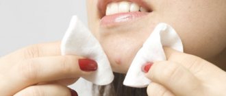
People often confuse dark circles with bags under the eyes, mixing these fundamentally different problems into one.
Bags under the eyes are formed due to the redistribution of so-called fat bags (hernias). Sometimes bags are formed due to ptosis of the skin in the lower eyelid area (read more about the formation of bags under the eyes).
Dark circles (bruises) under the eyes, on the contrary, appear when there is a deficiency of fat in the eyelid area. The cause may be associated with genetic, pathological and age-related factors. In the latter case, the problem is combined - the person develops both bruises and bags under the eyes.
It is important to understand that there is not only one reason for the appearance of circles under the eyes - it is almost always a complex of reasons.
15% discount on the first visit for any procedure with any doctor!
Diseases
Not always, swelling and bruising under the eyes are a sign of an unhealthy lifestyle. Often, the reason is the presence of certain diseases:
- Allergies. Accompanied by lacrimation, sneezing, redness of the eyes, difficulty breathing and coughing;
- Inflammation of the sinuses (sinusitis and sinusitis). They occur with painful sensations localized in the forehead and under the eyes, as well as copious discharge from the nose;
- Kidney diseases. When they occur, fluid retention occurs in the body, which causes not only swelling of the eyes, but also lower back pain and headaches;
- Diseases of the thyroid gland. Due to dysfunction of the thyroid gland, hormonal imbalance occurs - general weakness increases, body weight increases, the menstrual cycle changes, edema occurs;
- Heart failure. It is accompanied by shortness of breath and fatigue. Swelling and bruising under the eyes, almost invisible in the morning, appear in the late afternoon;
- Skin diseases - eczema and dermatitis.
Let's take a look at the anatomical structure of the eye area.
To understand the reason for the formation of circles, you need to imagine the structure of this area. Let's remember our school anatomy lessons.
Under the skin there is fat hidden - the so-called “fat packets of the face”. Below them are the orbicularis oculi muscles: they are responsible for squinting, squinting, blinking and other facial expressions.
Let's pay attention to the orbit of the eye and remember how many blood vessels and capillaries there are in this area.
It is very important to understand that blood vessels and muscle tissue are rather “dark” formations that are not very visible “on the face” only thanks to the skin and subcutaneous fat that cover these structures. Thus, skin and fat are the main concealers of dark circles under the eyes.
Thinning of the fat layer
The most common cause of dark circles. The skin of the lower eyelids is separated from the orbicularis oculi muscle and blood vessels by a thin layer of superficial fat.
With age, the superficial fat becomes thinner, “goes away,” and even through the thickest skin the orbicularis oculi muscle and blood vessels begin to show through. The muscle is dark red, the vessels are blue-scarlet.
The color of the circles depends on what is more visible - the blood vessels or the muscle. The color of dark circles has no connection with the kidneys, liver and other organs.
It happens that there is very little subcutaneous fat - this is a genetic feature. Then dark circles and semicircles can be seen from childhood.
Thin skin
The skin of the lower eyelids is the thinnest on the entire body. There is a thin layer of fat under the skin, but if there is little fat and the skin is thin, then the same vessels and muscles can be seen through the light. If your blood vessels are located close to the skin , and your skin is light, then it can show through even if there is fat!
Deep-set eyes
The lower eyelid is spatially located in the projection of the orbit of the skull. Anatomically deep-set eyes visually create a “retraction” of the skin relief in the area of the lower eyelid; when illuminated, a shadow part is formed in this area, which visually creates the effect of a dark circle.
To better understand this point, let's look at a visualization of another deficiency - the nasolacrimal trough depending on lighting.
Any age-related problem - be it a wrinkle, a bag, a hernia or bad skin - is noticeable thanks to only one thing: incident and reflected light.
The main essence of anti-aging aesthetic corrections is working with the distribution of light and shadow on our face. Any furrow, fold or bag is a play of light. Look at the illustrations to see how the visual appearance of the nasolacrimal trough varies depending on the direction of the lighting.
We all understand that both daytime natural and artificial light falls mainly on our face from above. It is this direction of lighting that most clearly emphasizes all the irregularities on the face!
It’s the same with deep-set eyes - the darkness of the lower eyelid depends to a greater extent on the lighting.
Dark circles from pigmented skin
Acquired and congenital pigment is the cause of the formation of dark circles in a normal anatomical structure. The peoples of the Caucasus region have a genetic predisposition to pigmented skin of the lower and upper eyelids.
Pigmentation can appear with age; usually such dark circles do not have a clear border at the bottom.
Diagnostics
To exclude diseases of internal organs, you should undergo comprehensive instrumental and laboratory diagnostics, including:
- general and biochemical blood test;
- hormone tests:
- thyroid gland (thyroid-stimulating hormone, free T4);
- produced by the adrenal glands (adrenocorticotropic hormone, cortisol);
- sexual (progesterone, estrogen);
- organs located in the abdominal region;
It is also necessary to consult with different specialists:
- endocrinologist;
- gynecologist;
- urologist;
- ophthalmologist;
- otolaryngologist;
- gastroenterologist;
- cardiologist;
- allergist.
How to get rid
Once the cause of the formation of dark circles is identified, it is necessary to take measures to get rid of them. We do not recommend self-medication. But there are several methods that will not harm your body:
- Drug treatment. It is prescribed when a patient has pathological diseases that require specific treatment. Often this may be related to the digestive organs or hormonal system.
- To refuse from bad habits. Smoking and alcohol negatively affect the general condition of the body and quitting them will help you avoid a large number of potential diseases.
- Active lifestyle. Running, morning exercises and just walking improve your blood circulation.
- Cosmetologist services. You can resort to the following procedures: mesotherapy, plasma lifting, biorevitalization and others that tone the vessels under the eyes.
- Using cosmetics or professional makeup.
- Hide. If you can’t get rid of the circles in the shortest possible time, you can simply disguise them. This task will be perfectly handled by: foundation, concealers and special creams that even out the skin color and nourish it. Choose a cream that contains vitamin C.
Other methods of dealing with bruises
Another effective method of cleansing the space around the eyes is the use of folk remedies.
| Main active ingredient | Mode of application | Time of action |
| Sauerkraut | Grind on a grater (in a food processor, blender). Apply to face. | 15 minutes (remove with a napkin, wash with cool water). |
| Cherry | Crush a few ripe cherries on your face | 15 minutes (after use, rinse with cool water). |
| Cottage cheese | Apply under eyes. Has an almost immediate effect | 15 minutes |
| Parsley and cucumber | The components are crushed and mixed in equal proportions | Apply on eyelids for 15 minutes. |
Do not forget that the body is capable of responding with an allergy to the use of one or another component. Therefore, you should use certain treatment methods with caution.
Folk remedies bring results only when used systematically. The course of procedures should last about two months.
[media=
https://youtu.be/JcvbA2MVR3g
]
Skin hyperpigmentation
Thyrotoxicosis
17891 May 21
IMPORTANT!
The information in this section cannot be used for self-diagnosis and self-treatment.
In case of pain or other exacerbation of the disease, diagnostic tests should be prescribed only by the attending physician. To make a diagnosis and properly prescribe treatment, you should contact your doctor. Skin hyperpigmentation: causes of appearance, what diseases it occurs with, diagnosis and treatment methods.
Definition
Skin hyperpigmentation is an excess deposition of melanin pigment of varying location, shape and size, leading to changes in skin color of varying intensity.
The surface layer of the skin (epidermis) is a multilayered (five layers) epithelium. The largest number of melanocytes, the main function of which is the production of melanin, are located in the basal layer of the skin (basal membrane)
Thanks to melanin, the skin protects a person from ultraviolet and infrared radiation.
The formation of age spots largely depends on the condition of the basement membrane.
Types of skin hyperpigmentation
Based on
the mechanism of occurrence,
the following types of skin hyperpigmentation are distinguished:
- melanocytic - pigmentation occurs due to an increase in the number of melanocyte cells;
- melanin - pigmentation is caused by an increase in the production and accumulation of melanin or a decrease in the rate of renewal of the stratum corneum of the epidermis.
The accumulation of pigment can be limited
or
widespread
, caused by
physiological
or
pathological
reasons, the process can be primary or secondary.
Possible causes of skin hyperpigmentation
Scientists identify several main causes of skin hyperpigmentation:
- genetic predisposition;
- exposure to ultraviolet radiation (in isolation or in combination with other reasons);
- burns (thermal, chemical, electrical);
- inflammatory processes;
- infectious diseases, including parasitic ones;
- endocrine disorders;
- metabolic disorders;
- use of herbal substances and drugs with photosensitizing effects
freckles for people
(ephelides) are a striking example of a realized genetic predisposition, which is based on an increase in the formation of melanin.
What diseases cause skin hyperpigmentation?
The group of melanocytic pigmentations includes various types of lentigines
.
Simple lentigo occurs at any age as a single (or multiple) formation up to 5 mm in size, brown in color. Doesn't change over time.
For xeroderma pigmentosum
Lentigo occurs before the age of 2 years on areas of the skin exposed to the sun (face, neck, back of the hands), then spreads throughout the body. Often combined with keratosis - thickening and peeling of the skin.
Solar lentigo
Appears, as a rule, after 40 years of age on areas of the skin previously exposed to sunburn. They appear as spots ranging in size from 1 to 3 cm, their color can vary from light yellow to dark brown.
Lentigo reticularis
resembles a black blot and is considered a type of solar lentigo. Most often occurs in individuals with skin phototypes I and II who have a history of severe sunburn with blistering.
Less common are other types of lentigo, occurring in isolation under the influence of solarium lamps, drug therapy (PUVA lentigo) or as part of syndromes affecting other internal organs (for example, with Peutz-Jeghers syndrome, lentigo of the oral mucosa is combined with intestinal polyps).
Skin hyperpigmentation can occur due to hormonal imbalance.
For example, during pregnancy the level of estrogen increases, and against the background of exposure to ultraviolet radiation, chloasma can form - round spots of different sizes and colors on the face.
In the later stages of pregnancy, existing moles, freckles, nipples and areolas of the mammary glands, the white line of the abdomen, and the skin around the navel may darken. Chloasma is often observed in women taking hormonal contraceptives, as well as with inflammatory or tumor pathologies of the ovaries. Chloasma is rarely recorded in men - as a rule, they have an increased level of luteinizing hormone and a decreased level of testosterone.
Hyperpigmentation of the skin throughout the body with darker areas exposed to sunlight is seen in primary or secondary chronic adrenal insufficiency (Addison's disease or syndrome) due to low levels of the hormone cortisol.
As a result of excessive thyroid function (thyrotoxicosis), secondary adrenal insufficiency occurs, and pigmentation can be diffuse or limited in the form of chloasma.
In diseases accompanied by extreme exhaustion (cachexia), the skin of the neck, abdomen, and genitals turns dirty brown.
The pigment can accumulate in places of thermal, chemical or electrical burns, injuries with damage to the skin. The pigment often remains after resolved boils, carbuncles, urticaria, lichen planus, psoriasis, as well as after scabies and lice.
In Riehl's melanosis, a bluish-brown reticulate pigmentation appears on the back of the hands and forearms, and the same occurs when in contact with synthetic clothing, rubber products or hydrocarbons, enhanced by ultraviolet exposure.
Some plants (legumes, figs, parsley, citrus fruits) contain photosensitizing substances - psoralens.
When present in food, they increase the photosensitivity of the skin. Such plants can be included in cosmetics; if you apply them to the skin and then go out into the sun, hyperpigmentation will occur at the site of application. Some medications also have a photosensitizing effect: sulfonamides, tetracyclines, antipsychotics, etc. Taking cytostatics slows down the rate of epidermal renewal, so the pigment is removed more slowly.
Which doctors should I contact for hyperpigmentation?
If hyperpigmentation occurs, you should consult a dermatologist. If the examination reveals pathologies of internal organs, you may need to consult an endocrinologist, therapist, gynecologist, urologist and other specialists, and if there is a risk of malignancy of the process, an oncologist.
Diagnosis and examinations for skin hyperpigmentation
Diagnosis of hyperpigmentation is based on clinical examination and patient interviews.
If necessary, the doctor can remove a pigmented formation (for example, lentigo reticularis) followed by histological examination to confirm its benignity.
Hereditary causes of dark circles under the eyes
Heredity is a key player in the list of causes of dark circles under the eyes. But the concept of “heredity” itself is very broad.
Hereditary factors that provoke the formation of bruises under the eyes include:
- Specific facial anatomy (deep-set or slightly bulging eyes);
- Thin skin;
- Close location of blood vessels to the skin in the eyelid area;
- Constitutional features – excessively thin physique, in which there is a general deficiency of subcutaneous fat;
- Less commonly, chronic genetic diseases (for example, heart or liver).
Dark circles under the eyes associated with hereditary causes appear early - sometimes even in childhood. Indirectly, those bruises that arose in youth - before 20-25 years old (normally this does not happen) can be attributed to “heredity”. If parents develop dark circles at a young age, it is possible that the same fate will befall their child. And pathological “components” are not necessary for this.
Hereditary reasons can also include the properties of the skin : if the skin is thin, then the vascular network of the periorbital region “shines through” through it. This is usually the case for those with fair skin - dark circles can form as early as the age of 20, when fat packets begin to thin out.
It happens that hereditary factors are combined - a person has thin skin in the periorbital area and problems with blood circulation. The approach to such problems must also be comprehensive . Fortunately, in modern cosmetology there are fillers and other drugs that act at different levels: for example, the American MesoEyeC17 has a positive effect on the processes of blood and lymph microcirculation.
How to deal with hereditary causes?
As you understand, heredity can hardly be cured, so there is only one correction option - correcting the lower eyelid area with fillers (contour plastic methods). Even surgery won't change the situation much.
The cost of such a correction is from 15,000 rubles, the duration of the effect is up to 10-14 months.
Treatment methods
If the diagnosis does not reveal internal diseases, then circles under the eyes are a cosmetic defect that appears as a result of an incorrect lifestyle or hereditary predisposition.
In such cases, the most popular and effective treatment methods are:
- Mesotherapy. Typically, meso-cocktails are used, which contain arbutin, ascorbic, kojic and phytic acids. The therapeutic course includes 6-10 procedures, between which there is a one-week break.
- Bioreparation with ascorbic acid (vitamin C). Thanks to the combination of ascorbic and hyaluronic acid, the skin around the eyes is well moisturized and noticeably brightened. The course of treatment consists of 2-4 sessions, with a 2-week break between them.
- Plasmolifting. The procedure restores metabolism, normalizes tissue respiration, activates blood microcirculation, thereby increasing local immunity. A similar effect will appear after 4-6 sessions at weekly intervals.
- Biorevitalization. The skin around the eyes shows a pronounced reaction to dehydration and, due to dryness, often takes on an unaesthetic appearance, creating the effect of “tired eyes”. A course of birevitalization, consisting of 2-4 procedures with a 2-week interval, will help solve the problem.
- Contour plastic. If the blood vessels are located close to the skin and “see through” through it, then the filler forms an additional layer, making the vessels almost invisible. In addition, the drugs smooth out the nasolacrimal groove.
- Laser peeling. Using a laser, the top layer of skin is removed, which stimulates the regeneration process and promotes rejuvenation. The treatment course involves 2-5 procedures with a one-month break.
Why do bruises appear under the eyes: anatomical features
The skin under the lower eyelid is several times thinner than on other parts of the body and contains a large number of blood vessels and small capillaries. And the vessels, capillaries, and muscles that are located under the epidermis are darker than our skin, so if they are visible, visually it will look like bruises under the eyes. The effect also occurs when the permeability of the walls of blood vessels and capillaries is disrupted.
The lower layer of the epidermis is subcutaneous fat. In some areas of our body (for example, the border of the lips) it is completely absent. Under the lower eyelid, the layer of subcutaneous fat is quite small, so any thinning of it leads to dark circles appearing under the eyes.
Thus, there are three anatomical causes of bruises under the eyes:
- very thin layer of dermis;
- thinning of the fat layer under the lower eyelids;
- violation of the permeability of vascular walls.
Age-related changes and dark circles under the eyes
The most common cause of dark circles is age-related changes.
Age-related changes in the eyelids occur according to an approximate “schedule”:
- 20-25 years – appearance of “crow’s feet”;
- 25-30 years – formation of a mild nasolacrimal groove;
- 35-40 years – appearance of dark circles under the eyes;
- 40-45 years – formation of fatty bags (hernias) of the lower eyelid;
- 45-50 years – growth of hernias (bags under the eyes);
- 45-55 years and beyond - ptosis of the upper and lower eyelids.
It is impossible to fit every person into this scheme - for some, age-related changes in the eyelids begin earlier, for others later. But there is an obvious fact: if you ignore professional care from the age of 30, the regression will be more rapid, and radical interventions will become relevant closer to the age of 40.
So, the main reason for the age-related manifestation of dark circles is the thinning of the fat packet of the lower eyelid and the weakening of the ligamentous apparatus.
The orbicularis oculi muscle and ligaments stretch and sag with age. Moreover, with age, the size of the orbit increases, and the zygomatic bone flattens.
The situation is aggravated by age-related slowdown of metabolic processes and lymphatic drainage in this area.
Combating impaired lymphatic drainage and redistribution of subcutaneous fat with the help of creams and ointments is frankly naive.
Even the most expensive cosmetics are not able to “resolve” fatty bags and redistribute the tissue evenly to compensate for its deficiency in areas with dark circles under the eyes - this is obvious. A good cream only serves as an addition to full-fledged procedures in aesthetic medicine, but will never replace them.
Age-related dark circles under the eyes are also often caused by impaired blood supply, so they are soon naturally supplemented by bags and puffiness. The stagnant processes themselves do not add beauty to the skin - it acquires looseness and an earthy tint - pigmentation.