Cyanosis is a symptom characterized by a blue tint of the skin and mucous membranes, which occurs due to an increased level of carbhemoglobin in the blood. Cyanosis, which is associated with the entry of chemical compounds into the body or the deposition of any substances in the skin, is considered to be false cyanosis.
Cyanosis resulting from hypoxemia is considered true. Capillary blood in the presence of true cyanosis contains more than fifty g/l of reduced hemoglobin.
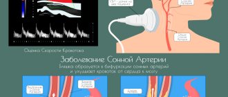
Cyanosis is a fairly common symptom of heart disease. The further a part of the body is from the heart muscle, the more intense its cyanosis.
Cyanosis of the fingers and toes, nasolabial triangle, and ears is called acrocyanosis. Acrocyanosis occurs when the level of reduced hemoglobin increases in the venous blood. Acrocyanosis occurs due to excessive absorption of oxygen by tissues when blood flow slows down. Central cyanosis is a widespread cyanosis. The cause of diffuse cyanosis is a lack of oxygen in the pulmonary circulation.
Patients with both peripheral and central cyanosis often come to the Yusupov Hospital. Cyanosis of the face brings aesthetic discomfort to patients, while cyanosis of the extremities may remain unnoticeable for a long time. By signing up for a consultation by phone or online, during an individual conversation you can find out the differences between cyanosis and acrocyanosis, learn about the possible causes of cyanosis, view photos of patients with cyanosis of the skin, photos of patients with cyanosis of the nasolabial triangle and calculate the financial side of the issue.
The Yusupov Hospital is engaged in scientific activities, diagnostics, treatment, rehabilitation and prevention. This is the only medical institution in Moscow of such a high level.
Causes of cyanosis
Whether constant cyanosis is characteristic of a particular disease or is transient in nature depends on the cause of its occurrence.
If after examination the cause of cyanosis of the skin cannot be determined, then it is called primary, or idiopathic.
Cyanosis may not have a pathological basis, but in order to protect the patient, it is necessary to undergo an examination and exclude organic pathology. Secondary cyanosis occurs as a result of some disease.
Central cyanosis most often occurs in respiratory failure, when there is high pressure in the pulmonary arteries, pathology of the bronchi, lungs, or if the patient has a congenital defect of the interventricular or interatrial septum. The most intense cyanosis will be in those areas where the skin is the thinnest; for example, cyanosis of the lips will be characterized by an almost purple color. A characteristic sign is also cyanosis of the nails.
Peripheral cyanosis is more often observed in patients with heart failure, as the speed of blood flow is significantly reduced. Areas of the body that are exposed to low temperatures have a bluish color.
Methemoglobinemia
A large number of drugs and chemical compounds, including nitrates, cause methemoglobinemia . In this case, hemoglobin iron is oxidized into trivalent iron, which is not able to firmly bind oxygen. The cyanosis that develops gives the skin a brownish rather than a bluish tint. The blood is dark in color and has a chocolate tint.
The severity of symptoms is determined by the level of methemoglobin in the blood: with a methemoglobin level of 15-20%, there is no cyanosis or there is mild cyanosis; at 24-45% methemoglobin, severe cyanosis, lethargy, dizziness, respiratory distress, and cardiac arrhythmias occur; at 45-55% methemoglobin, the effects of central nervous system depression increase; at 55-70% coma, convulsions, shock, arrhythmias develop; if the methemoglobin level exceeds 70%, death occurs.
Hereditary methemoglobinemia is associated with a hereditary defect in methemoglobin reductase, the latter ensures the reduction of ferric iron to ferrous iron. Blood test for children - medical.
Cyanosis in children
Cyanosis of the nasolabial triangle in an infant is not uncommon and can be observed both in healthy children and in children with diseases of organs and systems (nervous, cardiovascular, respiratory, etc.). In children, the level of oxygen in the blood decreases during strong screaming or crying, but normally it becomes no lower than ninety-two percent. Cyanosis in an infant occurs when the oxygen level drops below this level.
During the newborn period, cyanosis during screaming or crying is the norm, and occurs due to imperfect systems, but as the child grows, the cyanosis disappears. Cyanosis that persists several weeks after birth should be alerted and examined so as not to miss the pathology that causes hypoxia. If cyanosis is associated with extremely thin tissue, this is considered as a variant of the norm.
Sometimes cyanotic nasolabial triangle is observed during infectious diseases such as pneumonia, true croup, etc. In addition to cyanosis, they are accompanied by shortness of breath, difficulty breathing, intoxication, and increased body temperature. If cyanosis and shortness of breath occur instantly - this may be due to foreign objects entering the respiratory tract, parents should urgently call an ambulance.
Pathological conditions that often cause cyanosis in children are congenital heart defects, abnormal development of blood vessels, etc. Therefore, cyanosis in a child should be examined in any case. Carrying out an ultrasound examination of the heart, radiographic methods and an electrocardiogram will help the doctor confirm or exclude the diagnosis and plan further studies. The child should also be examined by a neurologist, because sometimes the cause of cyanosis is underdevelopment of the respiratory system.
Treatment of bluish skin
Cyanosis is a clinical sign of a number of pathologies in which the skin of patients acquires a blue color. The reason for such changes is the accumulation of deoxyhemoglobin in the blood - hemoglobin that gives oxygen to the tissues. Blood depleted of oxygen becomes dark, visible through the skin and makes it bluish. This is most clearly noticeable in places with thin skin - on the face and ears.
Cyanosis occurs in individuals with circulatory disorders leading to generalized or local hypoxemia.
With insufficient blood supply to the capillaries, acrocyanosis develops, which is manifested by cyanosis of the skin of the fingers and toes, and the tip of the nose. This term translated from ancient Greek means “dark blue limb.”
The severity of cyanosis varies from barely noticeable cyanosis to purple skin color. Temporary cyanosis occurs with excessive physical exertion, persistent cyanosis occurs with long-term cardiac or pulmonary diseases.
Classification
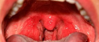
Central cyanosis is diffuse and of maximum severity. It develops with weak arterialization of the blood, leading to hypoxia. Gas exchange is disrupted in the lungs, excess carbon dioxide accumulates in the arterial blood, which is clinically manifested by blueness of the conjunctiva of the eyes, palate, tongue, mucous membranes of the lips and cheeks, and facial skin. Qualitative and quantitative changes in hemoglobin in the blood lead to disruption of its transport function and hypoxia. Acrocyanosis is localized on the feet, hands, tip of the nose, ears, lips. Peripheral cyanosis is considered a normal variant in the first days of a newborn’s life. Its origin can be easily explained by the incompletely eliminated germinal type of blood circulation, especially in premature infants. Blueness of the skin increases with swaddling, feeding, crying, and anxiety. When the infant fully adapts to the outside world, cyanosis will disappear.
Acrocyanosis in adults is a sign of varicose veins, heart failure, thrombophlebitis, vegetative-vascular dystonia, atherosclerosis, arteritis.
There are several types of cyanosis based on origin:
- The respiratory type is caused by an insufficient volume of oxygen in the lungs and a disruption in the transport chain of its supply to cells and tissues. It develops when there is a complete or partial disruption to the movement of air along the respiratory tract.
- Cardiac type - insufficient blood supply to organs and tissues leads to oxygen deficiency and bluish skin.
- The cerebral type develops when the blood loses its ability to attach oxygen to hemoglobin and deliver it to brain cells.
- The metabolic type develops when the absorption of oxygen by tissues is impaired.
- Respiratory cyanosis disappears 10 minutes after oxygen therapy; all other types persist for a long time. Massaging the earlobe helps get rid of acrocyanosis.
Symptoms
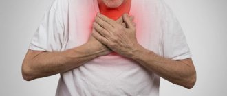
Cyanosis is a symptom of life-threatening diseases. With central cyanosis, the skin of the periorbital and perioral areas first turns blue, then it spreads to areas of the body with the thinnest skin. Peripheral cyanosis is most pronounced in areas distant from the heart. It is often combined with swelling and swelling of the veins of the neck.
Depending on the time of occurrence, cyanosis can be acute, subacute and chronic.
Cyanosis does not have a negative impact on the general well-being of patients, but in combination with other signs of the underlying pathology it becomes a reason to consult a doctor. If cyanosis occurs suddenly, increases rapidly and has a significant degree of severity, then it requires emergency care.
Cyanosis, depending on the etiology of the disease, is accompanied by various symptoms: severe cough, shortness of breath, tachycardia, weakness, fever and other signs of intoxication.
Cyanosis in bronchopulmonary diseases is manifested by a purple tint of the skin and mucous membranes and is combined with shortness of breath, wet cough, fever, sweating, and moist rales. These symptoms are characteristic of an attack of bronchial asthma, acute bronchitis and bronchiolitis, pneumonia. With pulmonary embolism, intense cyanosis develops against the background of chest pain and shortness of breath, and with pulmonary infarction it is combined with hemoptysis. Patients with similar symptoms require urgent hospitalization and respiratory resuscitation.
In heart disease, cyanosis is one of the main symptoms. It is combined with shortness of breath, characteristic auscultatory findings, moist rales, and hemoptysis. Cyanosis with heart defects is accompanied by secondary erythrocytosis, increased hematocrit, and the development of capillary stasis. Patients experience deformation of fingers like drumsticks and nails like watch glasses.
Cyanosis is not subject to special treatment. When it appears, oxygen therapy is carried out and the main treatment is intensified. Therapy is considered effective when the severity of cyanosis decreases and disappears.
In the absence of timely and effective treatment for diseases manifested by cyanosis, patients develop a disorder of the nervous system, the general resistance of the body decreases, sleep and appetite are disturbed, and in severe cases the person may fall into a coma. This condition requires emergency medical care in the intensive care unit.
Diagnostics
Diagnosis of diseases manifested by cyanosis begins with listening to complaints and collecting anamnesis. The patient is asked when the cyanosis of the skin appeared, under what circumstances cyanosis occurred, whether it is permanent or paroxysmal. Then the localization of cyanosis is determined and it is clarified how its shade changes during the day.
After a conversation with the patient, a general examination begins, the severity of his condition and the presence of concomitant diseases are established. The doctor performs auscultation of the heart and lungs.
Then they move on to laboratory and instrumental research methods
External manifestations
The cause of blue skin is pathological hemoglobin accumulated in the blood, depleted of oxygen. Blue discoloration of parts of the body does not affect health, but this phenomenon indicates that the patient needs emergency medical care.
Manifests itself in the following diseases:
- heart disease and respiratory diseases;
- venous insufficiency and Raynaud's disease;
- oncology, neuralgia, pancreatitis;
- with chest deformation;
- for food poisoning;
- plague, cholera, drug and alcohol overdose.
Blueness of the skin is often accompanied by the following symptoms:
- tachycardia;
- cough, shortness of breath;
- feverish condition;
- fever, weakness;
- varying degrees of pain.
Blue skin on certain parts of the body means:
- nasolabial triangle – congenital heart defect, heart disease;
- skin around the eyes – physical fatigue, exhaustion, nervous breakdown;
- genital organs - age-related changes;
- cervix - pregnancy;
- skin – poor circulation.
The blueness of the skin is affected by gas exchange between blood and oxygen, the speed of blood movement in the vessels and the body’s ability to bind oxygen to hemoglobin.
- Acrocyanosis: what is it, why and what diseases does it occur with?
Symptoms of the anomaly
The disorder has the ability to affect both the entire body and individual areas of the body. With a general anomaly, the following symptoms appear:
- blue color of tongue;
- discoloration of the epidermis;
- There is no change in temperature in the affected areas.
Cyanosis on the periphery looks like cyanosis of individual areas, while the tongue does not change color, and the temperature of the affected areas decreases.
Development mechanism
The pathogenesis of the pathological process is based on a group of factors. If we talk about them in a generalized format:
Breathing problems
They are especially common. In this case, the oxygen concentration itself decreases. During biochemical processes, carbon dioxide is released into the riverbed. This is a harmful, waste product. It needs to be brought out faster.
And this is where the problem arises. Not only are the lungs unable to deliver enough oxygen, but they are also unable to eliminate harmful gases that are toxic to the body.
The concentration grows, poisoning of all tissues and systems of the body begins. Need urgent medical attention.
Cardiovascular disorders
Another common factor in the development of the pathological process. There are a lot of diseases that cause cyanosis. Often we are talking about heart defects and functional disorders.
It makes sense to check this part with a cardiologist. The mechanism is based on a violation of blood transfer, its stagnation in the large and small circles.
Hence dysfunctional disorders. The heavier they are, the worse the situation is overall.
Blood abnormalities
In some cases, red blood cells themselves are not able to combine normally with oxygen and form full-fledged hemoglobin. This leads to the accumulation of carbon dioxide, which begins to poison the patient's entire body.
As a rule, the problem is acquired, but there are congenital types of the disorder.
It is necessary to approach the diagnosis carefully. A hematologist works with patients of this profile.
Endocrine disorders
Deviations from hormonal levels. They are extremely common. In this case, the tissues themselves are unable to normally receive and absorb oxygen. They produce carbon dioxide as usual.
The concentration of carboxyhemoglobin increases several times. Cyanosis is pronounced and clearly visible. Treatment is very difficult.
Intoxication processes
In case of poisoning with salts of heavy metals, vapors of acids and volatile compounds, cellular respiration is disrupted. Rapid ischemic processes occur that pose a danger to life and health. Urgent inpatient correction is needed.
These are the basic mechanisms of the problem. Typically they occur individually and not as a system. There are exceptions to what has been said.
Diagnostics
The examination is carried out under the supervision of cardiologists, pulmonologists and therapists. Subspecialty doctors are hired as needed.
A sample list of events would be:
- Questioning the patient about complaints and symptoms of the pathological process.
- Taking a history to better understand the origin of the disorder.
- ECG, ECHO-Kg. In order to investigate the condition of the heart. Anatomical (structural) and functional, detect defects and other disorders.
- Spirometry. Function of external respiration. To assess the condition of the pulmonary structures.
- General blood tests, biochemical, hormone tests. An expanded picture of lipids (lipid profile) is also needed.
- Allergy tests.
- Serological studies, bacteriological cultures, PCR, ELISA and other methods.
The question is put to rest by a specialized specialist or a group of doctors.
How to deal with cyanosis?
This pathology is not treated as a separate disease, since the symptoms indicate the presence of a more serious anomaly. Restorative measures are aimed at diagnosing the root cause and getting rid of the main pathological process. If the patient feels a constant lack of air, oxygen therapy is used. With regular use of the mask, the color of the fabrics is gradually restored, which indicates the effectiveness of the measure.
At risk are people who regularly consume ethanol-containing drinks, abuse inhalation of nicotine and combustion products, do not follow a diet, abuse fatty foods, are regularly in stressful situations, have an old post-traumatic condition of the lower extremities, diabetic syndrome, frostbite, varicose veins, maintain a regime of activity and proper rest.