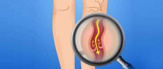
Viral hepatitis - symptoms and treatment
It is advisable to consider the detailed pathogenesis of each type of viral hepatitis in separate articles. It is enough to understand the options for normal functioning of the liver and the general mechanisms of its pathology.
The formation and metabolism of bilirubin is normal
Bilirubin is a product of the transformation of hemoglobin (Hb), a kind of “waste” that must be removed from the body. It is formed mainly from Hb, which is released from red blood cells (lifespan 120 days). The destruction of hemoglobin and the formation of bilirubin occurs in the cells of the phagocytic mononuclear cell system:
- Vysokovich-Kupffer cells, which capture and process old non-functional liver blood cells;
- splenic phagocytes - cells of the immune system that protect the body by absorbing harmful foreign particles or dying cells.
A brief scheme for the formation of bilirubin looks like this:
Red blood cells (in the spleen) - Hemoglobin (in the spleen) - Verdoglobin (a product of the enzymatic oxidation of the non-protein part of hemoglobin, formed in the cells of the macrophage system) - Biliverdin (an intermediate product of the breakdown of hemoglobin).
Stages of formation and excretion of bilirubin:
- First phase: formation of free (indirect) bilirubin in the spleen and hepatocytes (liver tissue cells).
- The second phase: its entry into the blood and transfer by albumins (simple water-soluble proteins) into hepatocytes.
- Third phase: binding of free bilirubin with glucuronic acid in hepatocytes (formation of bound, or direct, bilirubin).
- Fourth phase: release of direct bilirubin by hepatocytes as part of bile into the bile capillary.
- Fifth phase: entry of bilirubin-diglucoronide (direct bilirubin) into the duodenum in the form of bile.
Bilirubin diglucoronide can undergo two transformations:
- In the large intestine, it is converted into stercobilinogen, a substance created by colonic bacteria. Stercobilinogen is yellow in color and is further excreted in the feces in the form of stercobilin (provides the normal brown color of stool).
- In the small intestine it is converted into urobilinogen, a colorless product of the reduction of bilirubin, formed under the action of small intestinal bacteria. It is excreted through the kidneys with urine in the form of urobilin, and also enters the portal vein system into the liver with subsequent destruction.
Pathology of bilirubin metabolism in viral hepatitis
Damage to hepatocytes leads to a weakening of the uptake of free bilirubin and disruption of the process of addition of glucuronic acid to it (phase 3). As a result, the content of free bilirubin in the blood increases. Impaired transport of conjugated bilirubin into the bile capillary (phase 4) leads to an increase in conjugated bilirubin in the blood. At the same time, intrahepatic cholestasis is observed, i.e., the level of both free and bound bilirubin in the blood increases, and there is more of the latter (the skin turns yellow).
Bound bilirubin (the urine becomes dark in color) and bile acids penetrate from the blood into the urine, which reduce the surface tension of the urine and cause it to foam easily. Little bound bilirubin enters the intestines - the stool becomes discolored. Due to intrahepatic cholestasis and impaired bile secretion, little bile enters the intestine. Since bile is necessary for the digestion of fatty foods and the absorption of fat-soluble vitamins, due to its lack, a lot of fat remains in the stool (steatorrhea), and the absorption of vitamin K is impaired. Due to the low level of vitamin K, the synthesis of prothrombin in the liver decreases, as a result, blood clotting worsens .
General pathogenetic mechanisms in the liver during hepatitis
Cytolysis syndrome (cytolytic syndrome) is a set of signs indicating a disruption of the liver due to a violation of the integrity of liver cells and the release of liver enzymes into the blood. The degree of increase in the activity of aminotransferases (enzymes that reflect the functionality of the human liver) indicates the severity of the cytolytic syndrome, but does not directly indicate the depth of the dysfunction of the organ.
There is an empirical de Ritis coefficient - AsAT/AlAT. It reflects the ratio of the activity of serum AST (aspartate aminotransferase) and ALT (alanine aminotransferase). The de Ritis coefficient approximately indicates the predominant damage to one or another parenchymal organ. For hepatitis, its value is less than 1.33. In a healthy person it is in the range of 0.91-1.75.
Cholestasis syndrome is characterized by a violation of the outflow of bile. It lingers in the intrahepatic bile ducts, causing the liver tissue to swell and swell. There is an accumulation of bilirubin, cholesterol, β-lipoproteins and alkaline phosphatase in the blood. Also characteristic is the appearance of cholemia, a pathological syndrome characterized by the accumulation of bile acids in the blood. Normally, the acid content in the blood is 5-25 mmol/liter. If their number increases, cholemia develops, which is accompanied by itching, scratching, damage to the central nervous system (CNS) and staining of the skin through bile (80% bile acids + bilirubin).
Mesenchymal-inflammatory syndrome - damage to the liver parenchyma, connective tissue stroma, reticuloendothelium. Clinically expressed by enlarged liver and spleen, increased body temperature, acute phase parameters, as well as the level of autoantibodies, thymol test, β and γ proteins.
Immunosuppressive syndrome (secondary immunodeficiency) is a temporary or permanent depression of the immune system that develops under the influence of certain chemical and physical influences on the body, as well as due to certain infectious processes.
Biliary dyskinesia syndrome is a violation of their motility. It may occur due to changes in the innervation of the biliary tract due to the relative predominance of the tone of the vagus or sympathetic nerve.
Hepatic cell failure is a pathological process in which massive death of liver cells occurs. This condition is characterized by a decrease in albumin (manifested by edema syndrome) and a decrease in prothrombin (manifested by lethargy, fatigue, nausea, vomiting, hemorrhagic rash) [2][3][5][7][9].
Diagnosis of the disease
Diagnosis of chronic viral hepatitis is based primarily on laboratory methods, since in the early stages the disease is often asymptomatic.
First of all, to confirm the cause of the disease, a blood test for specific markers of viral hepatitis is necessary. These are antigens of the hepatitis B or C virus (viral particles) and antibodies produced by the body in the fight against it. In addition to the qualitative determination (detection) of the antigen, it is also necessary to study the level of viral load (the amount of antigen in a certain volume of blood). This makes it possible to judge the activity of virus reproduction and determine indications for antiviral therapy.
To assess liver function, a biochemical blood test is recommended, which includes determining the level of liver enzymes, bilirubin, protein, protein fractions and other indicators. The doctor gives more specific recommendations based on the individual picture of the course of the disease.
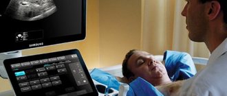
To assess the size and structure of the organ, abdominal ultrasound is recommended: this is the simplest method of visualizing the liver.
To determine the degree of fibrosis, fibroelastography or liver biopsy can be performed - sampling a small amount of liver tissue under ultrasound guidance for further examination under a microscope.
Flow
Low-active (persistent) hepatitis is asymptomatic or with minor symptoms, changes in laboratory parameters are also insignificant. Exacerbations of the process are uncharacteristic.
Chronic active recurrent (aggressive) hepatitis is characterized by severe complaints and clear objective clinical and laboratory signs. Some patients experience systemic autoallergic manifestations of the disease (polyarthralgia, skin rashes, glomerulonephritis, etc.). Frequent relapses of the disease are characteristic, sometimes occurring under the influence of even minor factors (errors in diet, overwork, etc.). Frequent relapses lead to significant morphological changes in the liver and the development of cirrhosis. In this regard, the prognosis for active hepatitis is more severe.
Liver puncture biopsy and laparoscopy make it possible to more accurately distinguish between these two forms of hepatitis, as well as to carry out differential diagnosis with other liver diseases.
A liver scan allows you to determine its size; with hepatitis, sometimes there is a reduced or uneven accumulation of the radioisotope drug in the liver tissue, in some cases there is an increased accumulation in the spleen.
Differential diagnosis in cases with a clear clinical picture of diffuse liver damage should first of all be carried out with liver cirrhosis. With cirrhosis, the symptoms of the disease are more pronounced, the liver is usually much denser than with hepatitis; it can be enlarged, but often reduced in size (atrophic phase of cirrhosis). As a rule, splenomegaly is observed, liver signs are often detected (vascular telangiectasia, hepatic tongue, hepatic palms), and symptoms of portal hypertension may occur. Laboratory tests show significant deviations from the norm in the results of the so-called liver tests; with a puncture biopsy - disorganization of the liver structure, significant proliferation of connective tissue.
Reasons for development
There is no generally accepted classification of all types of hepatitis. Depending on the reasons that caused the inflammation, there are:
- Infectious hepatitis: viral hepatitis A, B, C, D, E and others; hepatitis developing with yellow fever, cytomegalovirus infection, rubella, mumps, infection with the Epstein-Barr virus, herpes viruses, Lassa fever, HIV infection, etc.; bacterial hepatitis (for example, with syphilis, leptospirosis).
- Toxic hepatitis: alcoholic, drug-induced, and hepatitis caused by poisoning with other chemicals.
- Autoimmune hepatitis.
- Non-alcoholic steatohepatitis.
- Other types of hepatitis.
Liver fibrosis
unlike hepatitis, it is usually not accompanied by clinical symptoms and changes in liver function tests. Anamnesis (the presence of a disease in the past that could cause liver fibrosis), long-term observation of the patient and a puncture biopsy of the liver (if necessary) make it possible to differentiate it from chronic persistent hepatitis.
With fatty hepatosis, the liver is usually softer than with chronic hepatitis, the spleen is not enlarged, and a puncture biopsy of the liver is crucial in diagnosis.
Differential diagnosis
With functional hyperbilirubinemia is based on the features of their clinical picture (mild jaundice with hyperbilirubinemia without significant clinical symptoms and changes in the data of laboratory liver tests and puncture biopsy of the liver). Amyloidosis with a predominant hepatic localization, in contrast to chronic hepatitis, is characterized by symptoms of other organ localizations of the process, a positive test with Congo red or methylene blue; the diagnosis is confirmed by a puncture biopsy of the liver. With focal lesions (tumor, cyst, tuberculoma, etc.), the liver is unevenly enlarged, and scanning determines the focus of destruction of the hepatic parenchyma.
Patients with chronic hepatitis need employment (limitation of heavy physical activity, exemption from work associated with frequent business trips and which does not allow them to maintain a diet). Patients with aggressive hepatitis with rapid progression of the process are transferred to disability group III, and in some cases, group II.
Prevention of chronic hepatitis comes down to the prevention of infectious and serum hepatitis, the fight against alcoholism, eliminating the possibility of industrial and household intoxication with hepatotropic substances, as well as the timely detection and treatment of acute and subacute hepatitis.
Treatment. Patients with chronic persistent and aggressive hepatitis outside of exacerbation should follow a diet with the exclusion of certain dishes. In case of exacerbation of hepatitis, hospitalization, bed rest, and a stricter gentle diet with sufficient amounts of proteins and vitamins are indicated. In chronic cholestatic hepatitis, the main focus should be on identifying and eliminating the cause of cholestasis, in which case success can be expected from treatment measures.