What is a Baker's cyst?
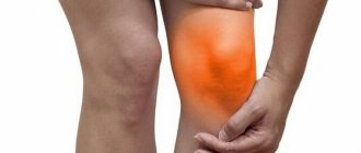
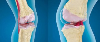
A Baker's cyst is an elastic, round-shaped protrusion located in the popliteal fossa (behind the knee joint). This formation is usually filled with synovial fluid, released as a result of any degenerative and inflammatory processes occurring in the knee joint, or injuries.
The body of the cyst forms in the mucous intertendinous bursa, where an excess amount of synovial fluid enters from the joint accumulates, which is why the gradual growth of this formation occurs. The presence of an intertendinous bursa, usually located among the tendons of the gastrocnemius and semimembranosus muscles of the leg, is observed in almost half of people, which is the norm of their anatomy.
Classification and symptoms
The neoplasm can be single-chamber, double-chamber and multi-chamber. Doctors have found that relapse of formation occurs in cases where there are multiple chambers in a cystic neoplasm. As the size of the bulge increases, the risk of it spreading to the lower leg increases. However, the considerable volume of the tumor provokes another consequence - rupture. A photo of a Becker cyst under the knee will help you understand the size of the formation that is considered threatening.
External sign.
The disease is classified into the following subtypes:
- Symptomatic form - the course of the pathological process is characterized by severe pain. Its localization is the knee joint.
- Idiopathic form - when the origin of the development of the disease is unclear, and it is not possible to establish the root cause even after conducting numerous types of research.
Absolutely asymptomatic development of popliteal bursitis is observed in no more than 28% of clinical cases . It is the absence of pronounced signs as atheroma forms and then develops that explains its late detection. Detection of neoplasms no earlier than stage 2 occurred when the inflamed areas were small in size.
Symptoms of a Becker cyst under the knee:
- Prolonged pain, often periodic, occurring inside the popliteal fossa immediately after physical activity.
- Feeling of discomfort, stiffness inside the limb, difficulty straightening the knee joint.
- Large formations compress the neurovascular bundle, causing swelling of the ankle joint.
- A rounded neoplasm can be felt under the knee - it is not fused to the skin.
- Performing several flexions and extensions helps to increase the size of the protrusion.
- Minor manifestations of neuropathy of the tibial nerve, when it, being large in size, pinched it.
The difficulty in identifying the etiological factor is associated with the anatomy of the knee joint, since synovial bursae, pockets, and inversions are located in this area. According to experts, the presence of multiple physiological structures complicates the early detection of the tumor. Also, this factor does not allow us to timely trace which particular anatomical part was the root cause of the development of the hernia.
The morphometric features of atheroma, regardless of the age and gender of the patient, are individual. Within the same age group, the volume of the hernia varies.
What are the causes of Baker's cyst formation?
The main reasons for the formation of this cyst are various inflammatory and metabolic-dystrophic diseases associated with the knee joint (especially due to excessive loads on it), injuries to the knee joint, and degenerative-dystrophic changes in cartilage tissue. Therefore, people most often at risk are those suffering from:
- any joint injuries
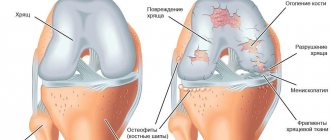
- pathological changes (including degenerative) of the menisci
- destruction of articular cartilage
- osteoarthritis
- osteoarthritis
- chronic synovitis
- rheumatoid arthritis
- patellofemoral arthrosis
Very rarely, Baker's cyst is diagnosed in patients without any apparent reason.
Features of treatment of the disease in children
Baker's cyst is most often found in children under 7 years of age. As a rule, pathology does not appear on its own, but against the background of inflammatory diseases of the joints or after injuries. The symptoms of the disease are practically no different from the clinical picture that develops in adults.
If the Baker's cyst does not cause discomfort in the child and does not grow rapidly, it is monitored for some time. Surgical treatment is prescribed only for severe impairment of the motor function of the knee joint. In children, good therapeutic results can be achieved through systematic physiotherapeutic procedures and exercise therapy.
What are the symptoms of Baker's cyst?
The initial stage of the disease, as a rule, is asymptomatic, and therefore is often ignored by the patient. Also, sometimes signs of Baker's cyst formation may disappear, but later appear again, even after a long time. The main symptoms indicating the presence of this disease are usually observed at the stage of formation of a larger cyst, when it begins to exert a compressive effect on the surrounding tissues. Then the disease may be accompanied by:
- discomfort (slight pain, especially during palpation, and an unpleasant feeling of pressure) that occurs in the popliteal fossa with sufficient physical exertion (the symptom is most pronounced in the straightened leg position)
- swelling in the knee joint, which prevents it from bending
- the presence in the popliteal fossa of a dense and elastic formation of a round shape
Diagnosis of Becker's cyst
To determine the disease, the doctor conducts a detailed examination of the patient’s knee. It is also necessary to exclude infectious inflammation. Then an ultrasound examination is performed, which is the “gold standard” for this pathology. Sometimes MRI or pneumoscintigraphy is prescribed. In case of combined pathology of the knee joint, an operation is often prescribed - arthroscopy, which is both a diagnostic and therapeutic procedure, including for this pathology, in cases where the cyst communicates with the joint cavity.
How dangerous is a Baker's cyst?
The main danger of Baker's cyst (in the absence of timely and effective treatment) is associated with its complications, which can manifest themselves:
- its rupture (the most common complication, characteristic of almost 10% of patients with this disease) - in this case, the synovial fluid of the cyst penetrates into the nearby calf muscle, filling the space between its fascia, which contributes to the formation of edema in the lower leg, which is often accompanied by general discomfort with pain , local hyperthermia and skin redness;
- deposition of calcium salts in the cyst - with more noticeable pressure of the compacted formation on the surrounding tissues;
- impaired venous circulation - the presence of a cyst often interferes with normal venous outflow in the lower leg, which, in turn, leads to the development of thrombophlebitis and thrombosis in the veins of the lower leg (which can be complicated by life-threatening pulmonary embolism);
- varicose veins located under the skin of the leg;
- compression of the tibial nerve - due to which the patient begins to experience a feeling of numbness or tingling (sometimes just weakness) in the lower leg;
- necrosis – due to compression of soft tissues;
- osteomyelitis - due to compression of the bone and its further suppuration.
What are the consequences of the disease?
If the hernia is not treated, it leads to thinning of the popliteal bursa, where the synovial fluid is collected. As a result, the capsule may rupture, and all the fluid begins to spread throughout the calf muscle. This complication is accompanied by severe swelling of the lower leg, redness of the popliteal cavity, increased temperature and the occurrence of acute pain in the joints.
Another rather dangerous complication is when the capsule does not rupture, but rapidly increases in size and begins to put pressure on the walls of blood vessels. This leads to their inflammation and the appearance of thrombophlebitis, varicose veins and thrombosis. It is worth noting that thrombosis is a fatal disease. A blood clot that breaks off can cause a stroke or block a pulmonary artery.
Baker's cyst treatment
To successfully treat this disease, depending on the size and duration of existence of the Baker's cyst, methods of conservative and surgical therapy can be used. In parallel, it is recommended to treat the underlying articular disease that provoked the development of the cyst.
Conservative therapy for Baker's cyst usually consists of puncture (to remove synovial fluid) and subsequent drug administration of glucocorticosteroid and non-steroidal anti-inflammatory drugs and painkillers into the intertendinous bursa.
Surgical therapy for Baker's cyst is indicated in the absence of effect from conservative treatment methods, as well as in large sizes of the formation and its pronounced destructive effect on surrounding tissues. The excision operation is performed using local anesthesia. Also, recently there has been a great demand for the removal of a Baker cyst using an arthroscope, which is less traumatic for the patient, since the outflow of synovial fluid is performed through a mini-puncture and this completely prevents the contents of the cyst from entering the joint cavity.
Clinical picture
Symptoms may include:
- Vague pain in the popliteal fossa.
- Swelling and formation of additional volume in the popliteal space.
- Tension in the area of swelling, which can change depending on physical activity.
- Limitations in the range of motion of the knee joint.
Most cysts are localized on the medial side of the popliteal fossa in the gastrocnemius-semimembranosus bursa. Also, a cyst can form in the popliteal bursa and then the protrusion will be located in the lateral part of the popliteal space. In addition, sometimes the popliteal cyst extends upward or anteriorly.
Cysts can range in size from small, clinically asymptomatic and non-palpable cysts, to large, space-occupying lesions that cause visible swelling in the popliteal area. The size of the cyst and the intensity of pain can lead to limited range of motion. In rare cases, there will be signs and symptoms of a meniscus tear, which can be checked with the McMurray test.
Popliteal cysts can put pressure on other anatomical structures. Compression of the popliteal artery or vein can cause ischemia or thrombosis, respectively, whereas compression of the tibial or peroneal nerves can cause peripheral neuropathy.
Prevention of Baker's cyst
The main preventive measures to prevent the occurrence of Baker's cyst usually include:
- timely and effective treatment of all pathologies associated with joints
- taking care of your joints and minimizing joint injuries
- normalization of weight (if overweight) to reduce stress on joints
- wearing special orthoses (if necessary) and knee pads in order to minimize the load on the knee joint (especially when performing heavy work and lifting heavy objects)
- maintaining an active lifestyle
- good nutrition
Have you started to worry about unpleasant sensations in the popliteal fossa or have you noticed a slight lump there? Experienced medical specialists are always ready to promptly determine the true cause of your ailment and return you to the joy of comfortable movement as quickly as possible!
Physiotherapy
Physiotherapeutic procedures are prescribed after relief of an acute inflammatory process or during the rehabilitation period after surgery. A pronounced effect is obtained during a series of sessions:
- mechanical traction - prevents the development of atrophy;
- massage of the knee joint and lower legs - to restore blood supply and nutrition of soft tissues;
- electrophoresis - activates tissue regeneration, normalization of metabolic processes;
- UHF, infrared laser therapy - to relieve pain, eliminate inflammation and swelling;
- magnetic therapy - restores metabolic processes and restores damaged tissue at the cellular level;
- hydrogen sulfide, radon baths - for the prevention of thrombophlebitis, varicose veins, stimulation of local blood circulation.
During physiotherapy, the condition of the damaged knee joint and the degree of its motor activity are constantly assessed. You can learn more about physiotherapy methods here.
Conservative therapy
An orthopedist is involved in eliminating the type of pathology in question; if the need arises, surgeons are involved in the implementation of the therapeutic process.
Conservative treatment of Becker cyst under the knee involves the following approaches:
- unloading the joint;
- physiotherapeutic treatment;
- puncture of the tumor, followed by evacuation of the contents and administration of medications.
For therapeutic purposes, sclerosing agents are administered, in particular, a 5% alcohol solution of iodine, hormonal, and cytostatic drugs are used.
- Physiotherapeutic treatment . Electrophoresis and UHF are recognized as the most effective methods. It is permissible to implement these treatment options only after excluding an oncological process.
- Puncture . Performed to evacuate the contents of the hernia. When the tumor reaches stage 2-3, the contents of its capsule have a jelly-like consistency. The compacted neoplasm is difficult to puncture and subsequent evacuation of the internal part. Also in this case there is no point in administering corticosteroids.
- A positive result is provided by hormonal treatment . Cortisone therapy is effective in 50% of all clinical cases. The scientific idea of using hormonal drugs was first developed and applied by rheumatologists. Patients with a hernia that occurred against the background of rheumatoid arthritis were subject to observation. Therapeutic tactics made it possible to evaluate that after intra-articular administration of glucocorticosteroids, the size of the tumor decreased.
Pumping out liquid.
Since then, hormones have been injected directly into the popliteal swelling. Hydrocortisone emulsion was actively used, combining it with antibiotics.
Important! If the patient has hormonal diseases - problems with the thyroid gland, diabetes mellitus, a disorder of the adrenal cortex - it is necessary to inform the doctor about the existing disorders.
of Becker cyst is not always possible
leads to complete elimination of the tumor. Statistics show that in 6 out of 10 cases, the atheroma has to be excised surgically. Such results serve as the basis for the search for new therapeutic approaches and the development of more effective conservative methods of therapy.
Development factors
Doctors have not yet established the exact causes of popliteal hernia. This tumor develops slowly and very rarely becomes malignant.
Reference. Rare cases of transformation of a knee hernia into a malignant formation are most often associated with an initially incorrect diagnosis. Doctors are mistaken, since many malignant formations are similar to Becker’s cyst.
Doctors identify the following alleged theories for the appearance of hernias of the knee joints:
- Inflammatory diseases. A popliteal cyst develops against the background of pathologies of bone joints and periarticular tissues (synovitis, vaginitis, tendovaginitis). These diseases lead to the fact that the knee joint is deformed and gradually collapses. As a result, a “weak spot” appears, which is subjected to daily physical stress, and later a protrusion forms.
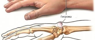
- Benign tumor process. Sometimes the cells of the synovial membrane begin to divide, and this is how the tumor process begins. More often, such cellular elements grow inside the articular membrane, then synovioma sarcomas (malignant tumors) are formed, less often they grow outward, as a result, hygroma develops in the knee.
- Violation of metabolic processes. As mentioned earlier, the cavity of the joints and tendon sheaths is filled with fluid produced by the synovial membrane. In autoimmune, endocrine, metabolic diseases, and paraneoplastic syndrome, the amount of synovial fluid produced exceeds the volume of secretion that is reabsorbed. Because of this, the pressure in the tendon sheaths and joint capsule increases. As a result, the weakest part of the connective tissue membrane protrudes and a hygroma appears.
The features of a popliteal tumor are inherent in all the above-described theories of its formation.
Pathological formation of the knee joint can be caused by the following factors:
- Hereditary predisposition in a child whose parents suffered from the disease.
- One-time severe injury or frequent minor injuries to the knee.
- Excessive physical stress of the knee joint associated with work or sports.
- Pathologies that are accompanied by inflammatory damage to the knee (arthritis, bursitis, periarthritis, etc.).
- Chronic diseases (arthritis, osteoarthritis).
Attention. Most often, a popliteal hernia is diagnosed in people who lead an active lifestyle. To prevent the pathological process, professional athletes and people who engage in heavy physical work must use protective devices (orthoses, bandages, elastic bandages).