Causes of atrial septal defect
The formation of an atrial septal defect is based on a violation of the development of the primary or secondary septum in the embryonic period.
- The primary defect is located in the lower part of the interatrial septum next to the fibrous rings of the atrioventricular valves (mitral and tricuspid); in fact, its lower edge is the fibrous ring itself.
- The secondary defect is located in the central part of the interatrial septum, and all its edges are represented by the septum itself.
- A venous sinus defect is a defect located in the upper part of the interatrial septum. This defect is often accompanied by abnormal drainage of the pulmonary veins (part of the pulmonary veins flows not into the left atrium, but into the superior vena cava).
Prevention
{banner_banstat10}
Prevention of ASD includes careful planning of pregnancy, prenatal diagnosis, and exclusion of exposure to adverse factors. To prevent the development of pathology in a child, a pregnant woman must follow the following recommendations:
- Eat a balanced and nutritious diet,
- Observe the work and rest schedule,
- Regularly attend antenatal clinics,
- Eliminate bad habits
- Avoid contact with negative environmental factors that have toxic and radioactive effects on the body,
- Do not take medications without a doctor's prescription,
- Receive immunization against rubella in a timely manner,
- Avoid contact with sick people,
- During epidemics, do not visit crowded places,
- Undergo medical and genetic counseling in preparation for pregnancy.
A child with a congenital defect and its treatment requires careful special care.
Hemodynamics
The meaning of the defect is the discharge of arterial blood from the systemic circulation (left atrium) into the pulmonary circulation (right atrium), which leads to overload of the pulmonary circulation with an excess volume of blood, primarily affecting the lungs. An extreme form of damage is the formation of pulmonary hypertension (high pressure in the vessels of the lungs). Pulmonary hypertension due to atrial septal defect is malignant, leading to severe heart failure and death of the patient.
How does pathology manifest itself?
Health complaints are mainly due to decompensation of blood flow. The stronger the overload of certain parts of the system, the more pronounced the symptoms will be. Patients begin to worry about shortness of breath, fatigue, and frequent colds, especially in children.
When listening to the heart, a systolic murmur is heard with a source above the pulmonary artery. The sounds are caused by difficulty when too much blood flows through the lumen of the pulmonary trunk. The ECG shows signs of overload on the right side of the heart with hypertrophy in this area.
Prices for services
| Name | Time | Price | |
| Appointment with a cardiologist, treatment and diagnostic, primary, outpatient | 1500 | ||
| Treatment and diagnostic appointment with a cardiologist, repeated, outpatient | 1000 | ||
| Preventive, outpatient appointment with a cardiologist | 550 | ||
| Comprehensive consultation with a cardiologist ECG with interpretation + Echo-kg | 3650 | ||
| Call a doctor to your home (Khimki, Starbeevo, Ivakino, Naberezhnye) | 15000 | ||
| Calling a doctor to your home (Skhodnya, Podrezkovo, Novopodrezkovo, Planernaya station, Firsanovka) | 15000 | ||
| Call a doctor to your home (Kurkino, Novogorsk) | 15000 | ||
| Call a doctor to your home (Left Bank, Moscow, within the Moscow Ring Road) | 15000 | ||
| Medical consultation | 3300 | ||
| Extract from outpatient card | 660 | ||
| Consultation (interpretation) with analyzes from third parties | 1300 | ||
| Consultation on treatment adjustments | 550 | ||
| Repeated reception of CMN | 1600 | ||
| Initial reception of CMN | 2000 | ||
| Express test for determining the level of Troponin in the blood. | 1900 | ||
| Expand all prices | |||
Symptoms
The first symptoms of the disease occur in the form of shortness of breath during physical exertion, frequent colds, and a feeling of interruptions in the functioning of the heart. At the same time, the symptoms are varied and depend on the location and size of the defect, its combination with other heart defects. Small defects do not have a characteristic clinical picture. Patients do not actively complain, their physical activity is not limited - the defect is discovered by chance. With large, hemodynamically significant defects, patients experience shortness of breath, increased fatigue, and exercise intolerance. After exercise, a cough may occur, sometimes accompanied by hemoptysis. Such patients are characterized by frequent pneumonia and bronchitis.
ASD and pregnancy
The issue of bearing a child with ASD deserves special attention. The presence of this defect requires mandatory consultation with a doctor at the beginning of planning. For example, you may need to stop taking certain medications.
In general, as a rule, patients with a small defect tolerate pregnancy quite easily. However, if there are complications of the defect and pronounced symptoms of ASD, certain problems may arise. In any case, the best solution in this situation is to consult a doctor.
Diagnostics
- Examination of the patient with auscultation (listening) of the heart reveals a heart murmur.
- An ECG reveals right ventricular myocardial hypertrophy, right bundle branch block, arrhythmias of varying severity, and a sharp deviation of the electrical axis of the heart to the left.
- X-ray of the chest organs reveals dilation of the pulmonary artery trunk, enlargement of the heart, and increased pulmonary pattern.
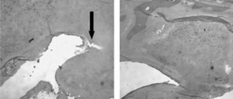
- Transthoracic echocardiography allows not only to visualize the atrial septal defect and clarify its nature (primary, secondary, venous sinus defect), but also to assess the direction of blood discharge through the defect and its hemodynamic significance (Qp/Qs).
- Transesophageal echocardiography in adults allows one to obtain detailed information about the edges of the defect, which is important for choosing a method of surgical treatment.
- Probing of the heart chambers and atriography is used when there is insufficient information from the research methods described above or as part of surgical treatment.
Publications in the media
Atrial septal defect (ASD) is a congenital heart defect with communication between the atria. Statistical data: 7.8% of all congenital heart disease detected in infancy, and 30% in adults; VSD of the ostium secundum type - 70%, ostium primum - 15%, sinus venosus - 15%; Lutembashe syndrome accounts for 0.4% of all cases of ASD, the combination of ASD with mitral valve prolapse - 10–20%; the predominant gender is female (2:1–3:1).
Etiology: factors that form congenital heart disease (see Tetralogy of Fallot).
Pathogenesis • The magnitude and direction of shunt depend on the size of the defect and the relative compliance of the ventricles • In adults, the right ventricle is more compliant than the left, as a result of which shunt occurs from the left atrium to the right • A small shunt leads to a moderate volume overload of the right heart, and pressure in the pulmonary artery remains normal • The severity of pulmonary hypertension may be insignificant even with a large shunt • Only in rare cases does severe pulmonary hypertension develop, leading to right ventricular failure and right-to-left shunt • Unlike VSD, with ASD the shunt is smaller and affects only the right side of the heart.
Variants of ASD • Ostium secundum (secondary defects) are localized in the area of the oval fossa, are often multiple, accompany many syndromes: Holt-Oram syndrome (ASD of the ostium secundum type in combination with digital hypoplasia), Lutembashe syndrome (combination of ASD with mitral valve stenosis), combination of ASD with mitral valve prolapse, etc. • Ostium primum (primary defects) are usually large in size, localized in the lower part of the septum, at the attachment point of the mitral and tricuspid valves, interatrial and interventricular septa. They are an integral part of the open AV canal and are often combined with abnormal drainage of the pulmonary veins, splitting of the anterior mitral valve leaflet, mitral regurgitation and Down syndrome • Defects of the sinus venosus type are localized near the mouth of the superior vena cava and the sinus node, often combined with sick sinus syndrome, AV nodal rhythm and abnormal pulmonary venous drainage.
Clinical picture
Complaints: shortness of breath, palpitations, fatigue during physical activity, retarded physical development, frequent infections, paradoxical embolisms.
Objectively • Pallor of the skin • Harrison's furrows - displacement of areas of the chest as a result of chronic shortness of breath • Splitting of the first tone with a pronounced component of the tricuspid valve • Pronounced fixed splitting of the second tone (pronounced - due to prolongation of the time of blood ejection from the right ventricle; fixed - due to - due to the fact that the dependence of venous return on the phases of breathing is leveled by discharge from the left atrium) • Ejection click and soft systolic murmur of relative pulmonary artery stenosis in the second intercostal space to the left of the sternum • Due to increased blood flow through the tricuspid valve, low-frequency diastolic murmur sometimes occurs above the xiphoid process of the sternum.
Instrumental diagnostics
• ECG. Signs of hypertrophy and overload of the left sections, and with pulmonary hypertension - also of the right. With ostium primum, there is a sharp deviation of the EOS to the left due to the displacement of the hypoplastic branch of the left leg of the His bundle forward. Various variants of sick sinus syndrome, AV block. With a sinus venosus type defect, there is an inferior atrial rhythm or an AV junction rhythm.
• Jugular venography: equal amplitude of A and V waves.
• X-ray examination of the chest organs. Strengthening the pulmonary pattern. Expansion and lack of structure of the roots of the lungs, bulging of the right atrium arch and upward displacement of the right cardiovasal angle. Fluoroscopy reveals increased pulsation of the roots of the lungs (a rather specific sign). The “Turkish saber” symptom with concomitant anomalous drainage of the right pulmonary veins into the superior vena cava.
• EchoCG. Hypertrophy and dilatation of the left sections, and with pulmonary hypertension - also of the right. Visualization of ASD in Doppler and B-mode. Differentiation from an open foramen ovale (the anatomical closure of the latter occurs no later than 2 years of life) is the inconsistency of visualization of the discharge in color Doppler mapping and the presence of a leaflet in the cavity of the left atrium. Diagnosis of associated anomalies (abnormal drainage of the pulmonary veins, valvular defects, etc.). The degree of discharge and the ratio of pulmonary minute blood flow to systemic blood flow (Qp/Qs) are determined. Adults undergo transesophageal echocardiography. With intravenous contrasting of the right parts of the heart, there is a negative contrast effect (displacement of the contrast agent by a stream of blood from the left atrium).
• Radionuclide angiocardiography (first pass method or equilibrium): registration of pathological discharge and its quantitative assessment, diagnosis of concomitant abnormal drainage of the pulmonary veins and ventricular dysfunction.
• Probing of the cardiac cavities •• Indicated for suspected pulmonary hypertension, before open-heart surgery and with conflicting clinical data •• If the catheter can be passed from the right atrium to the left, then this in itself cannot be a sign of an atrial septal defect: sometimes the catheter it is possible to carry out through the open foramen ovale •• Tests are carried out with aminophylline and oxygen inhalation to determine the prognosis regarding the reversibility of pulmonary hypertension •• The ratio of pulmonary minute blood flow to systemic blood flow (Qp/Qs) is calculated - a reference indicator of the amount of discharge.
• Right atriography, angiopulmonography: flow of contrast from the right atrium to the left; identification of concomitant abnormal pulmonary venous drainage.
Drug therapy. In uncomplicated ASDs of the ostium secundum type, infective endocarditis prophylaxis is usually not carried out. For ASDs of the ostium primum type, large defects of the sinus venosus type, and a combination of ASDs with mitral valve defects, antibiotics are prescribed before and for 6 months after uncomplicated surgical correction. For right ventricular failure, diuretics are prescribed.
Surgery
Indications: Qp/Qs ratio is 1.5 or more, defects of the ostium primum type, large defects of the ostium secundum type, concomitant hemodynamically significant anomalies (abnormal drainage of the pulmonary veins, mitral stenosis, etc.).
Contraindications: severe concomitant pathology that threatens the patient’s life; end-stage circulatory failure, irreversible pulmonary hypertension, the ratio of total pulmonary vascular resistance to peripheral vascular resistance is 0.9 or more.
Methods of surgical treatment. Endovascular correction with a button or two-patch Sideris device or an Amplatz device is feasible for central defects no larger than 2 cm in size. In the absence of experience in endovascular treatment, small defects are sutured under artificial circulation. In other cases, ASD repair with a synthetic or autopericardial patch under artificial circulation is recommended.
Specific postoperative complications • Sick sinus syndrome (after correction of sinus venosus defects) • AV block (after correction of ostium primum defects) • If mitral regurgitation existed before surgery, symptoms may worsen after correction of ASD • Atrial fibrillation that occurred before surgery , as a rule, persists after it.
Forecast. In early childhood the course is benign. In rare cases, severe circulatory disorders can lead to death in the first months of life. Spontaneous closure of the defect is possible before the age of 5. The average life expectancy without treatment is 40 years. 5–15% of patients die before age 30. 10-year survival rate - 90%, 20-year - 88%, 30-year - 67%; 40-year-old - 44%, 50-year-old - 25%, 60-year-old - 13%, 70-year-old - 7%. More than 75% of patients with large defects die from other causes. For uncomplicated defects of the ostium secundum type, perioperative mortality is less than 1%, it is slightly higher for defects of the ostium primum type, the latter also require mitral valve replacement or repair.
Abbreviations. Qp/Qs is the ratio of the pulmonary minute volume of blood flow to the systemic one.
ICD-10 • Q21.1 Atrial septal defect
Treatment of atrial septal defect
There are currently two types of surgical treatment:
- 1. Open heart surgery under artificial circulation with suturing of the defect or its plastic surgery with a patch from the pericardium.
- 2. Elimination of atrial septal defect using special devices (occluders) in the X-ray operating room through arterial punctures without sternotomy.
- It is necessary to understand that not every defect can be eliminated surgically, there are contraindications to surgery (small-diameter defects, severe pulmonary hypertension, right-left discharge through the defect), and not every defect can be eliminated using occluders (a combination of a defect with abnormal drainage pulmonary veins, primary defects with missing lower edge, large defects, combination of defect with another
- heart pathologies are an indication for open surgery).
- You can learn more about a specific case of the disease, indications and contraindications during a consultation with a cardiovascular surgeon (at an outpatient appointment).
How to get treatment in Israel
When deciding on treatment in Israel, a patient with developmental defects of internal organs, including the interatrial septum, receives many advantages, including the use of the latest scientific recommendations and innovative equipment, reasonable cost, rapid recovery of operated children.
Thanks to the coordinated work of specialists from different fields, surgeons cope with pathology even in very complex cases. Ignorance of the language will not become an obstacle to communication - each patient is accompanied by a Russian-speaking consultant.
To get into Hadassah, you need to fill out an electronic application. An employee of the international department will contact you, provide the necessary information and answer additional questions. By using our services, you will not have to overpay to intermediaries - all payments are made only to the cash desk of the medical center before hospitalization.