Exudative otitis media (ESO) is called a chronic form of otitis media, in which pathological fluid (exudate) accumulates in the middle ear as a result of damage to its mucous membrane. With ESO, the integrity of the eardrum is not compromised and in the absence of an inflammatory process in the nasal cavity, nasopharynx and oropharynx, infection of the middle ear does not occur, despite the fact that the formed exudate is a favorable environment for the development of viruses and bacteria, since it is a protein environment.
It is precisely because the exudate contains a large amount of protein that over time it changes its physical properties (thickens, becomes viscous), which leads to a long and severe course of the disease.
Pain is one of the signs of inflammation. The absence of inflammatory changes in the middle ear explains the fact that ESO is painless. This is the insidiousness of this pathology.
In the medical literature you can find other names for this common disease, namely: “ exudative otitis media ”, “ secretory ”, “ mucosal otitis ”, “ effusive otitis media ”, “gleu ear” - “sticky ear”.
Causes of exudative otitis media in children
The main reasons contributing to the development of ESO are:
- changes in the mucous membrane of the auditory tube against the background of acute and chronic inflammatory diseases of the nose, paranasal sinuses and nasopharynx as a result of decreased immunity,
- dysfunction of the auditory tube, due to dysfunction of the muscles that open it,
— obstruction of the mouth of the auditory tube by adenoid vegetations (growths), hyperplastic (enlarged) tubal tonsil, cicatricial changes, benign and malignant neoplasms of the nasopharynx,
- ineffective treatment of acute otitis media,
— anatomical and physiological features of the development of the auditory tube in childhood.
It should be noted that one of the predisposing factors to the occurrence of ESO is visiting a children's day care center. At the age of 2 to 7 years, the development of this pathology may be preceded by an acute viral infection, chronic adenoiditis, and acute catarrhal otitis media. In children aged 8 to 15 years, the development of exudative otitis media occurs against the background of vasomotor rhinitis or chronic rhinosinusitis.
Causes
The tympanic cavity is lined with a layer of epithelial cells that constantly produce small amounts of fluid. Normally, excess of this fluid is removed into the nasal cavity through the auditory tube, a violation of the drainage function of which is the main cause of exudative inflammation of the middle ear. The outflow of fluid can be obstructed by:
- Diseases of the auditory tube itself;
- Untreated acute otitis media with persistent edema;
- Swelling of the nasal mucosa in the place where the auditory tube opens (rhinitis);
- Mechanical obstruction to outflow (adenoids, benign tumors, deviated nasal septum);
Some medical interventions in the nasal cavity (septum and turbinate plastic surgery, tumor removal, nasal tamponade) can also lead to the accumulation of exudate in the ear.
Types of exudative otitis media in children
According to their duration, ESO is divided into three forms : acute (up to 3 weeks), subacute (3 to 8 weeks) and chronic (more than 8 weeks).
According to the nature of the changes occurring in the mucous membrane of the middle ear, four forms of ESO : initial exudative, secretory, productive secretory, degenerative-secretory (with a predominance of the fibrosclerotic process).
There is another classification of ESO, which is based on similar principles (physical parameters of the contents of the tympanic cavity: viscosity, transparency, color, density and duration of the pathological process). It distinguishes four stages of ESO: catarrhal (up to 1 month), secretory (from 1 to 12 months), mucosal (from 12 to 24 months), fibrous (more than 24 months).
What is needed for diagnosis?
To diagnose this disease, it is necessary to conduct a complete otorhinolaryngological examination. The most informative examination today is using video endoscopic equipment. During the examination, the doctor can observe the fluid level and air bubbles behind an unchanged or thickened, clouded eardrum. If necessary, to confirm the diagnosis, tympanometry is recommended, after which the otolaryngologist will be able to draw a complete picture of the condition of the tympanic cavity and middle ear as a whole.
During a traditional examination, the pathology may remain undetected and be diagnosed already at the stage of complications and hearing loss by an audiologist.
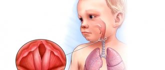
Exudative otitis media in children.
Both adults and young patients suffer from exudative otitis media. But diagnosing exudative otitis in a child can be more difficult than in adults, not only due to insufficiently clear symptoms, but also due to the difficulties that arise during the collection of complaints and anamnesis (young patients do not complain). Complaints and clinical picture directly depend on the stage of the disease. The initial stage of ESO is characterized by scant complaints and clinical manifestations.
In most cases, parents seek medical help for their child only when they notice a decrease in his hearing, that is, the child begins to speak loudly, does not immediately respond to calls, asks to increase the volume when watching children's television programs, which already corresponds to the productive secretory form of the disease. It is the absence of pain that leads to late detection of ESO. But sometimes young patients are able to describe their feelings. Adults consult a doctor with complaints of a sensation of fluid in the ear (“gurgling”), a feeling of fullness, autophony, and changes in hearing depending on the tilt of the head.
Prevention of diseases in the middle ear
Competent preventive measures significantly reduce the risk of developing pathology, which reduces both the ability to hear and the quality of life in general. To protect your baby from ESO, follow these recommendations:
- treatment of inflammation in the nasopharynx should be complete;
- make an appointment with an otolaryngologist after suffering a bacterial infection;
- ensure proper and regular nasal hygiene;
- make an appointment with the dentist at the first signs of caries development in your child;
- dress your baby in clothes that suit the season
These tips help prevent the development of any pathology in the middle ear. If the disease is detected, do not allow it to enter the chronic phase.
Diagnosis of exudative otitis media in children
To diagnose EOS, an audiological examination is carried out at the Federal State Budgetary Institution National Medical Research Center of the Federal Medical and Biological Agency of Russia in the Department of Pediatric ENT Pathology, which consists of acoustic impedance testing and tone threshold audiometry. For patients with this pathology, acoustic impedansometry is characterized by a tympanometric curve of type “B” and the absence of ipsi-reflexes, which reflects a violation of the transmission of the sound signal along the chain of auditory ossicles due to the pathological contents of the middle ear (exudate); on the audiogram - an increase in air conduction thresholds to 30-40 dB, mainly at low frequencies, bone conduction is not changed. In the case of a recurrent course of exudative otitis media, specialists from the Department of Pediatric ENT Pathology necessarily prescribe the patient a computed tomography (CT) scan of the temporal bones to obtain a reliable picture of the airiness of the middle ear cavity, the condition of its mucous membrane, the chain of auditory ossicles, the windows of the labyrinth and the bony part of the auditory tube, density and localization of pathological contents.
To identify the causes contributing to the occurrence of ESO, the patient in the Department of Pediatric ENT Pathology of the National Medical Research Center of the Federal Medical and Biological Agency of Russia undergoes an endoscopic examination of the nasal cavity and nasopharynx or an X-ray examination of the nasopharynx (in young children if endoscopy is not possible) and paranasal sinuses.
Diagnostics
An otolaryngologist diagnoses the disease. First of all, when a patient contacts, the doctor collects an anamnesis of the disease and. After listening to all the patient’s complaints, he examines the ear (otoscopy). During the examination, changes in the structure of the eardrum and its deformation are determined, and if the membrane is thinned, the volume of accumulated exudate is determined. The main examination methods used for suspected exudative otitis media are:
- audiometry – it is used to determine the patient’s hearing level, as well as his sensitivity to sounds of different frequencies;
- determination of the ventilation functions of the Eustachian tube;
- determination of the mobility of the eardrum, for which the Valsalva maneuver is used;
- endoscopic examination of the ear cavity and Eustachian tube - the same examination of the nasal cavity may be required;
- X-ray – prescribed if there are suspicions of cellular disorders;
- computed tomography is necessary in a situation where, after all the examinations, difficulties arise in making an accurate diagnosis due to the receipt of somewhat contradictory data.
In some exceptional cases, the patient may be referred for a general blood and urine test, as well as an ear smear. Usually it is quite easy for a doctor to make a correct diagnosis. Difficulties can arise only when pathology appears in a small child who cannot explain what is happening to them. Therefore, in children under three years of age, the disease most often becomes chronic, since there is no treatment, due to the fact that the disorder goes unnoticed.
Symptoms of exudative otitis media
It is important to consider that exudative otitis media is accompanied by painful sensations, usually only in complicated cases.
Typically, patients with exudative otitis media complain of decreased hearing and a feeling of fullness in both ears or one ear.
Hearing impairment with the development of exudative otitis is fluctuating, since the patency of the resulting mucus plug can decrease and increase.
In addition, the disease can go away naturally, but then the threat of its recurrence remains.
Introduction
The reason for once again turning our attention to the treatment of patients with exudative otitis media (EOM) was the multiple errors of specialists at different stages of providing medical care to this group of patients. The reason for the inappropriate actions was partly the significant difference in treatment methods for adults and children. As a rule, otolaryngologists working in adult hospitals deal with acute dysfunction of the auditory tubes, which is detected in the very first hours of the development of the disease against the background of acute rhinosinusopathy, requiring hospitalization. Inpatient treatment and early diagnosis in most cases make it possible to stop the process through a course of conservative treatment or laser tympanostomy with the formation of a short-term functioning tympanostomy. In children, dysfunction of the auditory tube is most often caused by chronic obstruction of the nasopharynx, chronic adenoiditis, recurrent rhinosinusitis and banal recurrent acute respiratory viral infections. There is no quick cure, early age does not imply independent complaints about the developed sensation of ear congestion, hearing loss, and accordingly, diagnosis is usually late. All of the above determines the need for a separate approach to the treatment of ESO in children and adults.
The choice of treatment method for severe dysfunction of the auditory tube in children is determined by the etiology and pathogenesis of the disease. It is generally accepted that ESO is a chronic inflammation of the middle ear, accompanied by the accumulation of exudate behind the eardrum without signs of acute inflammation [1]. Histologically, the process in the state of hypoventilation of the middle ear cavities manifests itself during the 1st month of the disease by an increase in the number of goblet cells and the formation of mucous glands in the middle ear, starting from the pharyngeal mouth of the auditory tube. The growth of the altered mucous membrane and the spread of the process throughout the tympanic cavity and into the cells of the mastoid process in the chronic form of the disease for 4 months or more is accompanied by persistent impairment of auditory function. Degeneration of the glands occurs with interruption of the vacuum state in the middle ear, and recovery is manifested by a gradual decrease in the frequency of goblet cell division and flattening of the epithelium of the mucous membrane [2, 3]. In accordance with the pathogenesis of the disease, since 1995 we have been using a classification that divides the disease into 4 stages: catarrhal, secretory, mucosal and fibrous, which is presented in detail in our methodological recommendations [4] and the National Guidelines [5].
Despite the high likelihood of spontaneous improvement, severe and prolonged cases of the disease cause persistent changes in the middle ear: atrophy, retraction and atelectasis of the tympanic membrane, adhesive otitis media, chronic otitis media with cholesteatoma, and in some cases sensorineural hearing loss. In young children, ESO can cause a temporary delay in speech development [6, 7].
The purpose of the study is to improve the efficiency of providing medical care to children with ESO.
results
All subjects had a chronic form of ESO; the main method of treatment was surgery. Sanitation of chronic diseases of the nose, paranasal sinuses, and pharynx is the first mandatory stage of treatment [8, 9].
To restore pneumatization of the middle ear cavities in preschool-aged patients with the secretory stage of ESO, it is initially possible to use Politzer's blowing of the auditory tubes, and in patients older than 6-7 years - catheterization of the auditory tube with the introduction of vasoconstrictors, antibacterial, corticosteroid and mucolytic agents for an average course of 10 days . In addition, oral mucolytics are useful to liquefy secretions in the middle ear cavities. Be sure to use vasoconstrictor and anti-inflammatory nasal drops for an average of 5-7 days. Among the means of physiotherapy, endaural phonophoresis with fluimucil, electrophoresis with potassium iodide on the mastoid process to liquefy secretions and prevent scarring in the cavities of the middle ear, amplipulse on the lateral surfaces of the neck to stimulate the work of the muscles of the pharynx (course of 8-12 procedures), as well as exercises for strengthening the muscles of the palatine curtain. The effectiveness of treatment should be monitored by tympanometry [6, 7].
As we presented previously, most patients underwent upper airway debridement and tympanostomy. In cases of primary ESO treatment, a coil tube was used for short-term ventilation (6–12 months). Patients who were candidates for revision tympanostomy were considered candidates for surgery using long-term T-tubes and balloon dilatation of the eustachian tubes. This group also included 4% of children with craniofacial anomalies and pharyngeal tumors.
Tympanostomy was performed under general anesthesia mainly in the anterioinferior quadrant (78.6% of cases). Viscous exudate was obtained in 86% of patients with the mucosal stage of ESO and 56% with the fibrotic stage. During the secretory stage of ESO and in other cases at other stages, the secretion in the cavities of the middle ear was liquid. Patency of the auditory tube was absent or reduced in 60.5% of cases (tested intraoperatively with transtympanic administration of corticosteroids). Hearing function returned to normal in 95.2% of patients and improved in 4.1%.
Balloon dilatation of the auditory tubes was performed in 11 patients (14 cases) with persistent dysfunction of the auditory tubes, who underwent repeated tympanostomy (Fig. 1, 2, Fig. 3 on color insert) . This type of ESO treatment was first used in 2010 and has now become available in our country.
Rice. 1. Histological confirmation of the mechanism of balloon dilatation of the auditory tube.
Rice. 2. Scheme of balloon insertion.
Rice. 3. Ballonization of the auditory tube of patient S.
According to the authors who developed and used balloon dilatation, its mechanism is the formation of micro-tears in the cartilage, followed by replacement of the resulting connective tissue defects and the formation of a wider lumen. No trauma to the mucous membrane of the auditory tube is noted [12]. Our experience also allows us to agree that there is no damage to the mucous membrane.
The longest follow-up period was 2 years. Relapse of the disease has now been registered in 2 patients: one underwent effective repeated balloon dilatation, the second underwent a short course of catheterization of the auditory tube on the affected side with the use of local corticosteroids, which led to the restoration of tympanometry parameters.
More than 20 years of experience in managing patients with ESO has shown that such patients require long-term follow-up by an otolaryngologist and audiologist, since the disease is prone to recurrence in 36% of cases [11]. We do not recommend removing short-term ventilation tubes in children. Their extrusion usually occurs independently towards the external auditory canal (EA) within 8-12 months. The appearance of mucous or purulent discharge in the NSP (in 6.8% of our observations versus 15% in foreign authors) was regarded by us as acute purulent otitis media, which required conventional treatment, but without removal of the ventilation tube [6]. T-shaped tubes do not spontaneously extrude; removal is carried out no earlier than 2 years after their installation in stationary conditions. We do not recommend leaving them for more than 3 years, as this leads to the formation of persistent perforation of the eardrum. During follow-up observation during otoscopy, foci of myringosclerosis form around the ventilation tube in a number of cases (22.3%), which do not impair hearing function by more than 5 dB HL [7]. The appearance of atelectasis of the tympanic membrane (17.7%) reflects ongoing dysfunction of the auditory tubes and late treatment with ESO [11]. Among our patients, only in 1% of cases did we observe persistent perforations after tympanostomy; in this case, tympanoplasty was performed (subject to stable normalization of the function of the auditory tube).
According to our observations, prolonged rhinitis of any etiology or inflammation of the middle ear can provoke an exacerbation of the disease; accordingly, the patient and his parents should be warned about a mandatory audiological examination after such episodes.
Treatment of exudative otitis media
An important step in treatment is eliminating the leading cause. Most often, grade 2-3 adenoid vegetations occur in children. The presence of exudative otitis media is a direct indication for adenotomy (removal of the pharyngeal tonsil), if there is severe hypertrophy. In the absence of hypertrophy of adenoid tissue, conservative treatment is carried out: treatment of rhinitis or sinusitis if present, rinsing the nasal cavity using the displacement method, Politzer blowing, catheterization of the auditory tube, pneumomassage of the eardrums, physiotherapy. Among the medications, antibacterial drugs can be prescribed systemically or nasally, anti-inflammatory and antihistamine drugs locally or systemically, drugs that reduce the viscosity of mucus (mucolytics), and nasal douche. If there is no effect from the treatment, surgical intervention is indicated - eardrum bypass or tympanostomy. With this intervention, a small hole of 2-3 mm in diameter is created in the eardrum, through which exudate is removed, then a short metal or polymer tube is installed there, through which the eardrum is ventilated and medications are administered into it. The shunt may remain in the eardrum for a long time. If during a bypass operation (especially if the duration of the disease is unknown) a very viscous exudate is detected that cannot be removed from the tympanostomy, then a tympanotomy with revision of the tympanic cavity is recommended. During this operation, a semicircular incision is made in the skin of the external auditory canal in the bony part, its detachment along with the tympanic membrane, which allows a good examination of the tympanic cavity and clearing it of exudate; after tympanotomy, a shunt is often left. With exudative otitis media in adults, which is especially difficult to respond to conservative treatment, it is important to exclude a neoplasm in the nasopharynx, which can lead to this disease. Often the cause of exudative otitis in adults is chronic inflammation of the nasal mucosa or paranasal sinuses, as well as deviated nasal septum, and accordingly, treatment of these diseases is required. If there is no effect from conservative treatment, tympanic cavity shunting or tympanotomy may be required.
Adhesive otitis media and tympanosclerosis are a morphological continuation of chronic inflammation in the middle ear and occur with untimely or incorrect treatment, as well as in the absence of treatment. They are also called fibrosing otitis due to the fact that connective tissue growth occurs in these diseases. Treatment, as a rule, is surgical, which consists of tympanotomy with revision of the tympanic cavity, with the removal of foci of tympanosclerosis or fibrous tissue from the auditory ossicles in case of adhesive otitis. Conservative treatment is also possible: the introduction of proteolytic enzymes during catheterization of the auditory tube or by electrophoresis, pneumomassage of the eardrums, and Politzer blowing.
Hearing examination for otitis media - why is it important?
Acute otitis media is harmless in most cases and, with proper treatment, almost always goes away without any consequences. Is there at least one child who has not suffered from otitis media? I think no. Under certain conditions, this disease, even without treatment, can end safely on its own. However, there are situations when acute otitis media occurs with complications. The audiologist is primarily concerned with the risk of sensorineural hearing loss.
Let's consider an ordinary case of treatment of acute otitis: the ear hurt and hearing was reduced, the person turned to an ENT doctor, who prescribed treatment for him, but did not order an audiogram (“test” of hearing), hoping that otitis would proceed without complications and after recovery hearing will be restored. 2 weeks pass, the ear no longer hurts, the ENT doctor sees upon examination that there is no trace of otitis left. But my hearing was not restored. Sends the patient for an audiogram and uses it to detect sensorineural hearing loss. The fact is that this complication requires specific treatment, and its results very much depend on the timing of its onset. If the acute sensorineural hearing loss is more than a month old, then the therapy is completely ineffective.
An audiogram for acute otitis media allows you to understand why hearing is reduced, whether there are complications in the form of sensorineural hearing loss, predict the results of treatment and, sometimes, adjust treatment.
There are no problems with “regular” audiometry in adults and children over 3-3.5 years old. But what about children who are younger? They are not always able to perform game audiometry. Of course, there are methods for diagnosing hearing without the participation of a child at this age, but they are very labor-intensive, require expensive equipment, take a lot of time, have a relatively large error and, as a rule, there is a waiting list. Therefore, in early childhood, they prefer to cure otitis media and then test their hearing using objective research methods. In fairness, it must be said that in early childhood complications in the form of sensorineural hearing loss are relatively rare.
Clinical picture
Patients with this diagnosis often complain of hearing loss, which occurs as a type of sound conduction, a feeling that liquid is pouring or squelching in the ear. When performing otoscopy, otomicroscopy, video otomicroscopy (special methods of examining the ear), the eardrum is thickened and looks somewhat “swollen”, but no noticeable hyperemia (redness) is observed. In some cases, the fluid level can be seen through the eardrum. During paracentesis (surgical dissection of the eardrum), viscous and viscous mucus is released, which, upon special examination, will show a high content of eosinophils. In case of perforation of the eardrum, ENT patients complain of discharge from the ear, which may be serous or mucopurulent in nature.
Friends! Timely and correct treatment will ensure you a speedy recovery!
ESO treatment
Treatment of patients diagnosed with ESO is aimed at eliminating the causes that led to dysfunction of the auditory tube, followed by restoration of hearing and preventing the development of morphological changes in the mucous membrane of the middle ear.
Treatment tactics directly depend on the stage of the disease. At the initial stages, conservative treatment methods are prescribed to relieve inflammatory processes in the nasal cavity and nasopharynx.
Next, wait-and-see tactics for 2-5 months + gymnastics for the auditory tubes (self-blowing, otovent ball).
If conservative methods of therapy are ineffective, the patient is indicated for surgical treatment aimed at eliminating the causes leading to blockage of the mouth of the auditory tube and disruption of its functions (adenotomy, surgery on the paranasal sinuses for the rehabilitation of chronic foci of infection) + shunting of the eardrum according to indications. When the child's hearing is normal, the shunt is removed.