Fungal infections (mycoses) are diseases that are caused by different types of microscopic fungi.
Mycoses of the skin, mucous membranes, and nails are the most common among all human fungal diseases. Systemic mycoses (affecting several body systems) are predominantly found in people with immunodeficiency. Most often these are HIV-infected patients, patients after organ transplantation, cancer patients, etc.
The main causative agents of skin mycoses: trichophyton fungi Trichophyton rubrum, Trichophyton mentagrophytes, var. interdigitale, Epidermophyton floccosum and candida Candida.
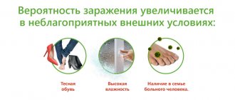
Infection with fungi occurs through direct contact with a patient, through shoes, clothing, when using general hygiene products (washcloths, manicure tools), when visiting gyms, baths, saunas, and swimming pools. The main factor of infection is the presence of abrasions and cracks in the skin, which occur with increased sweating, dryness, and poor circulation in the extremities. People with diabetes, immune disorders, blood diseases; Those taking antibiotics, glucocorticosteroids, and cytostatics for a long time are especially susceptible to the development of mycoses.
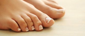
Most often, microscopic fungi cause damage to the skin of the feet and large skin folds (axillary, groin and others).
What it is? Terms and Definitions
Skin mycoses
– fungal infections of the skin and its appendages (hair, nails) by pathogenic fungi of various types. In fact, this is a large group of diseases united by a single criterion - the causative agent. Mycoses can manifest themselves in different ways, affecting only the skin or its appendages, some can simultaneously cause systemic lesions, that is, extend their effect to internal organs. Approaches to the treatment of different mycosis also differ: some require the use of local, topical drugs, such as ointments or creams for mycosis, some are supplemented with tablets for mycosis, oral forms of systemic drugs, in some cases doctors are forced to expand the range the drugs used against mycosis in order to first stabilize the patient’s condition and then rid him of the pathogenic fungus.
Types of deep mycoses
The classification of deep mycoses is based on the type of pathogen and includes the following forms:
- blastomycosis and cryptococcosis;
- histoplasmosis;
- coccidioidosis;
- sporotrichosis;
- mucorosis;
- aspergillosis;
- penicilliosis;
- chromomycosis;
- rhinosporidiosis;
- cephalosporiosis;
- cladosporiosis;
- keloid mycosis;
- mycetomas of a fungal nature.
Deep pseudomycosis includes actinomycosis and nocardiosis.
It is generally accepted that the causative agents of most of these diseases cause deep endemic mycosis, that is, they are found only in certain areas of the Earth. These include blastomycosis, coccidioidosis, paracoccidioidosis, histoplasmosis, penicillium, chromomycosis. They are found in some regions of America, Southeast Asia, Africa, and Australia. However, due to increasing population migration, these diseases may also occur in residents of other areas.
Symptoms of deep mycosis do not always depend on its cause. Patients develop cough, fever, pain in bones and joints, pain in the chest, right hypochondrium, and swollen lymph nodes. When the brain and its membranes are damaged, consciousness is impaired and coma develops.
Epidemiology
Fungal infections of the skin and nails
is a common global problem. The high prevalence of superficial fungal infections indicates that 20–25% of the world's population has mycoses of the skin, mycoses of the trunk, and mycoses of the feet, making them one of the most common forms of skin infections. Their distribution depends little on the average temperature or humidity in a particular country, geographical location and other reasons. Mainly - from background or previous diseases, from the state of immunity, including local, from previous treatment, especially antibacterial drugs, from age, personal and public hygiene and some other factors.
1.What are fungal diseases?
Fungal diseases (mycoses)
- infections caused by pathogenic fungal cultures. The methods of their transmission are different: by contact, through inhalation of spores, with food and drinks, through objects, from animals.
In a healthy state, human skin is protected from fungal infections, since the acid-base balance of its surface is slightly shifted towards acidity. However, under certain conditions (in closed shoes, with intense sweating in the heat), the natural balance is disrupted, and there is a danger of infection with skin fungal diseases.
A must read! Help with treatment and hospitalization!
Causes of the disease
Cause of mycoses
– various pathogenic fungi that can live both on the skin (feet, legs, hands, arms, head, torso) and on its appendages (scalp hair, body hair, fingernails and toenails).
In the host's body during mycosis, various pathological changes occur both due to the presence of the infectious agent and due to the products of its metabolism. All mycoses can be divided into three categories:
| Type of mycosis | Pathogen | Occurrence |
| Dermatophytosis Ringworm of the scalp, skin and nails | Dermatophytes (Arthroderma, Lophophyton, Microsporum, Nannizzia, Trichophyton, Epidermophyton) | Regular |
| Candidiasis of the skin, mucous membranes and nails | Candida, Debaryomyces, Kluyveromyces, Meyerozyma, Pichia, etc. | Regular |
| Dermatomycosis | Nondermatophytic forms of Neoscytalidium, Scopulariopsis | Rare |
Ecology of common human dermatophyte species:
| Kinds | Natural habitat | Occurrence |
| Epidermophyton floccosum | People | Regular |
| Trichophyton rubrum | People | Frequent |
| Trichophyton interdigitale | People | Frequent |
| Trichophyton tonsurans | People | Regular |
| Trichophyton violaceum | People | More rare |
| Trichophyton concentricum | People | Very rare |
| Trichophyton schoenleinii | People | Very rare |
| Trichophyton soudanense | People | Very rare |
| Microsporum audouinii | People | More rare |
| Microsporum ferrugineum | People | More rare |
| Trichophyton mentagrophytes | Mice and other rodents | Regular |
| Trichophyton equinum | Horses | Very rare |
| Trichophyton eriotrephon | Jerzy | Very rare |
| Trichophyton verrucosum | Cattle | Very rare |
| Microsporum canis | Cats | Regular |
| Nannizzia gypsea | The soil | Regular |
| Nannizzia nana | Soil, pigs | Very rare |
| Nannizzia fulva | The soil | Very rare |
| Nannizzia persicolor | Voles and bats | Very rare |
| Lophophyton cookei | The soil | Very rare |
| Lophophyton gallinae | Chickens and other poultry | Very rare |
Causes
The causative agents of systemic mycoses often live in the soil of endemic areas. Upon contact with dust and soil, they enter the human respiratory tract. With subsequent population migration, the infection spreads to more prosperous countries.
The respiratory route of infection into the body distinguishes deep mycosis from superficial one. With mycoses of the skin, nails or hair, infection occurs by contact. In this case, you can avoid infection by using means for the prevention of fungal diseases. These include the drug Mikospray, which you can buy in our online store fitosila.ru in Moscow at an affordable price.
A patient with deep mycosis himself does not pose an immediate threat to others. Infection occurs indirectly. In the patient’s body, pathogenic fungi multiply and transform into a special “tissue” form, which is safe if it comes into direct contact with the skin of another person. The tissue form of the fungus is excreted with the sputum and feces of the patient and enters the external environment (water, soil, air).
In the environment, this form of fungus transforms into mycelial one. If it enters through food, water or air, it can cause illness in another person.
In many cases, to activate fungi in the body, it is necessary to reduce the immune defense of the human body. These conditions arise with prolonged and unreasonable use of antibiotics, cytostatics, and anti-inflammatory hormones. Against this background, not only an infection that comes from outside can develop, but also internal saprophytic fungi that are harmless to a healthy person (actinomycetes, aspergillus, candida, mold fungi).
One of the main reasons for the development of systemic mycoses in our country is acquired immunodeficiency syndrome. In this case, the infection is very severe, difficult to treat and often causes an unfavorable outcome.
In addition, there are cases of infection of laboratory personnel working with fungal cultures.
Transmission routes
The main source of infection is humans
.
The main route of transmission is contact
. Activation of opportunistic flora is also possible when problems with the immune system occur. Fomites, that is, objects contaminated with pathogenic fungi, can be household items, shoes, clothing, and bedding of the sick person, if people who are sensitive to this fungus actively come into contact with them. Outbreaks are possible in organized groups, mainly in children's and army groups. Among public places, swimming pools, water parks and other structures where people can be barefoot inside are considered a common variant of fungal infection, plus a warm and humid microclimate is formed there, which contributes to the long existence of the pathogen outside the host.
In rare cases, for example, with microsporia, the source of infection can be a street or domestic animal, most often a cat. In common parlance, this disease is called “lichen.” “Ringworm” or, more precisely, “ringworm” in a child is microsporia. It is also possible to become infected with trichophytosis through contact in rural areas with hay or other substrates on which the secretions of sick rodents remain.
Methods for diagnosing mycoses
These infections are uncommon in general practice, so diagnosis is usually difficult. In addition, these diseases have similar external signs to other skin lesions, a variety of general manifestations, and are often combined with cutaneous tuberculosis and pyoderma.
They often occur “under the guise” of chronic ulcerative pyoderma. An in-depth examination of such patients reveals deep mycosis.
The basis of diagnosis is recognition of the tissue form of the fungus, which has characteristics for each pathogen. To do this, it is necessary to obtain a section of tissue, for example, during a biopsy, stain it with special substances and examine it under a microscope.
Obtaining a culture of the pathogen, that is, growing it on a nutrient medium, is extremely difficult and is carried out only in specialized laboratories.
Additional diagnostic methods:
- skin biopsy;
- determination of antigens and antibodies to fungus in blood serum;
- X-ray of lungs and bones;
- lumbar puncture;
- bone marrow puncture.
Types of disease
In the vast majority of cases, varieties of mycoses are named by the name of the pathogen: trichophyton causes trichophytosis, microsporum causes microsporia.
They differ primarily in their characteristic affected areas. Classification of mycoses:
| Type of mycosis | Affected area |
| Tinea pedis, athlete's foot | Feet |
| Tinea unguium, onychomycosis | Nails on fingers and toes |
| Tinea corporis | Skin of hands, feet and body |
| Tinea cruris, jock itch | Inguinal folds, armpits |
| Tinea manuum | Skin of the palms and back of the hands |
| Tinea capitis | Scalp |
| Tinea barbae | Scalp hair, including scalp and face |
| Tinea faciei | Skin on the face |
| Tinea versicolor, pityriasis versicolor, sun fungus | All skin |
Symptoms
The most common symptoms that lead to a visit to a doctor include nonspecific symptoms characteristic of the majority of mycoses. The following early signs may indicate this:
- Peeling and redness of the skin
- Itching, often unbearable
- Wetting in the folds of the skin
- Small vesicles (bubbles) that burst and dry on their own
Other symptoms of fungal diseases depend on the specific pathogen of mycosis.
Tinea pedis
There are four main clinical variants of tinea pedis; sometimes they overlap each other in manifestations. The most common variant is intertriginous, which is characterized by cracking, peeling, or maceration of the interdigital areas; unpleasant odor; itching; and a burning sensation. The infection often affects the lateral webbing of the toes and may spread to the sole or instep of the foot. Warm and humid conditions can worsen skin conditions. The second option is the chronic papulosquamous type, which often occurs on both feet. This type is characterized by mild inflammation and scattered peeling of the skin on the soles of the feet. The third option consists of small blisters or pustules on the instep and plantar surface. There is peeling of the skin in this area, as well as the membranes of the toes. The fourth option includes macerated, open, weeping ulcers on the sole of the foot with a characteristic odor. This option is often complicated by opportunistic gram-negative bacteria.
Tinea corporis
It can manifest itself in different ways and in different parts of the body. The lesions often appear as small, round, erythematous, scaly areas. In the center, cleansing occurs as the boundaries expand and vesicles or pustules develop. Tinea corporis can occur on any part of the body, depending on the type of dermatophyte infection. Animal-borne zoodermatophytes often infect exposed areas of skin, whereas anthropodermatophytes primarily infect closed or injured areas.
Tinea cruris
Jock itch occurs on the medial and upper thighs and groin area and is more common in men than women; the scrotum often remains intact. Symptoms such as unbearable itching, a feeling of constant weeping and burning are often present. Risk factors for this tinea pedis infection include tinea pedis infection, obesity, diabetes, and immunodeficiency.
Ringworm of the head
The exact incidence pattern of this form is unknown, but it most often occurs in children who come into contact with other children or pets. There are three types of tinea capitis: blackhead, gray spot and favus. Trichophyton often causes black dotted dermatitis on the scalp and is the predominant variant. Gray spotted dermatophytosis on the head occurs in epidemic and endemic forms, but the epidemic form is practically not registered in developed countries. The endemic form, caused by Microsporum, is often spread by cats and dogs. Favus, more common in Eastern Europe and Asia.
The blackhead variant is often asymptomatic at first. The erythematous scaly patch on the scalp enlarges over time and localized alopecia occurs. The hairs inside the spots break and a characteristic black dot appears (caused by the accumulation of detritus in the opening of the follicle). If left untreated, blackhead fungus can cause alopecia and scarring to become permanent. Sometimes the affected area may change and become raised, painful, severely inflamed, with nodules known as kerions. Kerion formation occurs due to an immune response to the fungus. Lymphadenopathy may occur when kerion occurs. The variant with gray plaques on the scalp consists of round patches of alopecia with noticeable flaking. Kerion formation can also occur in this case.
Onychomycosis
This type of mycosis is most often caused by dermatophytes, but it can also be caused by non-dermatophytes and Candida species. Affected nails often become thick, rough, yellow, opaque, and brittle. The nail may separate from the nail bed, and the dermis surrounding the infected nail may be hyperkeratotic. Risk factors include diabetes, trauma, family history, athlete's foot, smoking, prolonged exposure to water, and immunodeficiency.
Diagnostics
The diagnosis is confirmed by the detection of segmented hyphae in skin scrapings from the affected area with potassium hydroxide (KOH). When bubbles form, the top of the bubble can serve as an adequate sample. Alternative diagnostic procedures are dermatophyte culture test medium and culture method.
Patients who have significant erosion, ulceration, or malodor in the affected area should undergo Gram staining and culture to evaluate for secondary bacterial infection.
Differential diagnosis depends on clinical subtype:
Interdigital dermatitis of the foot
- Erythrasma
- Interdigital candidiasis
- Hyperkeratotic (moccasin) dermatitis of the foot
- Atopic dermatitis
- Chronic contact dermatitis
- Chronic palmoplantar (dyshidrotic) eczema
- Palmoplantar psoriasis
- Pitted keratolysis
- Juvenile plantar dermatosis
- Exfoliating keratolysis
- Keratoderm
Inflammatory dermatosis of the foot
- Acute palmoplantar (dyshidrotic) eczema
- Acute contact dermatitis
- Palmoplantar pustulosis
- Scabies
A positive KOH test demonstrating segmented hyphae distinguishes Tinea pedis from nonfungal diseases. With interdigital candidiasis, a test with the drug KOH will show budding yeast, pseudohyphae and hyphal septa.
Treatment and drugs
Treatment of mycoses
recommended to relieve symptoms (itching), reduce the risk of secondary bacterial infection, and limit the spread of infection to other areas of the body or to other people. Local antifungal therapy is the treatment of choice for most patients. Systemic antifungals are primarily intended for patients in whom topical therapy has failed. Also, systemic therapy comes to the fore for Tinea capitis, Tinea barbae, Tinea imbricata.
Topical medications effective for dermatophytosis include azoles, allylamines, butenafine, ciclopirox, tolnaftate, and amorolfine. A meta-analysis of randomized trials published up to February 2005 supports the effectiveness of topical therapy, finding strong evidence of superiority of topical antifungals (azoles, allylamines, ciclopirox, tolnaftate, butenafine, and undecanoate) over placebo. Allylamines may be slightly more effective than azoles; a meta-analysis of data from 11 studies that compared topical allylamines with topical azoles found a slightly higher cure rate for allylamines (hazard ratio for treatment failure 0.63, 95% confidence interval 0.42–0.94). Topical antifungal treatment is usually applied once or twice daily and continued for four weeks. Shorter courses of treatment may be effective; high cure rates were obtained with the use of 1% terbinafine cream for interdigital foot dermatitis for one week.
Patients requiring oral antifungal therapy are usually treated with terbinafine, itraconazole, or fluconazole. Typical treatment regimens for adults include:
- Terbinafine: 250 mg daily for two weeks.
- Itraconazole: 200 mg twice daily for one week.
- Fluconazole: 150 mg once a week for two to six weeks.
Griseofulvin can also be used to treat mycoses of the skin, but is likely to be less effective than other oral antifungals and will also require a longer course of therapy. A systematic review found that terbinafine was more effective than griseofulvin, while the efficacy of terbinafine and itraconazole was similar. Typical adult doses of griseofulvin for tinea pedis are 1,000 mg griseofulvin (microcapsule) per day for four to eight weeks or 660 or 750 mg griseofulvin (ultramicrocapsule) per day for four to eight weeks.
The dosage for children is based on weight, and the duration of treatment is similar to that for adults. Typical pediatric dosages for oral therapy include:
Terbinafine tablets:
- 10 to 20 kg: 62.5 mg per day
- From 20 to 40 kg: 125 mg per day
- Over 40 kg: 250 mg per day
Terbinafine granules:
- Less than 25 kg: 125 mg per day
- 25 to 35 kg: 187.5 mg per day
- Over 35 kg: 250 mg per day
Itraconazole: 3 to 5 mg/kg per day.
Fluconazole: 6 mg/kg once a week.
Griseofulvin (microcapsules) 10 to 20 mg/kg per day or griseofulvin (ultramicrocapsules) 5 to 15 mg/kg per day.
The exact regimens should be selected by the attending physician in accordance with the type of lesion, pathogen and clinical variant of mycosis.
Patients with hyperkeratotic dermatitis of the foot may benefit from combining antifungal treatment with a topical keratolytic such as salicylic acid. Wet Burow dressings (1% aluminum acetate or 5% aluminum subacetate) applied for 20 minutes two to three times daily or gauze or cotton between the toes may be useful as an adjunctive measure for patients with vesiculation or maceration. Interventions that may help reduce relapses include using dehydrating foot powders, treating shoes with antifungal powder, and avoiding tight shoes.
Mycoses of smooth skin
Among the widespread fungal diseases today, the most common are mycoses of smooth skin, such as microsporia, trichophytosis, lichen versicolor, mycosis of the feet (hands), and candidiasis. Sources of infection can be sick animals (cats, dogs, mouse-like rodents, cattle, etc.), as well as humans. In recent years, there has been an increase in the number of diseases caused by opportunistic fungi, among which superficial forms of candidiasis are most often recorded. Such a wide prevalence of these mycoses can be explained by the massive use of modern therapeutic agents, the environmental situation and other factors that reduce the body's defenses. One of the reasons for the significant prevalence of mycoses is the weakening of sanitary educational work in recent years. Due to insufficient awareness about the sources and ways of spreading infection, as well as adequate preventive measures, patients turn to the doctor late, and therefore mycoses become chronic, including in children suffering from mycoses of the scalp and smooth skin.
Microsporia is a fungal disease caused by various types of fungi of the genus Microsporum. In Russia, microsporia, which has spread over the last 50 years, is caused by a zoophilic fungus - fluffy microsporum (Microsporum canis), which parasitizes the skin of cats, dogs, and less often other animals. Infection from a sick person is observed in 2% of cases.
Epidemiology . Infection in 80-85% of cases occurs as a result of direct contact with a sick animal or through objects contaminated with the fur of these animals. Infection of children can also occur after playing in the sandbox, since the causative agent of microsporia is highly resistant to environmental factors and can remain viable in infected scales and hair for up to 7-10 years. Children often suffer from microsporia.
Clinic . After 5-7 days from the moment of infection, lesions appear on smooth skin, which can be observed on both open and closed parts of the body (children love to pick up animals and put them in bed with them). The lesions are round or oval in shape, pink or red, with clear boundaries, a raised ridge along the periphery, covered with blisters and thin crusts, with peeling in the center. The lesions are usually small, from 1 to 2 cm in diameter, single or multiple, sometimes merging. In 85-90% of patients, vellus hair is affected.
Treatment . If there are single foci of microsporia on smooth skin without damage to vellus hair, you can limit yourself to only external antifungal agents. The lesions should be lubricated with alcohol tincture of iodine (2-5%) in the morning, and sulfur-salicylic ointment (10% and 3%, respectively) should be rubbed in in the evening. You can rub the following antimycotics 2 times a day: mycozolon, mycoseptin, travogen or 1 time a day in the evening - mifungar cream, mycospor - until the clinical manifestations resolve. In case of multiple lesions of smooth skin and single lesions (up to 3) involving vellus hair, it is recommended to prescribe the antifungal antibiotic griseofulvin at the rate of 22 mg per 1 kg of the child’s body weight, in 3 doses after meals, in combination with exfoliating the stratum corneum of the epidermis in keratolytic lesions means (salicylic acid 3.0, lactic or benzoic acid 3.0, collodion up to 30.0). The lesions are lubricated with one of these products 2 times a day for 3–4 days, then 2% salicylic ointment is applied under compress paper for 24 hours, the detached scales of the stratum corneum of the epidermis are removed with tweezers and vellus hair is epilated. If during a control study carried out using a fluorescent lamp or microscope, affected hair is detected, the procedure is repeated. Detachment of the stratum corneum of the epidermis and manual hair removal of vellus hair can be carried out after using the “sealing” method. The lesions are sealed in a tile-like manner with strips of adhesive plaster for 2-3 days, this causes an aggravation of the process, which, in turn, facilitates hair removal.
The results of treatment for smooth skin microsporia are monitored using a fluorescent lamp or microscopic examination for fungi. The first control study is done after the resolution of clinical manifestations, then 3-4 days before the first negative test, and then after 3 days. The criteria for cure are resolution of the lesions, absence of luminescence and three negative tests on microscopic examination.
During the treatment, bed and underwear are disinfected: boiling in a soap-soda solution (1%) for 15 minutes (10 g of laundry soap and 10 g of caustic soda per 1 liter of water); ironing outerwear, furniture covers, and bedding five times with a hot iron through damp cloth.
Prevention. The main measure to prevent microsporia is compliance with sanitary and hygienic rules (you cannot use other people’s underwear, clothes, etc.; after playing with animals, you must wash your hands).
Trichophytosis is a fungal disease caused by various types of fungi of the genus Trichophyton. Trichophytons can be anthropophilic, parasitic on humans, and zoophilic, whose carriers are animals. Anthropophilic trichophytons include Trichophyton (Tr.) tonsuraus and Tr. violaceum, to zoophiles - Tr. mentagrophytes var gypseum and Tr. verrucosum.
Epidemiology. With superficial trichophytosis, caused by anthropophilic fungi, infection occurs through close contact with a sick person or indirectly through household items. Often children become infected from their mother, grandchildren from grandmothers suffering from a chronic form of the disease. The incubation period lasts up to a week. In zooanthroponotic trichophytosis, the sources of infection are sick animals: cattle, rodents. The highest incidence of this type of trichophytosis is recorded in the fall, which is associated with field work: it is at this time that the likelihood of infection through hay and straw increases. The incubation period ranges from 1–2 weeks to 2 months.
Clinic. On smooth skin with superficial trichophytosis, lesions can appear on any part of the skin - face, neck, chest, forearms. They have clear boundaries of a round or oval shape, with a raised ridge along the periphery of a bright red color; they are larger in size than in microsporia. The lesions are reddish-bluish in color, with peeling, nodules on the surface; in the chronic form, they develop on the skin of the buttocks, knee joints, forearms, less often the back of the hands and other parts of the body; the lesions do not have clear boundaries. Lamellar peeling is observed on the skin of the palms and soles. Vellus hairs are often affected.
With trichophytosis, caused by zoophilic fungi, the disease on the skin can occur in three forms: superficial, infiltrative and suppurative. Lesions are usually located on open areas of the skin. With a superficial form, they are round or oval in shape, with clear boundaries, a raised ridge along the periphery, on which bubbles, crusts, a pink center, and a bright red ridge are visible. The lesions are larger in size than with microsporia. Sometimes they are located around natural openings - eyes, mouth, nose. In the infiltrative form, the lesions rise above the skin level and are accompanied by inflammatory phenomena - infiltration. The suppurative form is characterized by the development of tumor-like formations, bright red in color, covered with purulent crusts due to the addition of a bacterial infection. When the lesion is compressed, pus is released from the hair follicles and pain is noted. The disease is accompanied by a violation of the general condition, sometimes the temperature rises. After the resolution of clinical manifestations, cicatricial atrophy of the skin remains at the site of former lesions. Clinical forms of zooanthroponotic trichophytosis can transform into one another.
Diagnostics. The diagnosis of trichophytosis is established on the basis of the clinic and when the fungus is detected during microscopy of pathological material, and the type of pathogen is determined using cultural examination.
Treatment. Treatment is carried out with antimycotics for external use. The lesions are lubricated with tincture of iodine (2-5%) during the day, and sulfur-salicylic ointment (10% and 3%, respectively) or mycoseptin is rubbed in in the evening. You can carry out monotherapy with ointment or cream (kanison, mifungar, mycozoral, mycospor (bifosin), exoderil, mycozoral, etc. In the infiltrative form, 10% sulfur-tar ointment is prescribed to resolve infiltration 2 times a day. Treatment of the suppurative form of trichophytosis begins with removing crusts from the affected area using bandages with 2% salicylic ointment, which are applied for several hours. After removing the crusts, vellus hair is epilated. Then apply lotions with solutions that have a disinfectant and anti-inflammatory effect (furacilin 1:5000, rivanol 1:1000 , potassium permanganate 1:6000, ichthyol solution (10%), etc.). As a result of this treatment, the hair follicles are freed from pus, inflammatory phenomena are reduced. Next, sulfur-tar ointment (5-10%) is prescribed for resorption of the infiltrate (5-10%) in the form of rubbing or under wax paper.After the infiltrate has resolved, antimycotics are used for external use (see superficial form of trichophytosis).In cases where vellus hair is affected in lesions on smooth skin, the stratum corneum of the epidermis is detached, followed by hair removal. To do this, you can use salicylic collodion (10-15%), milky-salicylic-resorcinol collodion (15%). If there is no effect, griseofulvin is prescribed orally at a daily dose of 18 mg per 1 kg of body weight, in 3 divided doses after meals daily until a negative test for fungi, then every other day. As an alternative method, terbinafine (Lamisil, Exifin) can be prescribed to adults 250 mg (1 tablet) once a day after meals every day, children weighing up to 20 kg - 62.5 mg, from 20 to 40 kg - 125 mg , over 40 kg - 250 mg in combination with antimycotics for external use.
The criteria for cure for trichophytosis are resolution of clinical manifestations and three negative fungal test results at three-day intervals.
Prevention. Prevention of trichophytosis depends on the type of pathogen. With superficial trichophytosis caused by anthropophilic fungi, the main preventive measure is to identify the source of infection, and it can be children suffering from superficial trichophytosis, or adults suffering from a chronic form of the lesion. In recent years, cases of chronic trichophytosis have been observed in middle-aged and older children. For suppurative trichophytosis, preventive measures are carried out jointly by medical workers, epidemiologists and veterinary services.
Mycosis of the smooth skin of the feet (hands). In a number of countries, up to 50% of the population suffers from mycosis of the feet. This disease is more common in adults, but in recent years it has often been observed in children, even infants.
Etiology. The main causative agents of mycosis of the feet are the fungus Trichophyton rubrum (T. rubrum), which is isolated in almost 90% of cases, and T. mentagrophytes var. interdigitale (T. interdigitale). Damage to the interdigital folds, which may be caused by yeast-like fungi, is recorded in 2-5% of cases. The anthropophilic fungus Epidermophyton floccosum is rarely isolated in our country.
Epidemiology. Infection with mycosis of the feet can occur in the family through close contact with a patient or through household items, as well as in a bathhouse, sauna, gym, or when using someone else's shoes and clothes.
Pathogenesis. The penetration of fungi into the skin is facilitated by cracks and abrasions in the interdigital folds caused by sweating or dry skin, abrasion, poor drying after water procedures, narrowness of the interdigital folds, flat feet, etc.
Clinic. Clinical manifestations on the skin depend on the type of pathogen and the general condition of the patient. The T.rubrum fungus can cause damage to the skin of all interdigital folds, soles, palms, dorsum of the feet and hands, legs, thighs, inguinal-femoral, intergluteal folds, under the mammary glands and axillary area, trunk, face, and rarely the scalp. The process may involve vellus and long hair, nail plates of the feet and hands. When the skin of the feet is affected, there are 3 clinical forms: squamous, intertriginous, squamous-hyperkeratotic.
The squamous form is characterized by the presence of peeling on the skin of the interdigital folds, soles, and palms. It can be flour-shaped, ring-shaped, lamellar. In the area of the arches of the feet and palms, an increase in the skin pattern is observed.
The intertriginous form is the most common and is characterized by slight redness and peeling on the lateral contact surfaces of the fingers or maceration, the presence of erosions, superficial or deep cracks in all folds of the feet. This form can transform into dyshidrotic, in which vesicles or blisters form in the area of the arches, along the outer and inner edges of the feet and in the interdigital folds. Superficial blisters open with the formation of erosions, which can merge, resulting in the formation of lesions with clear boundaries and oozing. When a bacterial infection is attached, pustules, lymphadenitis and lymphangitis occur. In the dyshidrotic form of mycosis, secondary allergic rashes are observed on the lateral and palmar surfaces of the fingers, palms, forearms, and shins. Sometimes the disease becomes chronic with an exacerbation in spring and summer.
The squamous-hyperkeratotic form is characterized by the development of foci of hyperkeratosis against the background of peeling. The skin of the soles (palms) becomes reddish-bluish in color, and pityriasis-like peeling is noted in the skin grooves, which extends to the plantar and palmar surfaces of the fingers. Pronounced ring-shaped and lamellar peeling may be detected on the palms and soles. In some patients, it is insignificant due to frequent hand washing.
In children, lesions of smooth skin on the feet are characterized by fine-plate peeling on the inner surface of the terminal phalanges of the toes, usually 3 and 4, or there are superficial, less often deep cracks in the interdigital folds or under the toes, hyperemia and maceration. On the soles, the skin may not be changed or the skin pattern may be enhanced; sometimes ring-shaped peeling is observed. Subjectively, patients are bothered by itching. In children, more often than in adults, exudative forms of lesions occur with the formation of blisters and weeping eczema-like lesions. They appear not only on the feet, but also on the hands.
Rubrophytia of smooth skin of large folds and other areas of the skin is characterized by the development of lesions with clear boundaries, irregular outlines, with an intermittent ridge along the periphery, consisting of merging pink nodules, scales and crusts, with a bluish tint (the color is bluish-pink in the center) . On the extensor surface of the forearms and shins, rashes can be located in the form of open rings. Lesions with nodular and nodular elements are often observed. The disease sometimes occurs as an infiltrative-suppurative trichophytosis (more often in men when localized in the chin area and above the upper lip). Foci of rubrophytosis on smooth skin can resemble psoriasis, lupus erythematosus, eczema and other dermatoses.
The fungus T. interdigitale affects the skin of the 3rd and 4th interdigital folds, the upper third of the sole, the lateral surfaces of the foot and toes, and the arch of the foot. This mushroom has pronounced allergenic properties. With mycosis of the feet caused by T. interdigitale, the same clinical forms of damage are observed as with rubrophytosis, but the disease is often accompanied by more pronounced inflammatory phenomena. In the dyshidrotic, less often intertriginous form, large blisters may appear on the skin of the soles and fingers, along with small blisters; in the case of bacterial flora, with purulent contents. The foot becomes edematous, swollen, and pain appears when walking. The disease is accompanied by an increase in temperature, deterioration of health, development of allergic rashes on the skin of the upper and lower extremities, torso, face, enlargement of the inguinal lymph nodes; the clinical picture is similar to that observed with eczema.
Diagnosis. The diagnosis is established on the basis of clinical manifestations, detection of the fungus by microscopic examination of skin flakes and identification of the type of pathogen by cultural examination.
Treatment. Treatment of mycosis of the smooth skin of the feet and other localizations is carried out with antifungal agents for external use. For squamous and intertriginous forms of lesions on the feet and other areas of the skin, medications are used in the form of a cream, ointment, solution, spray; you can combine a cream or ointment with a solution, alternating their use. Currently, the following medications are used to treat this disease: exifin cream, mycozoral cream, nizoral cream, canizon cream and solution, mycozon cream, mycospor (bifosin) cream, mifungar cream, lamisil cream and spray, mycoterbin cream. These drugs are applied to cleansed and dried skin once a day, the average duration of treatment is no more than 2 weeks. Antimycotics such as travogen, ekalin, batrafen, mycoseptin, mycozolon are used 2 times a day until clinical manifestations resolve, then treatment is continued for another 1-2 weeks, but once a day - to prevent relapse. In nodular and nodular forms of rubrophytosis, after relieving acute inflammatory phenomena using one of these ointments, sulfur-tar ointment (5-10%) is prescribed in order to further resolve the clinical manifestations. For intertriginous and dyshidrotic forms (the presence of only small blisters) of mycosis of the feet, drugs with a combined effect are used, which, along with an antifungal agent, include a corticosteroid, for example mycozolon, travocort, or a corticosteroid and an antibacterial drug - triderm, pimafucort.
In case of acute inflammatory phenomena (wetting, the presence of blisters) and severe itching, treatment is carried out as for eczema: desensitizing agents (intravenous or intramuscular administration of calcium chloride solution (10%), sodium thiosulfate solution (30%), calcium gluconate solution (10%) or calcium pantothenate orally; antihistamines. For external medications, at the first stage of therapy, lotions are used (2% boric acid solution, potassium permanganate solution 1:6000, 0.5% resorcinol solution), 1-2% aqueous solutions of methylene blue or brilliant green, fucorcin. Then they switch to pastes - boron-naphthalan, ichthyol-naphthalan, ACD paste - F3 with naphthalan, if complicated by bacterial flora - lincomycin (2%). At the 2nd stage of treatment after resolution of acute inflammatory phenomena, the above-mentioned antimycotic agents are used.
A drug such as Triderm, which contains, in addition to an antimycotic (clotrimazole 1%), a broad-spectrum antibiotic (gentamicin sulfate 0.1%) and a corticosteroid (betamethasone dipropionate 0), can quickly and effectively eliminate the symptoms of inflammation and itching in the presence of both fungal and bacterial infections. .05%). The presence of Triderm in 2 dosage forms - ointment and cream - makes it possible to use it for different types and at different stages of the pathological process.
If external therapy is ineffective, systemic antimycotics are prescribed: itraconazole in a continuous regimen of 200 mg per day for 7 days, then 100 mg for 1-2 weeks; terbinafine (Lamisil, Exifin) 250 mg once a day every day for 3-4 weeks; fluconazole (150 mg once a week for at least 4 weeks).
Prevention. To prevent foot mycosis, it is necessary to observe, first of all, the rules of personal hygiene in the family, as well as when visiting a bathhouse, sauna, swimming pool, gym, etc.; disinfect shoes (gloves) and linen during the treatment period. After visiting a bathhouse, swimming pool, sauna, to prevent mycosis of the feet, apply Daktarin spray powder to the skin of the interdigital folds and soles.
Tinea versicolor is a fungal disease caused by Malassezia furfur (Pityrosporum orbiculare), a yeast fungus. Lichen versicolor is quite widespread in all countries; young and middle-aged people suffer from it.
Etiology. Malassezia furfur as a saprophyte is found on human skin and, under favorable conditions, causes clinical manifestations.
Pathogenesis. Factors contributing to the development of the disease have not yet been precisely established, however, lichen versicolor is more common in people suffering from excessive sweating, changes in the chemical composition of sweat, diseases of the gastrointestinal tract, endocrine pathology, vegetative-vascular disorders, as well as immune deficiency .
Clinic. The disease is characterized by the presence of small spots on the skin of the chest, neck, back, abdomen, less often the upper and lower extremities, axillary and inguinal-femoral areas, on the head; the spots are initially pink in color and then become light and dark brown; Slight peeling is also observed, sometimes it can be hidden and can only be revealed by scraping. The rashes often merge, forming large areas of damage. After tanning, as a rule, white spots remain as a result of increased flaking. The disease is characterized by a long course with frequent exacerbations.
Diagnosis. The diagnosis is made on the basis of clinical manifestations, when the pathogen is detected in skin flakes during a microscopic examination and in the presence of a characteristic yellow or brown glow under a Wood's fluorescent lamp, as well as a positive iodine test.
Treatment. Currently, there is a sufficient selection of antifungal drugs for topical use that have a pronounced antifungal effect against the causative agent of lichen versicolor. These include imidazole and triazole derivatives, allylamine compounds. During the treatment of the disease, the following is used: exifin cream (applied to cleansed and dried skin in the affected areas 2 times a day for 7-14 days, if necessary, after a 2-week break, the course of treatment can be repeated), nizoral cream, mycozoral ointment, cream and canizon solution, mycozon cream, mifungar cream (prescribed once a day, duration of treatment is 2-3 weeks); lamisil cream and spray; nizoral shampoo (for three days, apply to the affected areas of the skin for 3-5 minutes and wash off in the shower). For common, often recurrent forms of lichen versicolor, systemic antimycotics are more effective: itraconazole (prescribed 100 mg once a day for two weeks, then take a two-week break, repeat the course of treatment if necessary), fluconazole (150 mg once a week within 4-8 weeks). During treatment, it is necessary to disinfect the patient’s clothes, hats, underwear and bed linen by boiling in a 2% soap-soda solution and ironing with a hot iron while wet. Family members of the patient should also be examined.
Prevention. To prevent recurrence of mycosis, it is necessary to use nizoral shampoo. Treatment should be carried out from March to May once a month for 3 days in a row.
Smooth skin candidiasis is a fungal disease caused by yeast-like fungi of the genus Candida.
Etiology. The pathogens are opportunistic fungi that are widely distributed in the environment. They can also be found on the skin and mucous membrane of the mouth, digestive tract, and genitals of a healthy person.
Epidemiology. Infection from the external environment can occur with constant fractional or massive infection with fungi.
Pathogenesis. Both endogenous and exogenous factors can contribute to the occurrence of candidiasis. Endogenous factors include endocrine disorders (usually diabetes mellitus), immune deficiency, severe somatic diseases and a number of others. The development of the disease is possible after the use of a number of modern medications: broad-spectrum antibiotics, immunosuppressive and hormonal drugs. The occurrence of candidiasis in the interdigital folds of the hands is facilitated by frequent contact with water, as this develops maceration of the skin, which is a favorable environment for the introduction of the pathogen from the external environment.
Clinic. On smooth skin, small folds on the hands and feet are more often affected, less often - large ones (inguinal-femoral, axillary, under the mammary glands, intergluteal). Lesions outside the folds are located mainly in patients suffering from diabetes mellitus, severe general diseases, and in infants.
In some patients, the disease begins in small folds of the skin with the formation of small, barely noticeable blisters on the lateral contacting surfaces of hyperemic skin, the process gradually spreads to the area of the fold, then peeling, maceration appears, or immediately shiny eroded surfaces of a deep red color with clear boundaries appear, with peeling of the stratum corneum of the epidermis along the periphery. The 3rd and 4th interdigital folds on one or both hands are most often affected. The disease is accompanied by itching, burning, and sometimes pain. The course is chronic, with frequent relapses.
In large folds, the lesions are dark red in color, shiny, with a moist surface, with a strip of exfoliating stratum corneum of the epidermis, occupying a significant surface, having clear boundaries and irregular outlines. New small erosions appear around large foci. In children, the process of large folds can spread to the skin of the thighs, buttocks, abdomen, and torso. Painful cracks sometimes form deep in the folds.
Candidiasis of smooth skin outside the folds has a similar clinical picture.
Diagnosis. The diagnosis is made on the basis of a typical clinic when a fungus is detected in scrapings from skin flakes during a microscopic examination.
Treatment. Limited and sometimes widespread acute forms of smooth skin lesions, especially those that developed during therapy with antibacterial drugs, as a rule, are easily treated with local antimycotic agents in the form of a solution, cream, ointment and can resolve even without treatment after discontinuation of antibiotics.
For candidiasis of smooth skin of large folds with acute inflammatory phenomena, treatment should begin with the use of an aqueous solution of methylene blue or brilliant green (1-2%) in combination with indifferent powder and continue for 2-3 days, then antifungal drugs are used until clinical resolution manifestations.
Among the antimycotic agents for candidiasis of smooth skin, the following are used: Canison solution and cream, Mycoson cream, Mifungar cream, Candida cream and solution, Triderm ointment and cream, Pimafucort, Pimafucin, Travocort, Travogen, Nizoral cream, Mycozoral ointment, Ekalin.
For common skin processes and in case of ineffectiveness of local therapy, systemic antimycotics are prescribed: fluconazole (Diflucan, Forkan, Mycosist) - adults at a dose of 100-200 mg, children at a dose of 3-5 mg per kg of body weight, itraconazole (100-200 mg), nizoral (adults 200 mg, children weighing up to 30 kg - 100 mg, over 30 kg - 200 mg) once a day daily, as well as the polyene antibiotic natamycin (adults 100 mg 4 times a day, children 50 mg 2–4 times a day). The duration of treatment is 2–4 weeks.
Prevention. Prevention of smooth skin candidiasis in adults and children consists of preventing its development in people suffering from underlying diseases, as well as in people receiving long-term antibacterial, corticosteroid, and immunosuppressive therapy. To prevent the development of candida infection in children hospitalized in somatic departments and receiving broad-spectrum antibiotics, it is necessary to prescribe fluconazole at the rate of 3 mg per kg of body weight once a day, treatment is carried out during the entire main course of therapy. Patients with intestinal candidiasis are prescribed nystatin 2–4 million units per day or natamycin 50 mg for children and 100 mg for adults 2 times a day for 15 days.
Zh.V. Stepanova, Doctor of Medical Sciences, TsNIIKV
Note!
- In recent years, there has been an increase in the number of diseases caused by opportunistic fungi, among which superficial forms of candidiasis are most often recorded.
- Due to insufficient awareness about the sources and ways of spreading infection, as well as adequate preventive measures, patients turn to the doctor late, and therefore mycoses become chronic
- 50% of the population suffers from mycosis of the feet. Adults get sick more often. Recently, there has been an increase in incidence in children, even infants.
- Treatment of mycosis of the smooth skin of the feet and other localizations is carried out with antifungal agents for external use.
- If external therapy is ineffective, systemic antimycotics are prescribed.
Prevention and hygiene
If anyone in your home has or has had a fungal skin infection:
- Get rid of combs, brushes, clips or other hair products that may have fungus on them. Do not use other things of the sick person that may have come into contact with his skin.
- Make sure your doctor checks everyone in the house for fungal infections.
- If the fungal infection may have been caused by a pet, contact your veterinarian.
Here are some more general tips to prevent fungal infections:
- Do not share unwashed clothing, sports equipment, or towels with other people.
- Always wear slippers or sandals when in the gym, swimming pool or other public places. This includes public showers.
- Wash with soap and shampoo after sports or exercise.
- Change your socks and underwear at least once a day.
- Keep skin clean and dry. Always dry yourself well after swimming or showering.
conclusions
Superficial fungal infections are most often caused by dermatophytes from the genera Trichophyton, Epidermophyton and Microsporum. These organisms metabolize keratin and cause a number of pathological clinical manifestations, including tinea pedis, dermatophytal dermatitis, dermatophytosis, etc. Based on clinical findings, the diagnosis of cutaneous dermatophyte infection can be strongly suspected. To confirm the diagnosis, potassium hydroxide should be used. Most dermatophyte infections can be managed with topical treatment. Examples of effective topical antifungal agents include azoles, allylamines, ciclopirox, butenafine, and tolnaftate. Oral antifungal therapy is used for widespread infections or infections that are resistant to topical therapy. Nystatin is not effective against dermatophyte infections.
List of literature / References
- Havlickova B, Czaika VA, Friedrich M. Epidemiological trends in skin mycoses worldwide // Mycoses. 2008 Sep;51 Suppl 4:2-15. doi: 10.1111/j.1439-0507.2008.01606.x. Erratum in: Mycoses. 2009 Jan;52(1):95. PMID: 18783559.
- Havlickova B, Czaika VA, Friedrich M. Epidemiological trends in skin mycoses worldwide // Mycoses 2008; 51 Suppl 4:2.
- Seebacher C, Bouchara JP, Mignon B. Updates on the epidemiology of dermatophyte infections // Mycopathologia 2008; 166:335.
- Ameen M. Epidemiology of superficial fungal infections // Clin Dermatol 2010; 28:197.
- El-Gohary M, van Zuuren EJ, Fedorowicz Z, et al. Topical antifungal treatments for tinea cruris and tinea corporis // Cochrane Database Syst Rev 2014; :CD009992.
- Alston SJ, Cohen BA, Braun M. Persistent and recurrent tinea corporis in children treated with combination antifungal/ corticosteroid agents // Pediatrics 2003; 111:201.
- Greenberg HL, Shwayder TA, Bieszk N, Fivenson DP. Clotrimazole/betamethasone diproprionate: a review of costs and complications in the treatment of common cutaneous fungal infections // Pediatr Dermatol 2002; 19:78.
- Rosen T, Elewski BE. Failure of clotrimazole-betamethasone dipropionate cream in treatment of Microsporum canis infections // J Am Acad Dermatol 1995; 32:1050.
- Hawkins DM, Smidt AC. Superficial fungal infections in children // Pediatr Clin North Am 2014; 61:443.
- Crawford F, Hollis S. Topical treatments for fungal infections of the skin and nails of the foot // Cochrane Database Syst Rev 2007; :CD001434.
- Korting HC, Tietz HJ, Brautigam M, et al. One week terbinafine 1% cream (Lamisil) once daily is effective in the treatment of interdigital tinea pedis: a vehicle controlled study. LAS-INT-06 Study Group // Med Mycol 2001; 39:335.
- Gupta AK, Cooper EA. Update in antifungal therapy of dermatophytosis // Mycopathologia 2008; 166:353.
- Bell-Syer SE, Khan SM, Torgerson DJ. Oral treatments for fungal infections of the skin of the foot // Cochrane Database Syst Rev 2012; 10:CD003584.
- Adams BB. Tinea corporis gladiatorum // J Am Acad Dermatol 2002; 47:286.
- van Zuuren EJ, Fedorowicz Z, El-Gohary M. Evidence-based topical treatments for tinea cruris and tinea corporis: a summary of a Cochrane systematic review // Br J Dermatol 2015; 172:616.6.
- Bourlond A, Lachapelle JM, Aussems J, et al. Double-blind comparison of itraconazole with griseofulvin in the treatment of tinea corporis and tinea cruris // Int J Dermatol 1989; 28:410.
- Cole GW, Stricklin G. A comparison of a new oral antifungal, terbinafine, with griseofulvin as therapy for tinea corporis // Arch Dermatol 1989; 125:1537.
- Panagiotidou D, Kousidou T, Chaidemenos G, et al. A comparison of itraconazole and griseofulvin in the treatment of tinea corporis and tinea cruris: a double-blind study // J Int Med Res 1992; 20:392.
- Faergemann J, Mörk NJ, Haglund A, Odegård T. A multicentre (double-blind) comparative study to assess the safety and efficacy of fluconazole and griseofulvin in the treatment of tinea corporis and tinea cruris // Br J Dermatol 1997; 136:575.
- Elewski BE, Hughey LC, Sobera JO. Fungal diseases. In: Dermatology, 3rd ed, Bolognia JL, Jorizzo JL, Schaffer JV (Eds), Elsevier Limited, Philadelphia; London 2012. Vol 2, p.1251.
- Voravutinon V. Oral treatment of tinea corporis and tinea cruris with terbinafine and griseofulvin: a randomized double blind comparative study // J Med Assoc Thai 1993; 76:388.
- Farag A, Taha M, Halim S. One-week therapy with oral terbinafine in cases of tinea cruris/corporis // Br J Dermatol 1994; 131:684.
- Smith KJ, Neafie RC, Skelton HG 3rd, et al. Majocchi's granuloma // J Cutan Pathol 1991; 18:28.
- Gill M, Sachdeva B, Gill PS, et al. Majocchi's granuloma of the face in an immunocompetent patient // J Dermatol 2007; 34:702.
- Cho HR, Lee MH, Haw CR. Majocchi's granuloma of the scrotum // Mycoses 2007; 50:520.
- Tse KC, Yeung CK, Tang S, et al. Majocchi's granuloma and posttransplant lymphoproliferative disease in a renal transplant recipient // Am J Kidney Dis 2001; 38:E38.
- Kim ST, Baek JW, Kim TK, et al. Majocchi's granuloma in a woman with iatrogenic Cushing's syndrome // J Dermatol 2008; 35:789.
- Akiba H, Motoki Y, Satoh M, et al. Recalcitrant trichophytic granuloma associated with NK-cell deficiency in a SLE patient treated with corticosteroid // Eur J Dermatol 2001; 11:58.
- Ilkit M, Durdu M, Karakaş M. Majocchi's granuloma: a symptom complex caused by fungal pathogens // Med Mycol 2012; 50:449.
- Novick NL, Tapia L, Bottone EJ. Invasive trichophyton rubrum infection in an immunocompromised host. Case report and review of the literature // Am J Med 1987; 82:321.
- Feng WW, Chen HC, Chen HC. Majocchi's granuloma in a 3-year-old boy // Pediatr Infect Dis J 2006; 25:658.
- Gupta AK, Prussick R, Sibbald RG, Knowles SR. Terbinafine in the treatment of Majocchi's granuloma // Int J Dermatol 1995; 34:489.
- McMichael A, Sanchez DG, Kelly P. Folliculitis and the follicular occlusion tetrad. In: Dermatology, 2nd ed, Bolognia JL, Jorizzo JL, Rapini RP (Eds), Elsevier Limited, St. Louis 2008.
- Gupta AK, Groen K, Woestenborghs R, De Doncker P. Itraconazole pulse therapy is effective in the treatment of Majocchi's granuloma: a clinical and pharmacokinetic evaluation and implications for possible effectiveness in tinea capitis // Clin Exp Dermatol 1998; 23:103.
- Burg M, Jaekel D, Kiss E, Kliem V. Majocchi's granuloma after kidney transplantation // Exp Clin Transplant 2006; 4:518.
- Liao YH, Chu SH, Hsiao GH, et al. Majocchi's granuloma caused by Trichophyton tonsurans in a cardiac transplant recipient // Br J Dermatol 1999; 140:1194.
- Bonifaz A, Vázquez-González D. Tinea imbricata in the Americas // Curr Opin Infect Dis 2011; 24:106.
- Cheng N, Rucker Wright D, Cohen BA. Dermatophytid in tinea capitis: rarely reported common phenomenon with clinical implications // Pediatrics 2011; 128:e453.
- Romano C, Rubegni P, Ghilardi A, Fimiani M. A case of bullous tinea pedis with dermatophytid reaction caused by Trichophyton violaceum // Mycoses 2006; 49:249.
- Al Aboud K, Al Hawsawi K, Alfadley A. Tinea incognito on the hand causing a facial dermatophytid reaction // Acta Derm Venereol 2003; 83:59.
- Veien NK, Hattel T, Laurberg G. Plantar Trichophyton rubrum infections may cause dermatophytids on the hands // Acta Derm Venereol 1994; 74:403.