The formed cells that circulate throughout the body are represented by a wide group of structures. The following elements can be roughly distinguished:
- Leukocytes. White bodies. Participate in all immune reactions without exception. They themselves are heterogeneous. They exist to protect the body. This is a kind of defense force.
- Red blood cells. Red blood cells. They act as transporters of carbon dioxide and oxygen. Without them, there can be no tissue respiration, and, consequently, life itself as such. Any deviations in the red blood cell count are accompanied by ischemia and disturbances in the functioning of the body.
- Platelets are red blood platelets. They do not have a core, they act as temporary building material. They close the wound surface and do not allow liquid connective tissue to leave the vessels.
PLT in blood testing is the abbreviation for platelets. In the general study (GSC), this is how the number of formed elements is designated.
Deviations clearly speak in favor of bone marrow diseases, inflammatory processes, liver disorders and other conditions.
The role of platelets in the body
If we talk about the functions of these formed cells, they can be represented by the following list:
- Creation of a primary plug that prevents blood from leaving the body. This process takes place in several stages. It all starts with adhesion, that is, attachment to a damaged vessel.
Then aggregation is observed. The clumping of platelets, the creation of the actual plug itself. Fibrinogen takes an active part in the process. A special protein that ensures the connection of platelets.
- Regeneration of damaged tissues. As it turned out recently, the plates are also capable of producing special substances that increase the intensity of cell division in place. Thus, normal tissue restoration is accelerated. This is another powerful feature.
- Nutrition of the vascular wall. A close task. The point is to restore normal blood circulation at the local level. This happens due to the release of special substances.
Red blood cells
Erythrocytes, or red blood cells, ensure the delivery of oxygen to all cells of the body. In English-language literature, red blood cells are called “red blood cells,” which is where the international abbreviation RBC .
To transport oxygen, these cells contain hemoglobin, which can combine with it through chemical reactions. The analyzer displays the following indicators regarding these processes:
- RBC is the number of red blood cells per liter of blood. An increase in this indicator in humans can be observed with various neoplasms, hormonal disorders, but much more often it is the result of blood thickening due to dehydration, burns, taking diuretics and some other reasons. A decrease in RBC is observed with blood loss, anemia, disorders of their formation or increased destruction. A slight decrease in the number of red blood cells during pregnancy is normal.
- HGB - total hemoglobin content. The indicator grows following an increase in RBC and decreases for the same reasons. A decrease in HGB is also observed during hemodilution—blood dilution, for example, against the background of intensive intravenous administration of a large volume of solutions. Hemoglobin levels in women are usually lower than in men due to physiological blood loss during menstruation.
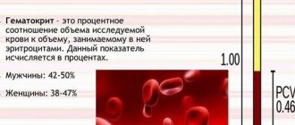
- HCT - hematocrit. This is a value that reflects the ratio of all blood cells to its liquid part - plasma. Since 99% of these cells are red blood cells, the indicator is used to estimate their volume.
Red blood cell indices
In addition, the so-called erythrocyte indices are assessed:
- MCV - average erythrocyte volume. Before the introduction of new analyzers, an accurate assessment was not carried out, and the result was designated as microcytosis, normocytosis or macrocytosis - depending on the size of the cells. An increased level of MCV may indicate a deficiency of vitamin B12, folic acid, hemolytic anemia, a decreased level may indicate iron deficiency and sideroblastic anemia.
- MCH is the concentration of hemoglobin in one red blood cell. In older analyzers, for this purpose, the color indicator was assessed, which is 0.03 of the MCH.
- MCHC is the average hemoglobin concentration in the erythrocyte mass, which reflects the degree of saturation of RBC with hemoglobin. It is the most stable indicator and can serve as an indicator of a correctly performed analysis.
All of the above indicators are closely related to each other and are assessed as a whole. For example, bleeding is accompanied by the loss of a large number of red blood cells. Accordingly, the total hemoglobin content also decreases, but MCH may remain normal. With a disease such as iron deficiency anemia, a slight deficiency of red blood cells occurs with a decrease in hemoglobin and various changes in erythrocyte indices. It makes no sense to list all possible changes in the body related to the content of red blood cells, and the doctor deciphers the result based on knowledge of those processes in the body in various diseases.
A separate indicator associated with red blood cells is their sedimentation rate - ESR (in modern analyzers - ESR) . This is a completely non-specific indicator, an increase in which indicates some problems in the body that the doctor needs to look for. In some cases, for example during pregnancy, an increase in ESR is physiological and is considered normal.
Tables of norms by age
The abbreviation PLT (short for platelets) in a general blood test refers to platelets.
The rate of red plates, regardless of gender, is highest in the first days after birth and steadily decreases with age.
Among women
| Age | Indicator in thousands U/μl |
| Up to 17 | 160-382 |
| 17-25 | 170-380 |
| 26-36 | 175-400 |
| 37-60 | 170-350 |
| 60 years and older | 175-310 |
The PLT norm in women is in the range of 160-400 thousand units/μl of blood and may vary slightly during one age interval, deviating from the presented calculations.
In men
| Age (years) | Norm in thousands of units per µl |
| Up to 17 | 175-380 |
| 17-25 | 175-390 |
| 26-36 | 185-400 |
| 37-60 | 190-395 |
| 60+ | 175-370 |
In children
| Age | PLT level thousand units/µl |
| Newborns | 180-490 |
| First month | 185-400 |
| Up to a year | 180-400 |
| 1-2 years | 155-390 |
| 2-3 years | 160-385 |
| 3-10 years | 160-380 |
During pregnancy
| Trimester | Normal value (thousand units/μl) |
| I | 170-340 |
| II | 155-321 |
| III | 145-310 |
The PLT norm in a blood test is a dynamic level, we are talking about ranges from 145 to 490 thousand units/μl of blood. Natural factors can also affect the results: physical activity, stress, habits. Also past illnesses. This needs to be taken into account.
Decreased platelet levels
Low platelet levels, the norm of which differs between men and women, provoke the development of a condition known as thrombocytopenia. Very often it occurs against the background of uncontrolled use of medications: antidepressants and antibiotics.
The reasons for a decrease in the level of platelets in the blood can be various infectious diseases: ARVI, hepatitis, herpes, etc. Thrombocytopenia can be observed when a large number of blood thinning products are included in the diet. These are ginger, cherries, garlic, onions, etc.
Non-infectious factors that reduce the level of platelets in the blood include pregnancy, vitamin deficiency, alcohol or heavy metal poisoning.
Thrombocytopenia can be suspected based on the following signs:
- Heavy menstruation.
- Frequent nosebleeds.
- The appearance of hematomas.
With a constant pathological decrease in the level of platelets in the blood, the risks of developing severe bleeding and stroke conditions, which are life-threatening, increase.
Reasons for the increase
An increase in platelet concentration or PLT is associated with pathologies of the excretory, hematopoietic or digestive systems. Other problems are also possible.
Let's try to summarize the list of pathological processes.
Inflammatory conditions
Multiple in character and properties. We are talking about a variety of disorders. This can include infectious and autoimmune diseases.
Particularly common are:
- Respiratory problems.
- Respiratory system lesions.
- Lupus erythematosus.
- Rheumatoid arthritis.
- Autoimmune disorders of the heart, myocardium.
- Crohn's disease.
And others. There are actually dozens of options. Just look at the ICD classifier to get an idea of the number of possible diseases.
Symptoms depend on the specific pathological process. If we talk about a general clinic, there will always be the following symptoms:
- Uncomfortable sensations.
- Increased body temperature. Systematic or rare. Indicators are also determined by the disease.
- Weakness, nausea, drowsiness.
For the rest, you need to start from a specific diagnosis. Therapists and rheumatologists treat pathological processes.
Correction includes the use of antibiotics, anti-inflammatory drugs, and antiviral agents. If there is an autoimmune process, glucocorticoids cannot be avoided. If they are ineffective, immunosuppressants are prescribed.
The list of medications, as well as treatment regimens, is determined by the doctor. PLT values will return to normal on their own as soon as the underlying disease is treated.
Sepsis
It stands apart from other pathological processes. We are talking about generalized inflammation. All body systems are involved. The disorder is always infectious in nature. It is provoked by a variety of agents.
The condition is accompanied by several typical symptoms.
Among them:
- Pain of unclear localization.
- Serious increase in body temperature.
- Impaired consciousness. If nothing is done, the patient will very quickly go into shock and die.
Sepsis is a deadly disorder. The concentration of platelets in this pathological process increases rapidly. This is due to the body's attempts to compensate for the disorder.
Treatment is carried out strictly in intensive care. Loading doses of antibiotics are prescribed. Hardware methods of blood purification. The goal is to return the patient to an adequate state.
The primary disorder is then treated. The one that provoked the development of generalized inflammation.
Oncological processes
Simply put, cancer. Benign tumors do not cause an increase in PLT concentration. Localization doesn't matter much. However, the process is especially aggressive during the degeneration of bone marrow cells. Which is quite understandable: this is where the formed elements of blood are produced and mature.
For cancer, especially advanced cancer, a typical group of symptoms is:
- Increased body temperature. Up to 37-38 degrees. The condition is stable, because the body is poisoned by the decay products of neoplasia.
- Weakness, drowsiness. Loss of performance.
- Loss of body weight. The person is not getting better. This is due to the increased need of altered tissues for nutrition.
The clinic is also focal. Depends on the type and location of the formation. Its size.
The therapy is carried out by oncologists. The goal is to remove the tumor. It is excised surgically. Chemotherapy and radiation treatment are provided as needed.
An increased concentration of PLT means that the cancer continues to develop. Based on the dynamics of the indicator, it is quite possible to talk about the progress of the process.
Consequences of surgical interventions
This includes almost any surgical treatment that affects the spleen. This organ acts as a kind of storage facility for blood cells.
If damage occurs, platelets (among others) are released into the bloodstream, PLT in the blood is increased steadily, although there is a temporary increase in the indicator.
Then, after a few weeks, everything returns to normal. It is possible that the process of change will end many times faster.
A pathological condition is also observed with injuries to the spleen. For example, its increase and further rupture. Deposited reserves of blood cells circulate throughout the body and are recorded using objective laboratory methods.
There is no need for correction as such. After surgery, you just need to wait. After some time, everything will recover on its own. If we talk about injuries, surgical treatment is necessary. Splenectomy.
Anemia
Most often, this reason for an increase in platelet count is observed in younger patients. From approximately 3 to 12-15 years. Although changes are also possible in adults.
Elevated PLT indicates anemia due to insufficient iron intake or absorption. This is a classic case. Other options are also possible: megaloblastic forms, congenital pathological conditions.
The clinical picture, especially in the early stages, is atypical or completely absent.
Typical symptoms include:
- Weakness, drowsiness.
- Increased sweating.
- Decreased performance.
- Nausea.
- Brittle hair and nails.
- Paleness of the skin.
- Decreased exercise tolerance.
- Perversion of taste preferences.
Bleeding is possible despite a formally high platelet count. They do not perform their function sufficiently, although there are many of them.
Treatment is carried out under the supervision of a hematology specialist. Therapy involves external administration of a deficient substance. Whether it's iron or vitamins.
If the problem is in the absorption of a beneficial compound, the cause must be treated. Gastritis, inflammation of the digestive tract.
Blood levels of PLT will remain high until the anemia resolves. Therapy may take more than one month. As a help, a diet is prescribed. Balanced, fortified diet.
Read more about the causes and treatment methods of iron deficiency anemia (IDA) here.
Colitis
Isolated inflammatory process. We are talking about intestinal damage. Thin or thick, that's a different question. The point is different.
The disease is accompanied by bleeding. Even if not so noticeable at first glance. The body is forced to produce more formed cells to replace those that are eliminated, this leads to an increase in the concentration of PLT. Until the condition is stabilized, the disorder will not stop.
Colitis is accompanied by typical manifestations, so it is difficult not to notice it.
Among the clinical signs of the disorder:
- Stomach ache. Intense, cramping.
- Nausea.
- Vomit.
- Weakness.
- Diarrhea. Including interspersed with mucus, blood, pus.
- Disorders of normal digestion.
- Increased body temperature.
Treatment rests on the shoulders of a gastroenterologist. The goal is to stop the inflammatory process. It almost always has an infectious origin.
Antibiotics and drugs are prescribed to restore microflora and protect it. Therapy requires several weeks.
During this entire time, be sure to adhere to a gentle diet. The diet is developed individually or a standard treatment table is used for patients with colitis.
Blood clotting disorders
The platelet concentration may change if coagulation (blood clotting) occurs directly within the vessel itself. This is the so-called DIC syndrome.
The disease is associated with profound fundamental changes in the body. In this case, a paradoxical situation is observed.
On the one hand, platelets stick together and die, breaking down on their own. On the other hand, the body produces them in inadequately high concentrations, which is why an abnormally high level of PLT is recorded.
The condition does not have any specific symptoms. However, it causes a lot of discomfort to the patient.
Among the clinical manifestations:
- The appearance of hematomas and bruises on the body for no apparent reason.
- Thrombosis. They are more likely to be considered complications. The formation of clots can lead to tissue necrosis. The condition is often urgent and requires hospitalization and intensive treatment.
Therapy is carried out under the supervision of a hematologist. As a rule, this is not a quick matter. Up to several months of close correction is required.
The problem is solved with the help of anticoagulants and antiplatelet agents. The prognosis is mostly favorable.
Genetic abnormalities
{banner_banstat9}
In some cases, the reason lies deeper than it seems at first glance. The pathological process may well be congenital.
In this case, the bone marrow works too intensively. Produces many shaped cells. And not necessarily only platelets. Complex disorders also occur.
Diseases are caused by mutations and are established before birth. If nothing is done, life-threatening complications are likely.
There are no symptoms as such. Thrombosis is possible. The abundance of red plates makes the blood thicker, this is a serious risk factor.
Treatment as such makes no sense. At least radical ways. All that remains is symptomatic correction. The problem is that the violation is encoded at a genetic, deep level. There's nothing you can do about it.
As for methods that will alleviate the condition, anticoagulants and antiplatelet agents are prescribed. They help prevent thrombosis and serious complications.
An increase in indicators indicates in favor of diseases.
How does a blood clot form? Why is a test for induced platelet aggregation prescribed?
Normally, platelets in the blood are in an inactive state; the cells have a discoid, slightly elongated shape, which is why in old textbooks they are called “blood plates.” When bleeding begins, platelets are activated: they acquire a spherical shape and form special outgrowths - pseudopodia. With their help, they can connect with each other (aggregate) and stick to the site of damage to the vascular wall (adhere). These two processes provide the basis for thrombus formation.
To assess the quality of platelet aggregation and find out whether it is reduced or, conversely, does not occur too intensely, an analysis for induced platelet aggregation is prescribed. To do this, they take blood from a vein, add special substances (activation inducers) to it and evaluate the process.
When preparing for the study, it is important to comply with certain conditions - for 3 days, follow a special diet prepared by a doctor, 24 hours before, avoid taking stimulants (coffee, alcohol, nicotine, garlic) and immunostimulant drugs, 8 hours before, stop taking medications and fatty foods.
Low platelet activity occurs in diseases of the hematopoietic system, constant use of antiplatelet drugs, in this case the duration of bleeding increases. Increased aggregation, on the contrary, increases the risk of thrombosis: venous thrombosis, heart attack, stroke. You may ask, why order an induced activation test if the risk of bleeding/thrombosis can be assessed by the total platelet count? Alas. Even with normal numbers, most of the cells may turn out to be “defective”, so we are talking about severe platelet deficiency with their normal concentration in the blood.
Reasons for the decline
{banner_banstat10}
A similar condition occurs no less often. Among the development factors.
Hepatitis
Inflammatory liver damage. Accompanied by acute dysfunction of the organ. In the chronic phase it is not so dangerous.
Among the symptoms:
- Pain in the right side of the abdomen.
- Nausea.
- Vomit.
- Change in color of stool.
- Diarrhea, constipation.
- Digestive disorders.
- Bitter taste in the mouth.
- Increased body temperature (not always).
The disease is progressing. Not at the same speed, but steadily. If nothing is done, necrotic changes will begin.
The condition is corrected under the supervision of a hepatologist. When such a narrow specialist is not available, a gastroenterologist is involved. The goal is to slow down, and ideally stop, change altogether. Protective drugs are prescribed.
Be sure to prescribe a gentle diet low in animal fat. If hepatitis is of infectious origin, you cannot do without special antiviral medications.
The prognosis for systematic treatment is positive, PLT concentration will recover quickly.
Cirrhosis of the liver
Necrotic process. Characterized by the death of organ tissue. In the acute phase, days and even hours count. It is necessary to transport the patient to the hospital. Then carry out urgent correction.
The clinical picture corresponds to that of hepatitis, only more severe. Problems begin with the nervous system and hematopoietic structures.
Among the manifestations:
- Pain in the right side.
- Impaired consciousness.
- Nausea.
- Vomit.
- Decline in cognitive level and intellectual abilities.
- Discoloration of stool.
- Diarrhea or constipation.
- Bleeding.
- The formation of spider veins on the body.
Chronic forms allow more time for treatment. Therapy consists of using protectors. We must do everything we can to stop the progression of the disease.
At the initial and middle stages of the pathological process, there is a chance of a liver transplant. The operation can only be successful during these periods.
In the decompensated stage, transplantation will make little sense. Bleeding begins and surgery is no longer possible. The disease is serious and must be dealt with quickly.
PLT decreases because cells die and new ones are not produced in sufficient quantities.
Thyroid disorders
Pathologies in which too much hormones T3 and T4 are synthesized. The lifespan of platelets in this case is significantly reduced. Hematopoiesis slows down. This is the reason for the deficiency of red PLT plates.
Pathology is treated under the supervision of an endocrinologist. The task is not quick, it takes several months. Iodine preparations are prescribed, as well as a diet low in this element.
Radiation sickness
As such, it inhibits the normal functioning of the bone marrow. This just ends with insufficiently rapid platelet maturation.
Attention:
The same is possible with a systematic, but weak influence of radiation. For example, when performing X-rays, CT scans too often.
Interpretation of the blood test on PLT
Decoding the analysis is within the competence of the attending physician. Thrombocytosis or thrombocytopenia may be the first signs of various pathologies.
The platelet level is determined during a general blood test
If the level of platelets in the blood exceeds normal values, the development of the following diseases can be assumed:
- Blood poisoning.
- Lymphogranulomatosis.
- Stomach cancer.
- Malignant blood diseases.
- Consequences of severe blood loss.
- Consequences of surgery to remove the spleen (thrombocytosis persists for two months).
- Chronic diseases: colitis, tuberculosis, rheumatoid arthritis and others.
Thrombocytopenia may indicate the following pathological conditions:
- Leukemia (chronic and acute).
- Werlhof's disease, or thrombocytopenic purpura.
- Systemic connective tissue diseases: scleroderma, lupus erythematosus, dermatomyositis.
- Folate deficiency anemia.
- Increased activity of the spleen in hepatitis (chronic, viral), liver cirrhosis.
- Taking certain medications.
- Viral infections: influenza, rubella, chickenpox, measles.
- Endocrine diseases: hypothyroidism, thyrotoxicosis.
- Metastases in the bone marrow in cancer.
- Aplastic and hypoplastic conditions of unknown origin, in which there is a decrease in platelet formation in the bone marrow.
Additional examinations
Ancillary measures are aimed at establishing the origin of the pathology.
Among the techniques:
- Oral interview with the patient.
- Anamnesis collection.
- Ultrasound of the digestive tract.
- MRI as needed.
- Blood and urine tests: general, biochemical. For hormones.
- Coagulogram.
This is a basic diagnostic checklist. Based on its results, specialized examinations are prescribed at the discretion of the doctor.
Consequences of low platelet count
In a state of low platelet count, disorders subsequently develop, which include frequent bleeding (menstrual and nosebleeds), while the bleeding time increases significantly and becomes difficult to stop. Sudden gum bleeding may develop (not everyone does). Blood clots may be found in urine or stool. Petechiae (red dots resulting from damage to the capillary wall) appear on the skin of the lower extremities, most often the legs. Even minimal damage leads to the formation of ecchymoses (bruises), when normally a bruise would not appear.
The severity of symptoms directly depends on the platelet count. The fewer platelets, the more severe the clinic. If the platelet count is very low, internal bleeding into the digestive tract or even bleeding into the brain may occur.
Even in the presence of a mild clinic, it is recommended to consult a general practitioner in order to prevent this condition.
Treatment
Regarding symptomatic correction:
With elevated platelets
It is necessary to eliminate the primary cause of the pathological process. These are the diseases that were described earlier. There is no need to fight the platelets themselves. The task is to remove the primary factor. It is better to address this question to a specialized specialist.
Symptomatic correction is to prevent the formation of blood clots. Anticoagulants or antiplatelet agents are prescribed.
At low
The same. Etiotropic treatment is necessary. Fighting the primary cause.
Decoding and data of normal indicators
The result of the analysis is a conclusion, which is usually a table about the content of chemicals and biological agents in the blood. The first column indicates the indicator, the second - the identified value, and the third - the normal range. Only a specialist, assessing the indicators, can determine the presence of disturbances in water-salt metabolism, identify inflammatory processes and infections, and also assess the performance status of all the patient’s organs.
As for normal indicators, the data for adults looks like this:
| Analysis | Men | Women |
| Total protein | 64-84 g/l. | 64-84 g/l. |
| Hemoglobin | 130-160 g/l | 120-150 g/l. |
| Haptoglobin | 150-2000 mg/l | 150-2000 mg/l |
| Glucose | 3.30-5.50 mmol/l. | 3.30-5.50 mmol/l. |
| Urea | 2.5-8.3 mmol/l. | 2.5-8.3 mmol/l. |
| Creatinine | 62-115 µmol/l | 53-97 µmol/l. |
| Cholesterol | 3.5-6.5 mmol/l. | 3.5-6.5 mmol/l. |
| Bilirubin | 5-20 µmol/l. | 5-20 µmol/l. |
| AlAT (ALT) | up to 45 units/l. | up to 31 units/l. |
| ASAT (AST) | up to 45 units/l. | up to 31 units/l. |
| Lipase | 0-190 units/l. | 0-190 units/l. |
| Alpha amylase | 28-100 units/l. | 28-100 units/l. |
| Pancreatic amylase | 0-50 units/l. | 0-50 units/l. |
Decoding the main indicators of the LHC
Total protein.
A biochemical blood test determines the total concentration of various proteins, which consist of amino acids. Protein takes an active part in the processes of coagulation, processing and transport of nutrients to organs and tissues.
Hemoglobin.
This specific protein from the red blood cell system is responsible for transporting oxygen in the body
Haptoglobin.
A blood plasma protein that binds hemoglobin and is responsible for preserving iron in the body. Also involved in the control of local inflammatory processes.
Glucose.
An important component responsible for carbohydrate metabolism. Its content in arterial blood is higher than in venous blood.
Urea.
This is the main product of protein breakdown, in which nitrogen unnecessary by the body is removed in the urine.
Creatinine.
This, like urea, is the end product of protein metabolism. The content of creatinine in the blood depends on gender, age, and muscle mass.
Cholesterol
. A component of fat metabolism, which is also involved in the construction of cell membranes and the synthesis of vitamin D and sex hormones. Another name is cholesterol. There are: total cholesterol, low-density lipoprotein (LDL) and high-density lipoprotein (HDL) cholesterol.
Bilirubin
. The breakdown product of hemoglobin, a toxic substance of two types: direct and indirect. They form a “general” one and the norm when deciphering a biochemical blood test is indicated specifically for it.
AlAT (ALT).
Alanine aminotransferase is an enzyme contained in the cells of the liver, kidneys and heart, so its presence in the blood indicates the destruction of the cells of these organs.
AST (AST).
Aspartate aminotransferase is a cellular enzyme that is involved in the metabolism of amino acids and is found in the liver cells of the heart and kidneys.
Lipase.
An enzyme that promotes the breakdown of fats.
Amylase.
It breaks down carbohydrates from food and ensures their digestion. There are alpha-amylase (diastase) and pancreatic amylase.
It is important to remember that only a specialist can evaluate the results of a blood test, decipher a biochemical analysis, make a diagnosis and prescribe treatment!
Prevention
Preventive measures are extremely simple. It is enough to follow simple recommendations:
- Regularly undergo inspections. Every six months or a little less often.
- Eat well. To prevent the development of anemia and deficiency diseases.
- Treat pathologies of any profile in a timely manner. Even an old source of infection can present an unpleasant surprise.
- Eliminate alcohol from your life.
- Do not self-medicate.
PLT stands for platelet, which means platelets in English. The indicator is informative, but it needs to be clarified by other methods. This way you can quickly detect health problems.