Author: Sozinova A.V., obstetrician-gynecologist, has been in continuous practice since 2001. January, 2022.
Synonyms: platelets, platelet count, PC, plt.
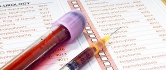
Platelets are blood cells, and their concentration is determined during a general blood test. On the CBC form, platelets are designated as platelet count or PC. Blood for studying its composition, including counting the number of platelets, is taken from a finger (capillary) or from a vein. The unit of measurement for platelets is the number (N) of cells multiplied by 109 per liter.
Platelets, like other formed elements, are formed in the red bone marrow. Their lifespan in the bloodstream is about 7–12 days. Destruction and breakdown of cells occurs in the spleen and liver tissues.
The main function of platelets is to carry out hemostasis, that is, to stop bleeding. Contact with damaged tissues causes transformation of cells, up to 10 processes are formed in them, and when they spread, the wound is closed with a platelet mass (thrombus). Thus, platelets prevent large amounts of blood from leaking out of damaged soft tissues.
Another equally important function of platelets is to protect the injured area from the penetration of pathogenic microorganisms (they secrete lysozyme and B-lysine).
Platelets also participate in the processes of hemostasis (maintaining the internal environment of the body), nutrition of the capillary endothelium (the inner layer of the walls of blood vessels) and regenerative processes of damaged tissues due to the release of growth factors that stimulate cell division.
A 2022 scientific paper provides evidence showing that in addition to the above functions, platelets also play an important role in the human immune system.
Indications
Prescription of an OAC with a study of platelet concentration is indicated for all patients who visit the clinic, undergo a routine medical examination, or receive a referral for inpatient treatment. Also, platelet determination is prescribed to all patients requiring surgery (emergency or planned). The main indications for platelet testing include:
- suspicion of disturbances in the hemostatic system (bleeding, formation of subcutaneous hematomas);
- suspicion of a malignant process;
- immunity disorders;
- bronchopulmonary pathology;
- diseases of the digestive tract and urinary system (stomach ulcer, glomerulonephritis);
- cardiovascular pathology;
- endocrine disorders (obesity, diabetes).
Symptoms that should alert you
- red, purple bruises on the skin
- frequent nosebleeds, bleeding gums
- prolonged bleeding of even small wounds or cuts
- heavy menstrual bleeding in women
How is the platelet count calculated?
The number of blood platelets is determined by different methods: using smears (according to Fonio) and chamber (in the Goryaev chamber). Recently, automatic blood cell counters have been increasingly used. It is especially convenient to use the Fonio method with this form of calculation. Reagents required for this procedure:
- 14% magnesium sulfate (sterile solution);
- Leishman fixative or Main-Grunwald fixative;
- Romanowsky-Giemsa stain.
To count the number of platelets using the Fonio method, a finger is treated, then wiped dry and a puncture is made. Then magnesium sulfate (1 drop) is dripped onto the finger and mixed with the blood released from the puncture. The resulting mixture is used to make smears, which are dried and painted.
Determination of the number of blood cells is carried out using automatic counters
In the stained smear, 1000 red cells and all the platelets that come across during this time are counted. A relative amount is obtained, which is measured in ppm. To find out the absolute level of blood platelets, the resulting value is multiplied by the number of red blood cells contained in 1 μl of blood, and then divided by 1000.
Preparing for the study
Before taking the OAC, you should stop drinking alcoholic beverages, fatty, spicy, excessively salty and fried foods the day before the procedure. Blood is donated on an empty stomach, the patient is advised to maintain physical and emotional peace (refrain from sudden movements, climbing stairs, avoid stressful situations) for half an hour before the test. Also, platelet testing is not recommended immediately after recovery from a long-term illness, which may distort the results due to weakened immunity.
Detailed instructions for preparing for a general blood test are here.
How is the analysis carried out?
The platelet level is determined during a general blood test simultaneously with the calculation of other blood parameters. Blood is taken from a finger in the morning - before 10-11 o'clock. Before the procedure, you should not eat or drink sugary drinks - only clean water. Smoking is not allowed for three hours before the test. It is not recommended to drink alcoholic beverages the day before. The doctor should be warned about undergoing physical treatment and taking medications.
Important! During clinical analysis, only the total number of platelets is determined, but not the percentage of different forms. You should know that the absolute value may correspond to the norm, while the level of mature forms is reduced, and the level of young and degenerative forms exceeds acceptable limits. In this case, the effectiveness of platelets is significantly reduced. To assess their activity, it is necessary to examine their morphology, that is, size, structure, shape. The most complete picture of the state of the hemostatic system can be obtained using a regular or detailed coagulogram.
Platelets normally have a certain shape and size. During diseases, small or large tailed forms are found in the blood, their internal structure and color change. Using a microscope, their functionality is studied, that is, the ability for aggregation and adhesion. Blood obtained from a vein is better suited for assessing activity. The study of functional fitness is of great importance in the diagnosis of certain pathologies, such as von Willebrand disease, congenital thrombocytopenia, Bernard-Soulier disease and others.
Platelet norms
Important! Standards may vary depending on the reagents and equipment used in each particular laboratory. That is why, when interpreting the results, it is necessary to use the standards adopted in the laboratory where the analysis was carried out. You also need to pay attention to the units of measurement.
The list shows the reference values of platelet cells adopted in the Invitro laboratory:
| Age | Platelet concentration, thousand/µl (103 cells/µl) | |
| Children | boys | girls |
| less than 2 weeks | 218 — 419 | 144 — 449 |
| 2 weeks — 1 month | 248 — 586 | 279 — 571 |
| 1 - 2 months | 229 — 562 | 331 — 597 |
| 2 — 6 | 244 — 529 | 247 — 580 |
| 6 months - 2 years | 206 — 445 | 214 — 459 |
| 2 years - 6 years | 202 — 403 | 189 — 394 |
| Adults | men and women | |
| over 6 years old | 150 — 400 | |
In the Helix laboratory there is a slightly different gradation of values:
| Age | Reference values 109/l |
| Less than 10 days | 99 — 421 |
| 10 days – 1 month | 150 — 400 |
| 1-6 months | 180 — 400 |
| 6 months – 1 year | 160 — 390 |
| 1-5 years | 150 — 400 |
| 5-10 years | 180 — 450 |
| 10-15 years | 150 — 450 |
| More than 15 years | 150 — 400 |
It should be noted that the platelet count decreases slightly in women during the menstrual period, but returns to normal after bleeding stops. A slight decrease (up to 150) in platelet concentration during the gestational period is also possible, which is explained by blood dilution due to an increase in BCC (circulating blood volume) and due to insufficient nutrition of the expectant mother.
Important! The interpretation of the results is always carried out comprehensively. It is impossible to make an accurate diagnosis based on only one analysis.
Diagnostic value
Unlike the total number of platelets in a unit of blood, MPV reflects qualitative characteristics, that is, by this indicator one can judge their usefulness. Old cells are smaller in size, while young forms are large in size and have a structureless structure. Their activity, tendency to stick together, and the content of biologically active substances in them depend on the size of the plates.
The physician can obtain important information based on the MPV. If this indicator is elevated, it means that young cells are present in the blood. The higher the value, the more immature forms there are. If this indicator is low, this indicates that small forms are present in the bloodstream. MPV may be increased or decreased if the platelet count is normal.
By assessing the average platelet volume in the blood, your doctor may find the following:
- increased adhesion of platelet plates to each other, development of thrombosis;
- blood loss in people with iron deficiency anemia when large forms of platelets are detected;
- This test does not make it possible to determine myeloproliferative disease.
Drugs that affect platelet levels
Long-term use of these drugs may change platelet levels:
- aspirin
- pain relievers such as ibuprofen and naproxen
- antihistamines
- asthma medicine
- sildenafil (Viagra)
- drugs used to prevent blood clots, such as clopidogrel
- antibiotics
- antidepressants and antipsychotic drugs
- Cholesterol-lowering drugs (statins)
- calcium channel blockers (verapamil).
Interpretation and normal values of the main blood test parameters
| How is it designated? | What does it mean | Norm for women | Norm for men |
| R.B.C. | Red blood cells | 3,5-4,5 | 4,0-5,5 |
| WBC | Leukocytes | 4-9 | |
| PLT | Platelets | 180-320 | |
| HGB | Hemoglobin | 120-140 | 130-170 |
| MCV | Average erythrocyte volume | 82-98 | 81-95 |
| MCH | Average HGB level in erythrocyte | 26-32 | |
| MCHC | Average concentration of red blood cells in HGB (%) | 31-38 | |
| HCT | Hematocrit (in%) | 35-44 | 40-50 |
| RET | Reticulocytes (%) | 0,2-1 | |
| ESR | ESR (mm/h) | 2-15 | 1-10 |
| CPU | Color | 0,85-1,05 | |
Platelets are reduced (thrombocytopenia)
When the concentration of platelets in the CBC decreases, they speak of the development of thrombocytopenia, which is accompanied by a blood clotting disorder and a tendency to bleed (gums bleed, nosebleeds or intestinal bleeding often occur, menstruation becomes long and heavy). Thrombocytopenia develops in a number of serious diseases due to the loss of elasticity of the vascular wall, their fragility and fragility and the risk of internal bleeding.
Reasons that provoke a decrease in platelet levels include:
- hemolytic-uremic syndrome or Gasser's disease (a combination of hemolytic anemia, thrombocytopenia and acute kidney failure);
- thrombocytopenic purpura or Werlhof's disease (one of the widows of hemorrhagic diathesis);
- allergic thrombocytopenia (taking a number of medications: procainamide, heparin, co-trimoxazole);
- DIC syndrome (second stage);
- severe liver damage (hepatitis, cirrhosis);
- alcoholism;
- malaria;
- enlarged spleen;
- bone marrow diseases, some leukemias;
- megaloblastic anemia;
- pathology of the thyroid gland (hypothyroidism, thyrotoxicosis).
It is important to note that a decrease in platelet concentration (75 – 150) is observed in pregnant women, which is not regarded as a pathology.
Activation
To perform their main function - repairing damage in the vessel wall - platelets must enter an active state. Like most cells in our body, this process proceeds according to the following scheme: signal - receptor - intracellular signal - amplifier - regulator - response (Fig. 3). The signal for activation is the appearance in the bloodstream of an agonist - a special signaling molecule, which should appear only when necessary and bind to a specific molecule that penetrates the platelet membrane (receptor). The agonist interacts with one “tail” of the receptor, protruding from the outside, and this leads to a change in the other, on the cytosol side, where the next signaling molecule, the secondary messenger, appears. It triggers the synthesis of several more messengers, which, in turn, triggers several more, and so the signal propagates in the cytosol and is amplified through a cascade of intracellular reactions, which ultimately leads to a complex platelet response. It is important that in the platelet there are special regulatory systems that modulate the concentrations of intracellular messengers at different stages of activation, so that, for example, there is no reaction to trace amounts of the agonist.
Rice. 3.
Platelet activation scheme
How is this scheme implemented in our body? In blood vessels, platelets are pushed out of the main flow by red blood cells and move along the walls, conducting a kind of monitoring of their condition. One of the first signals for platelet activation is collagen, the main protein of connective tissue, exposed when a vessel is damaged. Having detected collagen, they bind to it through special receptors, simultaneously becoming activated and firmly attaching to the site of damage. The interaction of a platelet with collagen leads to the launch of the mentioned intracellular signaling cascade and the appearance of a secondary messenger in the cytosol - inositol triphosphate (IP3). This small water-soluble molecule is able to move quickly in the cytosol and serves as a signal for the release of calcium ions from intracellular stores. And an increase in its intracellular concentration can lead to various responses of the platelet: splashing out the contents of granules (secretion), changing shape, attaching to the vessel wall (adhesion), bonding with other platelets (aggregation), and the appearance of procoagulant activity (Fig. 4). After the circulatory system has already recognized the damage to the vessel, three more natural platelet activators appear in the blood - thrombin, ADP and thromboxane A2. The thrombin protein is formed from its precursor, prothrombin, in the blood plasma, but in large quantities - already on the membranes of activated platelets. When their dense granules are secreted, large amounts of ADP (a small molecule that primarily performs energy functions in cells) are released, and much less ADP is released from damaged endothelial cells lining the inner surface of blood vessels. Thromboxane A2 is synthesized from arachidonic acid, located in the membranes of activated platelets. The binding of these three activators to their receptors on the platelet membrane leads, as in the case of collagen, to the appearance of IP3 in the cytosol and an increase in the calcium concentration in it (Fig. 4). Thus, all three soluble activators and collagen act through the same pathway, but induce different platelet responses. For example, thromboxane A2 provokes the release of dense granules, but ADP does not. Activation separately by collagen or thrombin causes all of the above responses simultaneously, and together leads to the appearance of a group of procoagulant platelets and the synthesis of thrombin on their membranes. Apparently, there are still insufficiently studied differences in the signaling triggered by different agonists. To prevent accidental activation from turning a platelet into a real “bomb”, carried in the bloodstream and triggering the entire coagulation system, intact endothelial cells in the body constantly secrete prostacyclin and nitric oxide, which block cell activation, preventing an increase in calcium concentration in them.
Rice. 4.
Scheme of the main pathways of platelet activation and its responses:
ADP
- adenosine diphosphate,
IP3
- inositol triphosphate,
ER
- endoplasmic reticulum
Signaling is one of the most complex and poorly understood areas in platelet research. There are many questions about the structure of each receptor and signaling pathway, and the simplest of them is: why are there so many activators at all?
Platelets are increased (thrombocytosis)
An increase in platelet count (thrombocytosis) is observed when:
- excessive physical stress
- chronic inflammatory processes (rheumatoid arthritis, tuberculosis, sarcoidosis);
- myeloproliferative diseases (primary erythrosis, chronic myeloid leukemia, myelofibrosis, myelosclerosis);
- some hemolytic anemias;
- hemolysis or severe blood loss;
- carcinoma, lymphoma;
- after removal of the spleen.
Sources:
- Eugenio D. Hottz. Platelets in Immune Response to Virus and Immunopathology of Viral Infections. — Front Med (Lausanne). Apr 2018.
- Data from the independent laboratory Invitro.
- Data from Helix laboratory.
- Steven Kim, MD. Acquired Platelet Function Disorder. — Healthline, Jan 2016.
- Douglas B. Cines. Thrombocytopenia in pregnancy. - Blood. 2017 Nov 23; 130(21): 2271–2277
- Danilova L.A., Doctor of Medical Sciences, Prof. Analyzes of human blood, urine and other biological fluids at different age periods, SpetsLit, 2014.
What to do in case of bleeding
If you start bleeding at home, take the following measures:
- Apply direct pressure to the bleeding area. If you have a nosebleed, pinch the bridge of your nose and apply ice to it.
- After applying pressure to the bleeding area, call your Memorial Sloan Kettering (MSK) doctor. If you cannot reach your doctor and are unable to stop the bleeding, go to your nearest emergency room.
When should you call your doctor?
Call your doctor right away if you have any of the following:
- black stools, blood in the stool, or bleeding from the rectum;
- blood in urine;
- persistent headache;
- blurred vision;
- dizziness;
- any persistent bleeding, including coughing up blood, vomiting blood, or bleeding from the nose.
to come back to the beginning