The first description of hypospadias dates back to the second century AD in the work of Galen, who first used the term. During the first millennium, the only treatment for this pathology was amputation of the penis distal to the external opening. Since then, there have been great changes in the surgical treatment of this defect for the better. More than 300 different methods for correcting this pathology are described in the medical literature. Although most of the techniques used today have been described over the past 60 years, the basic principles were proposed more than a century ago.
Modern methods of anesthesia, microsurgical instruments, sutures, dressings and antibacterial therapy improve the results of treatment of hypospadias and, in most cases, allow reconstruction to be performed in one stage during the first year of the child’s life.
History of surgical treatment of hypospadias
The earliest description of this pathology and treatment comes from the works of Celsius (25 AD) and Galen (second century AD).
In 1874, Duplay published his work, which began the modern stage of development of urethral reconstruction techniques. Currently, more than 300 methods of operations are described. Most techniques are multi-stage, in which the first stage eliminates stenosis of the meatus (external opening of the urethra), if necessary, and the second stage involves excision of the fibrous chord and correction of the curvature. The third stage is the actual urethroplasty using various techniques.
Many cosmetic and functional problems are associated with multi-stage treatment methods: they require multiple operations, the external opening of the urethra is not brought to the top of the head of the penis, or its retraction (downward displacement) is noted due to multiple manipulations of tissues and resulting scars.
To reduce the incidence of complications, Hinderer proposed a one-stage treatment method in 1960. Numerous modifications and new techniques were proposed by various surgeons in the following decades. These studies confirmed the advantage of using scar-free tissue for urethral reconstruction while reducing the number of surgical procedures.
Stages of urethral formation. Classification
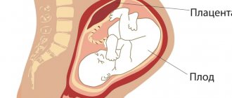
Hypospadias is a congenital defect that occurs due to impaired development of the urethra between 8 and 20 weeks of pregnancy. The external genitalia are identical in male and female fetuses up to the 8th week of gestation. Mainly under the influence of testosterone and its active metabolite - dihydrotestosterone - the genitals develop according to the male type. As the phallus grows, the urethral track develops from the base to the head of the penis.
The classical theory is that the urethral ridges fuse at the anterior urethra to form the tubular penile urethra and midline suture. The formation of the capitate part of the urethra occurs towards the already formed stem part. This explains the high incidence of coronary hypospadias.
The foreskin normally forms as a growth of skin around the coronal sulcus, which grows circularly and merges around and above the glans. Disruption of the fusion of the urethral ridges prevents the process of closing the foreskin; as a result, the prepuce in hypospadias is located along the dorsal surface in the form of a hood. In rare cases, there may be preserved foreskin with a split meatus - megalomeatus.
Notochord (ventral curvature of the penile shaft) is often combined with distal hypospadias (up to 90% of cases) and in almost all patients with proximal forms. This occurs due to a disproportion in the growth of the corpora cavernosa and the altered urethra.
Hypospadias is classified depending on the location of the external opening of the urethra. Although several classifications have been proposed, most surgeons use the one described by Barcat and modified by Duckett, who assessed the forms of hypospadias after straightening the corpora cavernosa.
Hypospadias is divided into:
- Anterior (capitate and coronal);
- Middle (distal-trunk, mid-trunk and proximal-trunk);
- Posterior (memberoscrotal, scrotal and perineal).
Capitate hypospadias is characterized by the location of the external opening of the urethra on the head of the penis, the foreskin is dysplastic, and the penis is often not curved. A one-step correction is performed.
In coronal hypospadias, the external opening of the urethra is located under the head of the penis. This is the most common form. The foreskin is split, and curvature is more common than with capitate. A one-step correction is performed.
Trunk hypospadias - the hole is located somewhere on the shaft of the penis, the fibrous chord is more pronounced, the curvature has a greater degree. More often, a one-stage correction is performed.
Scrotal hypospadias - the meatus is located in the scrotum area, it is split, there may be transposition of the penis into the upper or middle third, the corpora cavernosa are curved, the corpus spongiosum of the urethra is dysplastic, its removal and reconstruction is required, female-type urination is required. With this form of hypospadias, corporoplasty is necessary. Two-stage methods are preferable.
Perineal hypospadias is even more proximal. Pronounced fibrous tissue is noted along the ventral surface of the genital receptacle with its curvature. Large defect of missing urethra requiring reconstruction. Reconstruction of the corpora cavernosa is necessary. Two-stage methods are preferable.
Anterior hypospadias occurs in 50% of cases, middle hypospadias in 20%, and posterior hypospadias in 30%.
Pathogenesis
The development of the pathology is based on a violation of embryogenesis at 10–14 weeks of gestation, which leads to disruption of the normal process of differentiation of the embryonic epithelium. In this case, underdevelopment of the peripheral part of the urethra is noted and the urethral groove is closed. Deformation of the penis develops due to the inability of the urethral groove to transform into a tubular organ. Accordingly, a short/dense inelastic connective tissue cord (notochord) is formed in this area, which shortens the ventral surface of the genital organ, which leads to its bending down.
Combined pathology
Hypospadias is often combined with a narrowing of the external opening of the urethra. Undescended testicles and inguinal hernias are the anomalies most often associated with hypospadias. In 1981, Khuri's review of over 1000 patients with hypospadias reported that the incidence of cryptorchidism and inguinal hernia was about 9%. In more severe forms of hypospadias, the frequency of undescended testicles exceeded 30%, and the combination with inguinal hernia reached 20%. A combination with such pathologies as: cryptorchidism - 9%, inguinal hernia - 9%, hydrocele, urethral fistula, testicular hypoplasia, anomalies of the upper urinary tract and vesicoureteral reflux was also noted. Cardiovascular and craniofacial malformations are extremely rare.
The combination of hypospadias with undescended testicles may be associated with a violation of sex formation and requires a more complete endocrinological and genetic examination before surgery. In a 1999 study by Kaefer et al., sex formation disorder was identified in approximately 30% of patients with hypospadias and cryptorchidism. Moreover, the more proximally located the external opening of the urethra, the higher the likelihood of disorder of sex formation (DSD). If one gonad is not palpable, then the possibility of DSD increases to 50%, if both testicles are palpable, then 15%.
Frequency of occurrence
The incidence of this anomaly is 1 in 250 boys born on average in the world and is the most common disease of the urinary tract. In some countries there is a trend towards an increase in the birth of children with this problem. In general, the birth rate is fairly constant - 0.26 per 1000 children in Mexico and Scandinavia, 2.11 per 1000 births in Hungary.
The incidence of hypospadias is higher in whites than in blacks, and is more common in Jews and Italians. Familial inheritance of hypospadias is about 7%.
Symptoms
The clinical symptoms of hypospadias are determined by the location of the meatus and the degree of its narrowing. The more proximal the meatus is located and the smaller its diameter, the more pronounced the specific symptoms. So, with a mild form of (capitate) hypospadias, there is no severe urination disorder, and such patients in most cases do not need reconstructive surgery. With a small diameter of the meatus, the patient complains of slow/difficult urination in a thin stream.
In cases of coronary hypospadias, there is a partial curvature of the genital organ, against which there is difficulty urinating. In this case, the patient is forced to take a comfortable position for the penis so that the stream is not directed towards the legs.
With trunk hypospadias, there is a sharp deformation of the penis. At the same time, the patient experiences difficulty when it comes to going to the toilet: it is necessary to strongly lift the penis or urinate while sitting. The narrowing of the urethra prevents free urination.
With perineal/scrotal hypospadias, the patient's external genitalia are modified: a bifurcated scrotum and a deformed, critically small penis. The process of urination in this pathology can be carried out exclusively in a sitting position. Urine excretion is significantly difficult. Hypospadias in boys is often combined with various developmental anomalies of the genitourinary system with characteristic symptoms.
Causes
The following factors lead to the occurrence of hypospadias: genetic, endocrine and environmental.
Genetic factors. Among monozygotic twins, the incidence of hypospadias is 8 times higher than in the general population. This situation may arise due to insufficient production of human chorionic gonadotropin by the placenta during critical periods of urethral development.
Familial inheritance of hypospadias also occurs. The probability of having a child with this pathology from a father with hypospadias is about 8%. Inheritance is most likely polygenic.
Endocrine factors. Decreasing androgen concentrations during pregnancy or blocking their effects on tissue can lead to hypospadias. In 1997, Aaronson et al. it was found that 66% of boys with distal hypospadias and 40% with proximal forms had a defect in testosterone biosynthesis.
Mutations in the gene responsible for the production of the enzyme 5-alpha reductase, which converts testosterone into its active metabolite dihydrotestosterone, have been associated with the occurrence of this pathology. A report published in 1999 by Silver et al. showed that nearly 10% of boys with isolated hypospadias had at least one affected allele for the production of the enzyme 5-alpha reductase. Despite the fact that androgen deficiency leads to hypospadias, there are other causes that lead to impaired formation of the penis and urethra.
Some authors have noted that in countries with cold climates, the incidence of children with hypospadias is higher. Theoretically, this may be due to the effect of daylight and sun on pituitary function, which in turn affects the hormonal milieu of the mother and fetus. However, other authors have not noticed this connection.
With in vitro fertilization (IVF), there is a fivefold increased risk of having a child with an anomaly of the genital organs compared to the control group. This is due to the effects of progesterone, which is used in IVF protocols. Progesterone is a substrate for 5-alpha reductase and acts as a competitive inhibitor of the conversion of testosterone to dihydrotestosterone. Other endocrinopathies or fetal endocrine abnormalities also play a role.
Environmental factors. In many animal studies, estrogens have been implicated as the cause of this defect. Chemicals with significant estrogenic activity are observed throughout industrialized societies. Found in pesticides used on fruits and vegetables, endogenous plant estrogens through animal milk, plastic linings on metal cans, and pharmaceuticals.
The Hadziselimovic study reported increased estradiol concentrations in placental basal syncytiotrophoblasts in boys with undescended testes compared to the healthy population. Undescended testes and hypospadias are likely related, but increased estradiol concentrations have not been shown to cause hypospadias.
Combined theory. A growing body of evidence suggests that hypospadias is a multietiological disorder involving genetic predisposition and environmental factors.
Androgen deficiency
The cluster of causes for the development of the disease is hormonal disorders: insufficient production of male androgen hormones by the placenta and ovaries. Absolute (reduced concentration) or relative (decreased tissue sensitivity) deficiency is the main cause of developmental abnormalities.
Many enzyme defects are known, such as 5α-reductase and androgen receptor deficiency. In 10-70% of cases of severe proximal hypospadias, an enzymatic defect in androgen synthesis or a hormonal disorder affecting hormonal balance is found.
Forecast
The use of modern anesthesia, suture material, microsurgical instruments, magnifying equipment, and antibacterial therapy makes it possible to perform surgical treatment of hypospadias in just one stage with minimal risk of complications and obtain an excellent cosmetic result. Compliance with the optimal timing of surgical treatment allows you to eliminate the psycho-emotional component in the child. The desire to perform surgical treatment before 2 years of age is also due to the fact that the child will not remember the very fact of treatment and hospital stay.
Studies conducted on patients who underwent surgery for hypospadias showed that they were more satisfied with their sex lives than those who did not have surgery.
It is worth noting that treatment methods for hypospadias continue to evolve. New methods of tissue adhesion are being developed: tissue adhesives and the use of laser for adhesion, which leads to improved wound healing and a reduced risk of fistula formation.
Cellular technologies are also being actively developed and will soon make it possible to create an artificial urethra, especially in patients with severe forms of hypospadias. The identification of factors and understanding of the causes leading to hypospadias continues, which allows us to develop an approach to the prevention of this condition and carry out correction in utero.
Postoperative period
Patients are discharged after one day in the hospital, but require long-term post-operative care. Support measures include:
- Use of a catheter. Used to empty urine contained in the bladder for the first 10 days after the procedure. Without it, urination would be impossible.
- Bandage on the wound. Ensures that the penis is immobilized for several days to prevent infection and allow the tissue to heal.
After surgery, it is normal to experience some loss of blood and urine. It is necessary to take measures to prevent infection. If pus, fever and rash appear, you should consult a doctor.
Within 2-3 months, it is advisable to exclude mechanical trauma to the affected area. After the operation, it is necessary to visit the doctor regularly: after a week, 3 and 6 months, and also a year after the correction. At the same time, the specialist will once again examine the appearance of the penis. Unfortunately, no hypospadias surgery is complete without scars. However, the surgeon tries to limit the incisions to the midline of the lower part of the penis. These cuts heal with the best possible cosmetic results.
External manifestations
Although the diagnosis of hypospadias can be established either by fetal ultrasonography or by magnetic resonance imaging (MRI), the diagnosis is usually made during examination of the newborn or during a clinical examination.
The foreskin is located on the dorsal surface in the form of a hood, and is absent on the ventral surface. The anterior wall of the urethra is absent, and the external opening of the urethra is displaced proximally. It rarely happens that the foreskin is preserved and has a normal appearance. In this case, hypospadias is discovered during ritual circumcision or due to phimosis or at an older age, when the head of the penis begins to protrude beyond the foreskin.
The external opening of the urethra (meat) can be located on the head (60%), shaft of the penis (35%), in the scrotum or perineum (5%). 75% of patients experience a narrowing of the meatus. Curvature of the penis to varying degrees is observed in more than 60%.
If hypospadias is detected during circumcision, the procedure should be stopped and the patient referred to a qualified urologist for consultation.
The fibrous chord leading to the curvature may be visible to the naked eye, or may only be detected during an erection. Proximal forms of hypospadias are usually combined with scrotal splitting and penoscrotal transposition.
Indications for surgery
Meatotomy is indicated at any age if the narrowing of the external opening of the urethra causes a disturbance in the process of urination (dysuria).
Correction of hypospadias at a young age is indicated for the following reasons:
- — ensuring normal emptying of the bladder (male type) without splashing the stream of urine and its deflection;
- — ensuring normal erection and sexual intercourse in the future;
- - prevention of urinary tract infection;
- — correction of the possible occurrence of impotence and infertility;
- - achieving normal structure of the external genitalia;
- — ensuring psychosocial adaptation in society.
Timing of the operation
Until 1980, correction of pathology was carried out in children over 3 years of age due to the larger size of the organ. However, genital surgery at this age (children's awareness of genitals and self-identification as a particular gender occurs around 18 months of age) can result in significant psychological trauma, including abnormal behavior and gender identity disorder. Late surgical treatment during puberty and older leads to more frequent complications, first of all, the occurrence of fistulas occurs more often, with a probability of more than 50%. Some works by foreign authors indicate a higher number of complications in 5-year-old patients than in one-year-old patients.
Currently, most urologists try to operate on hypospadias at the age of 4-18 months, with a tendency towards earlier dates. This is associated with improved emotional and psychological outcomes. Tissue healing also occurs faster at an earlier age.
List of sources
- Marchenko A.S., Smirnov I.E., Zorkin S.N., Apakina A.V., Sukhodolsky A.A., Shakhnovsky D.S. Treatment of children with hypospadias. Pediatric surgery. 2013. No. 5. P. 40-44.
- Akranov N. R., Sharabidze G. G. Parasurgical aspects of the treatment of boys with hypospadias (literature review) // Reproductive health of children and adolescents. 2010, no. 5, p. 39–48.
- Fayzulin A.K., Prokopyev V.M., Demin N.V. Modern methods of treating hypospadias // Andrology and genital surgery. 2009. No. 2. 158 p.
- Volodko E.A. Main characteristics of disorders of sex formation in hypospadias in children // Materials of the VI Russian Forum “Mother and Child”, M., 2004. - P.554-555.
- Akranov N. R., Sharabidze G. G. Parasurgical aspects of the treatment of boys with hypospadias (literature review) // Reproductive health of children and adolescents. 2010, no. 5, p. 39–48.
Treatment of hypospadias
There is no conservative treatment for hypospadias.
Surgical treatment of hypospadias has the following goals:
- an increase in the diameter of the external opening of the urethra;
- correction of curvature of the cavernous bodies;
- reconstruction of the missing part of the urethra;
- restoration of the normal appearance of the external genitalia.
Operation techniques
The general principles of the main stages are common to all methods. First, the corpora cavernosa is exposed from the skin of the penis. The fibrous cords and “notochord” present on the ventral surface are excised as much as possible. An “artificial erection” test is performed by injecting saline into the corpora cavernosa to detect any deviation of the penis. If the curvature is less than 45⁰, dorsal plication corporoplasty is performed under the neurovascular bundle. When the curvature is more than 45⁰, it is advisable to use patch corporoplasty or by applying multiple incisions on the ventral surface of the tunica albuginea of the corpora cavernosa. It is also possible to correct the curvature of the corpora cavernosa by intersecting the tunica albuginea at the point of maximum curvature and fixing a free graft (buccal mucosa, preputial skin or other grafts) to the resulting defect. This technique leads to the elimination of curvature and lengthening of the penis, in contrast to the plication method of correction.
If there is a pronounced fibrous “chord” or dysplasia of the corpus spongiosum of the urethra, it may require its intersection or excision. Various methods of urethroplasty can be used: tubularization of the urethral site itself, the use of local blood-supplied flaps, tissue grafts or urethral advancement procedures (GAP procedures).
The most commonly used technique for the correction of distal and mid-trunk hypospadias is TIP (tubularized incised plate, Snodgrass). This method allows you to create a urethra from local tissues according to the Duplay principle, but a distinctive feature is the dissection of the posterior wall of the urethra, which creates the opportunity for the formation of an artificial urethra of larger diameter. Current research shows that this technology is used by the majority of urologists worldwide to treat distal hypospadias.
Next, the wings of the head are mobilized to cover the formed urethra and give the head a more conical shape. Excess skin on the dorsal surface is mobilized to close the defect on the ventral part.
Various suture materials are used for urethroplasty, the most suitable for the requirements of modern plastic genital surgery are PDS 6-7/0 and Monocryl 6/0, as they are quite strong and quickly absorbable.
Many studies have shown that the use of additional layers of tissue (the foreskin, vaginal tunica, etc.) between the urethra and the skin reduces the risk of developing fistulas. The issue of using short urethral stents in combination with cystostomy and long-term catheterization of the bladder remains controversial. A retrospective study in 2015 showed that longer catheterizations of up to 3 weeks resulted in fewer complications than catheter placements of less than 1 week.
Repeated plastic surgeries for hypospadias, performed in conditions of insufficient plastic material, constitute a separate problem group of patients with a higher percentage of complications. To correct hypospadias in these patients, various free grafts are used: the mucous membrane of the cheek, lips, lower surface of the tongue, skin of the postauricular area, etc.
Correction of penis-scrotal transposition is usually performed as a separate stage, since additional incisions can cause ischemia of the skin flap from which the urethra is formed. It is usually performed six months after the main stage of the operation, when peripheral blood supply is restored.
Most forms of hypospadias can be corrected in one operation, but in case of severe curvature and removal of the dysplastic corpus spongiosum of the urethra, it is advisable to use a step-by-step approach. In this case, at the first stage, the penis is straightened, the urethral platform of the required diameter is created, and the urethra is formed at the second stage after 6 months. The diagram shows an algorithm for the treatment of proximal hypospadias:
Hormonal therapy before surgery
Many specialists use hormonal therapy to increase the size of the glans penis. Preoperative injections of testosterone or ointments (creams) based on testosterone or dehydrotestosterone, as well as injections of human chorionic gonadotropin are used. In a study of 182 children with distal hypospadias (mean age 30 months), Asgari et al. It has been shown that the use of parenteral testosterone can be effective in reducing the incidence of complications from 12 to 5%.
In our practice, we use Andractim Gel ointment or Androgel for preoperative preparation in patients with a small glans penis.
Complications
Early complications include bleeding, inflammation, dehiscence, skin flap necrosis, and swelling. Late complications - urethral fistula, narrowing of the urethra, secondary curvature of the penis. The number of complications in the treatment of proximal forms of hypospadias is higher than with distal ones. Postoperative bleeding rarely occurs and is usually controlled by a compression bandage on the penis.
Urethral fistulas are long-term complications and are most often detected after removal of the catheter draining the urinary tract. The probability of fistula formation for most one-stage operations is about 10% according to world literature. When reconstructing proximal forms of hypospadias, the probability of fistula formation approaches 40%. Fistulas rarely close spontaneously and most often require reoperation after 6 months. The probability of fistula recurrence is about 10%.
Another complication is meatal stenosis, or narrowing of the urethra. Urethral strictures can occur in the long term and require bougienage; if ineffective, surgical treatment. Urethral diverticula may appear after urethral reconstruction using preputial skin. The created urethra does not have a frame base, therefore, when resistance to the flow of urine appears in the distal sections, a diverticulum-like expansion of the urethra occurs. This complication usually requires removal of excess skin tissue and suturing of the urethral tube onto an age-appropriate catheter. It is optimal to create the same diameter throughout.
One of the complications is the growth of hair in the urethra when using skin bearing hair follicles. As a result, stones appeared in the urethra, which required multiple repeated operations. Despite the fact that skin flaps with hair follicles have ceased to be used, such patients are still encountered in urologist practice.
Consultation with an endocrinologist
If a violation of sex formation is suspected in a patient with hypospadias, or if hypospadias is combined with cryptorchidism or micropenia, consultation with an endocrinologist is indicated.
Prevention
No specific preventive measures have been developed for patients with hypospadias. Taking into account the risk factors for the development of this pathology, one should try to avoid them.
- Late birth.
- Avoid neuropsychic stress during pregnancy.
- Avoid infections, including influenza , and intrauterine infection of the fetus in the first trimester.
- Examine the father for spermatogenesis disorders.
- Avoid uncontrolled use of progesterone before pregnancy and oral contraceptives during pregnancy.
- Avoid alcohol, smoking and exposure to toxic substances, including pesticides/herbicides during pregnancy.
- Avoid using hairspray during pregnancy (it contains flalate, which is harmful to the body).
Parents
The optimal age for hypospadias correction is 10 months.
Correction of hypospadias is most often performed in one operation.
You may need to use hormonal ointment before surgery. The ointment is applied 3 weeks before surgery in the morning and evening. The ointment should be applied to the penis, the size of a drop is the size of the nail plate of the parent’s thumb.
Before the operation, you need to collect the necessary tests.
In the postoperative period, 2 diapers are used, the catheter is removed into the second (external) diaper. This technique allows the child to be active throughout the entire period of hospitalization.
Antibacterial therapy is prescribed for the entire postoperative period.
After the operation, a special compression bandage is applied to the penis, and a urethral catheter is installed for 10-12 days. After this period, the bandage is removed and the catheter is removed.
To evaluate the result of the operation, it is necessary to videotape the process of urination.
Use of anesthesia or general anesthesia
During surgery, the most modern method is considered to be combined anesthesia, when local conduction anesthesia is used. The drugs “Marcain” and “Naropin” make it possible to anesthetize the child from 4 to 9 hours after the operation.
If such a need arises, traditional analgesics can be used in the future. The use of local anesthesia can reduce the load on the nervous system, since the amount of general drugs administered is reduced.