How to avoid preeclampsia during pregnancy?
There is no sure way to avoid preeclampsia, but there are steps you can take to reduce your risk of getting it or developing severe complications:
- rest, lack of excessive physical activity;
- bed rest for high blood pressure;
- reducing salt intake throughout pregnancy;
- taking calcium and vitamin D supplements if they are deficient.
If preeclampsia is indicated or at risk, your doctor may also prescribe medications to lower blood pressure, diuretics (diuretics), magnesium sulfate, and other medications. If a woman is at high risk for preeclampsia, or has had it in previous pregnancies, her doctor may recommend taking low-dose aspirin on a regular basis to prevent new complications.
Aspirin and preeclampsia
Preeclampsia can occur in any woman, but there are risk factors that greatly increase the chance. An obstetrician-gynecologist will recommend taking aspirin for prevention if:
- the woman has already had a pregnancy with preeclampsia;
- have autoimmune diseases, diabetes, chronic hypertension or chronic kidney disease;
- pregnancy is multiple.
You should also take aspirin if there are at least two of the following factors:
- obesity;
- age over 35 years;
- the presence of preeclampsia in the mother or sister;
- a large number of births;
- the presence of complications during a previous pregnancy.
If there is a risk of preeclampsia, aspirin is taken from the end of the first trimester (12-14 weeks of pregnancy) until 36 weeks. The dosage is determined by the obstetrician-gynecologist.
Also, for prevention, it is important to undergo regular examinations with a gynecologist, measure blood pressure, take a urine test, and be attentive to any changes in well-being⁶. Timely diagnosis of late gestosis is an important component of ensuring the survival of women and their unborn children.
Preparing for a future pregnancy
If you had preeclampsia during pregnancy or postpartum preeclampsia, you are at high risk of complications in future pregnancies. But it is possible that you will not have complications during your next pregnancy. Your doctor will likely monitor you more closely throughout your future pregnancy for signs of complications.
Your doctor may also recommend preventative treatment to reduce the risk of complications
Translation by Svetlana Sheremeteva
Photos from free sources
Symptoms and signs of preeclampsia
Preeclampsia manifests itself in three characteristic signs:
- high blood pressure;
- swelling;
- proteinuria – the presence of protein in the urine (Fig. 1).
Figure 1. Main signs of preeclampsia.
Source: VectorMine / Depositphotos Systolic blood pressure above 140 mmHg is considered elevated. Art. and/or diastolic blood pressure above 90 mm Hg. Art. Measurements are taken at least twice every 4-6 hours. If blood pressure is elevated two or more times, hypertension is diagnosed. If it is necessary to clarify the diagnosis, daily monitoring of blood pressure is carried out.
Edema is not a mandatory sign of preeclampsia, but its severity allows us to assess the severity of the condition. If the swelling is generalized (appears in several areas or throughout the body), appears from time to time, this can be a dangerous symptom indicating the development of preeclampsia due to other diseases.
Proteinuria is a high level of protein in the urine. It is diagnosed by assessing the amount of protein in a portion of urine (normally up to 30 mg). After 22 weeks of pregnancy, during each appointment at the antenatal clinic, the protein content in the urine is assessed using indicator strips or by submitting morning or daily urine for analysis. If the content turns out to be high, the doctor looks for the cause.
Often, signs of preeclampsia begin to develop unnoticed, and the woman’s health remains normal for a long time. Even when symptoms appear, they can easily be confused with normal pregnancy ailments. In order to identify complications in a timely manner, it is important to tell the obstetrician-gynecologist in charge of the pregnancy in detail at each appointment about your health. Symptoms that occur with preeclampsia may include:
- swelling;
- rapid weight gain;
- decreased visual acuity;
- the appearance of “floaters” before the eyes or an aura around objects;
- sensitivity to light;
- noise in ears;
- heartburn, nausea and vomiting;
- pain in the shoulder girdle or in the right upper abdomen;
- shortness of breath, feeling of lack of air;
- coughing or dry cough;
- difficulty breathing through the nose;
- rare urge to urinate (daily urine volume up to 500 ml per day).
If the condition worsens, confusion and signs of neuromuscular irritability may appear. You may also experience pregnancy seizures (eclampsia), a deadly complication of pregnancy characterized by generalized seizures that occur as one long seizure or a series of seizures. In this case, it is possible to develop apnea (stop breathing), dilated pupils, and foam at the mouth. This condition is very dangerous, and after convulsions a woman can fall into a coma⁴. Severe gestosis also threatens the child: the development of convulsions, hypoxia, damage to the cardiovascular, central nervous, immune, respiratory and other systems is possible.
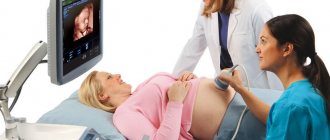
During 24-hour blood pressure monitoring, a woman is fitted with a monitor and a cuff, and her blood pressure is measured every 15-30 minutes. During the measurement, you should be at rest, the arm with the cuff should be relaxed. Source: BTL Medical Solutions / YouTube
Diagnosis of preeclampsia
The risk of preeclampsia begins to be assessed in the first trimester. This is usually done during routine screening. To do this, the obstetrician-gynecologist measures blood pressure and prescribes a blood test. The doctor also collects medical history data. He needs to be told about existing and past diseases, health status, well-being and complaints. Already in the first trimester, Doppler testing is prescribed. This is an ultrasound examination that evaluates blood flow in both uterine arteries.
In the future, additional ultrasound examination may be prescribed if preeclampsia is suspected. During such a study, fetal fetometry is performed (its size, anatomical structure, position, etc. are analyzed), and the condition of the placenta and amniotic fluid is assessed².
Important!
Already in the first trimester, diagnostics are carried out, which helps to assess the risk of developing preeclampsia. Early identification of this risk allows for the prevention of severe complications, so it is important for pregnant women to register with the antenatal clinic in a timely manner - the sooner the better.
At every doctor's appointment, a pregnant woman's blood pressure is measured. The doctor also regularly orders a urine test. This is necessary to control the risk of preeclampsia. When blood pressure rises to 140/90 mm Hg. Art. (or more) and the appearance of protein in the urine, the woman will be referred for additional examination.
Blood test results for the levels of sFlt-1, PlGF proteins and their ratio can predict preeclampsia 4 weeks before the onset of symptoms.
If preeclampsia is suspected, your doctor may order the following tests:
- blood test to determine the level of total hemoglobin and estimate the hematocrit;
- blood test to determine platelet levels;
- biochemical blood test to assess the level of total protein, blood glucose, creatinine, total bilirubin, urea, uric acid, determination of ALT, AST.
The quality of blood flow in the uterine arteries indicates whether the fetus is adequately supplied with oxygen and nutrients.
Source: olesiabilkei / Depositphotos During diagnosis, blood pressure is measured at least twice, with an interval of 4-6 hours (except in emergency cases). Proteinuria is diagnosed by a 24-hour urine test (loss of 300 mg of protein or more). Diagnosis is also possible by the ratio of protein and creatinine or by the result of a test with an indicator test strip (1+ and above).
Preeclampsia can be diagnosed even in cases where protein is absent in the urine, provided that new-onset hypertension is combined with any of the following conditions:
- thrombocytopenia;
- renal failure or impaired liver function;
- pulmonary edema;
- visual impairment;
- dysfunction of the brain.
Edema can accompany preeclampsia due to impaired renal function, but during diagnosis they are not considered as a symptom, since they can also occur during the normal course of pregnancy.
It is important to distinguish preeclampsia from other conditions that occur during pregnancy due to high blood pressure (Fig. 2):
- Chronic arterial hypertension - observed up to 20 weeks of pregnancy.
- Gestational hypertension occurs in the second half of pregnancy and is not accompanied by proteinuria or other symptoms of preeclampsia.
After the studies are completed, the doctor determines the severity of preeclampsia and prescribes appropriate therapy.
Preeclampsia. Preparation and management of pregnancy after preeclampsia
Preeclampsia is a severe complication of the second half of pregnancy, accompanied by increased blood pressure and the appearance of protein in the urine. The main reason for the development of preeclampsia during pregnancy is a disruption in the formation of the placenta before 16 weeks of gestation. Preeclampsia is the fourth most common cause of maternal mortality and a risk factor for the development of diseases of the cardiovascular system, kidneys, eyesight, and metabolic disorders in the mother after childbirth. Developed preeclampsia during pregnancy in its severe forms is an indication for early delivery of a woman, the birth of a very premature child, increasing the incidence of perinatal morbidity and mortality. Currently, there are no effective treatments for preeclampsia other than delivery, so prevention of this pathology is extremely important. The main method of preventing preeclampsia is taking low-dose aspirin by a woman from the stage of preconception and early pregnancy in women at high risk, as well as in women from 12-13 weeks of pregnancy with an increased risk of preeclampsia according to the first prenatal screening.
Our center’s specialists have many years of experience working with high-risk pregnant women, including those following preeclampsia.
Definition
Preeclampsia is a pathological condition that occurs during pregnancy (after 20 weeks), childbirth and the postpartum period and is characterized by the presence of arterial hypertension and significant proteinuria.
The incidence of preeclampsia during pregnancy is quite high and is approximately 3-4%. At the same time, developed preeclampsia threatens the health and life of the mother and fetus.
The main cause of the development of preeclampsia, especially early preeclampsia, which develops before 34 weeks of pregnancy, is a violation of the processes of placentation, that is, the formation of the placenta.
“
Arterial hypertension ”
This is a persistent increase in systolic blood pressure above 140 mm Hg, and diastolic blood pressure above 90 mm Hg.
«Proteinuria»
Presence of protein in urine.
Preeclampsia is characterized by significant proteinuria > 0.3 g in 24-hour urine or 0.3 g/L in two urine samples taken more than 6 hours apart.
Currently, the classification of the American Society of Obstetricians and Gynecologists (ACOG) is accepted, which includes two degrees of severity of preeclampsia - moderate and severe preeclampsia, as well as eclampsia (development of seizures). In the literature you can find another name for this complication - “preeclampsia”, which is not currently used.
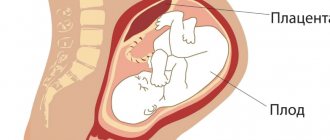
Placentation
The placenta is an organ that is formed only during pregnancy and allows the fetus to develop in the mother’s body throughout the entire period of pregnancy.
The placenta performs a huge number of functions during pregnancy, including:
- Nutritional:
- transport of nutrients to the fetus.
- Respiratory:
- transport of oxygen to the fetus.
- Transport:
- transport of exchange products.
- Hormonal:
- synthesis of hCG, which ensures the maintenance of the function of the corpus luteum and the production of progesterone;
- synthesis of placental lactagen;
- synthesis of serotonin, relaxin and other biologically active substances.
- Protective:
- ensuring immunological tolerance to the fetus (a fetus that has half the genes from the father is not rejected by the mother’s body);
- transport of protective antibodies from mother to fetus, protection from foreign microorganisms.
The placenta is formed sequentially in 3 stages:
- Trophoblast.
- Chorion.
- Placenta.
Trophoblast is the outer layer of embryonic cells, which is formed at the late blastocyst stage on the fifth to seventh days after fertilization and ensures the process of implantation of the blastocyst into the endometrium.
During implantation, the trophoblast divides into cytotrophoblast (inner layer) and syncytiotrophoblast (outer layer).
Subsequently, the trophoblast forms protrusions into the endometrium - villi, after which it begins to be called the villous chorion. Subsequently, blood vessels are formed inside the villi, which connect with the vessels of the embryo.
The section of the chorion facing the uterine cavity loses villi by the eighth week of pregnancy and is called the smooth chorion.
During immersion into the mucous membrane of the uterus, the chorionic villi actively grow and branch.
The mother's blood does not mix with the blood of the fetus, and nutrition occurs due to the diffusion of nutrients and oxygen from the mother's blood flowing through the spiral arteries into the intervillous space.
Also in the placenta there is a so-called extravillous cytotrophoblast (villous cytotrophoblast lines the inside of the chorionic villi). Extravillous cytotrophoblast plays a huge role in the process of placenta formation. Extravillous cytotrophoblast cells penetrate the walls of the spiral arteries and cause the so-called remodeling of the spiral arteries.
The depth of extravillous cytotrophoblast invasion (relative to the thickness of the myometrium - the muscular middle layer of the uterus) determines the degree of remodeling of the spiral arteries.
Placentation disorders
If the remodeling of the spiral arteries does not occur fully, the arteries retain their ability to contract, which in late pregnancy can lead to a decrease in blood supply to the fetus - placental insufficiency and, as a consequence, intrauterine growth retardation.
In this case, the fetus, due to a decrease in the supply of oxygen to it, sends certain signals to the maternal body, in response to which the woman’s body reacts by increasing blood pressure. With an increase in blood pressure, the blood supply to the fetus should normally increase, which does not happen if the spiral arteries of the uterus in the first and early second trimesters of pregnancy have not lost their ability to contract.
Thus, if the processes of remodeling of the spiral arteries occur correctly before sixteen weeks of pregnancy, the risks of developing pregnancy complications such as intrauterine growth restriction and preeclampsia are minimal. If the processes of placenta formation have been disrupted, this will most likely lead to these complications of gestation.
Symptoms of preeclampsia
Preeclampsia comes in two grades – moderate and severe. Depending on the severity of the disease, various symptoms may occur:
- Increased blood pressure above 140/90 mm Hg. Art.;
- Proteinuria (presence of protein > 0.3 g in daily urine);
- Edema;
- Headache;
- Visual impairment;
- Nausea, vomiting;
- Pain in the epigastrium, right hypochondrium;
- Oliguria (decrease in urine output to less than 500 ml per day);
- Nasal congestion;
- Dry cough.
Complications of preeclampsia for the mother
Preeclampsia is the fourth most common cause of maternal death.
High blood pressure (>160/110 mmHg) significantly increases the risk of hemorrhagic stroke. Severe preeclampsia can be accompanied by multiple organ failure, cerebral edema, massive bleeding, threatening the life of the mother.
Long-term consequences for maternal health also exist. Preeclampsia suffered by a woman increases the risks of cardiovascular (strokes, heart attacks) complications, kidney diseases (chronic renal failure), vision (retinal pathology), and metabolic disorders (type 2 diabetes mellitus) throughout life. Considering the fact that preeclampsia is a life-threatening condition for the mother, and therapy during pregnancy is ineffective, doctors unfortunately have to resort to surgical delivery by cesarean section even when the prognosis for the fetus is extremely unfavorable (less than 25-26 weeks of pregnancy ).
Complications of preeclampsia for the fetus
Preeclampsia is a leading cause of perinatal morbidity and mortality.
The main cause of fetal death during pregnancy (antenatal fetal death) in the case of developed preeclampsia is decompensation of placental insufficiency, that is, a profound dysfunction of the placenta, leading to severe fetal hypoxia.
A significant cause of death and morbidity in newborns is extreme prematurity. Children born at 22-25 weeks have an extremely unfavorable prognosis; at this period, the survival rate of newborns is minimal. With increasing gestational age, the mortality and morbidity of newborns decreases and largely depends on the capabilities of the medical institution to provide care to premature babies, both in the immediate hours and days after birth in the neonatal intensive care unit, and in the second stage of nursing - in the neonatal pathology department.
«Prematurity»
Preterm birth is birth between 22 weeks and 36 weeks 6 days. Newborns born before 37 weeks are considered premature.
Some of the consequences of prematurity include:
- Diseases of the organ of vision (retinopathy of prematurity);
- Hearing impairment (hard of hearing);
- Bronchopulmonary dysplasia (lung pathology);
- Pathology of the nervous system (CP);
- Delayed psychomotor development.
Thus, it is extremely important to identify women at risk of developing preeclampsia at the stage of pre-conception preparation (preparation for pregnancy) and in the early stages of pregnancy (up to 12 weeks).
Preventing preeclampsia
At the moment, there is the only effective method of preventing preeclampsia - taking aspirin (acetylsalicylic acid) in a dose of 75-162 mg, started before 16 weeks of pregnancy. But the sooner a woman starts taking the drug, the lower the likelihood of complications associated with impaired placental function.
We can distinguish categories of women for whom taking aspirin is indicated from the stage of preconception preparation or immediately upon pregnancy.
Aspirin is indicated for women at high risk of preeclampsia:
- preeclampsia during a previous pregnancy;
- chronic kidney disease;
- autoimmune diseases (systemic lupus erythematosus, antiphospholipid syndrome and others);
- diabetes mellitus type 1 or 2;
- chronic arterial hypertension;
- obesity of 2 or more degrees (BMI > 35 kg/m2);
- preeclampsia in female relatives (mother, grandmother, sisters);
- multiple pregnancy;
- Leiden mutation (F5);
- age over 35 years;
- connective tissue diseases;
- The duration of sexual activity before pregnancy is less than 6 months.
Assessing the risk of preeclampsia
The group of women at high risk of developing preeclampsia can also be identified in the first trimester at the stage of the first prenatal screening at 11 weeks - 13 weeks 6 days.
The risks of preeclampsia are calculated based on:
- Medical history (past PE, family history, age, presence of chronic diseases, etc.);
- Mean arterial pressure;
- Pulsatility index of the uterine arteries (PI MA);
- Determination of the concentration of placental growth factor in the blood (PlGF) and pregnancy-associated protein A (PAPP-A).
A high risk of preeclampsia is characterized by:
- high values of mean arterial pressure;
- high values of the pulsatility index of the uterine arteries;
- low levels of placental growth factor (PlGF).
It should be noted that so far in many medical institutions, during screening of the first trimester, ultrasound does not assess the pulsatility index of the uterine arteries, and biochemical screening includes the assessment of only two markers - pregnancy-associated protein A (PAPP-A) and free subunit of beta-hCG (free beta-hCG), which does not allow us to accurately identify a group of pregnant women with a high risk of placental disorders!
An additional but less sensitive marker than placental growth factor is pregnancy-associated protein A (PAPP-A). A decrease in PAPP-A, especially in combination with a decrease in PlGF, is typical for pregnant women at high risk of developing preeclampsia.
Additionally, the level of these markers can be assessed at an earlier stage of pregnancy. Assessment of the level of pregnancy-associated protein A (PAPP-A) is possible from 8 weeks of gestation, and placental growth factor (PlGF) from 10 weeks. These tests can be taken at the CIR laboratory.
Of great importance in the management of pregnant women with a high risk of placental disorders is the assessment of feto-placental and utero-placental blood flow using Dopplerometry. In the period starting from 16-17 weeks of gestation in case of a high risk of early preeclampsia, and in the period 19-21 weeks in all pregnant women, it is mandatory to assess the pulsation index of the uterine arteries and fetal umbilical cord arteries. The normal values differ at different stages of pregnancy and are assessed using special tables by ultrasound doctors. The intervals and frequency of examinations are individual in each case.
Doppler measurements of the uteroplacental and fetal placental blood flow in combination with fetometry during ultrasound examinations make it possible to timely identify pregnant women with a tendency to intrauterine growth restriction (or fetal growth restriction syndrome).
To assess the risk of developing preeclampsia after 20 weeks, it is possible to determine the ratio of soluble fms-like tyrosine kinase-1 to placental growth factor (sFlt-1/PlGF). Soluble fms-like tyrosine kinase-1 (s1Flt) is a protein produced by the placenta.
During a normal pregnancy, the level of soluble fms-like tyrosine kinase-1 remains stably low until 33–36 weeks of pregnancy, after which it may increase. The level of placental growth factor in a successful pregnancy is, on the contrary, high. The ratio of soluble fms-like tyrosine kinase-1 to placental growth factor (sFlt-1/PlGF) is low.
On the contrary, in preeclampsia, the level of soluble fms-like tyrosine kinase-1 is increased, the value of placental growth factor is low, and the sFlt-1/PlGF ratio is higher than in normal pregnancy. A ratio of soluble fms-like tyrosine kinase-1 to placental growth factor of less than 38 excludes the development of preeclampsia within the next 7 days. An increase in the ratio of more than 85 at less than 34 weeks of gestation and more than 110 at more than 34 weeks of gestation is associated with a high risk of developing preeclampsia.
Treatment of preeclampsia
There is currently no effective drug treatment for preeclampsia. Treatment of preeclampsia at the outpatient (polyclinic) stage is symptomatic and is possible only in the case of moderate preeclampsia.
Therapy includes:
- Salt-restricted diet (
- Antihypertensive therapy.
Treatment of severe preeclampsia is possible only in a hospital and, as a rule, includes:
- Multicomponent antihypertensive therapy (2-3 antihypertensive drugs);
- Anticonvulsant therapy;
- Magnesium therapy;
- Prevention of fetal respiratory distress syndrome (FRS) before 34 weeks of gestation.
Drug therapy for severe preeclampsia usually lasts no more than 48 hours and is aimed at:
- Stabilization of the mother's condition;
- Prophylaxis of fetal and newborn respiratory distress syndrome (RDS).
The only effective treatment for severe preeclampsia is still delivery.
Planning a second pregnancy after preeclampsia
The recommended interval between birth and the onset of the next pregnancy is 2 years, but this period can be reduced to 6–12 months in some cases.
At the stage of preparation for the next pregnancy, a woman with a history of severe pregnancy complications (pre-eclampsia, intrauterine growth restriction, antenatal fetal death) is recommended to undergo a more in-depth examination to assess risk factors for the development of these conditions.
First of all, a woman should visit doctors of the following specialties in order to eliminate the consequences of preeclampsia and compensate for existing diseases:
- Therapist. The general practitioner will rule out chronic arterial hypertension and, if necessary, recommend 24-hour blood pressure monitoring (ABPM). When the diagnosis is confirmed, the therapist recommends antihypertensive therapy, taking into account preparation for the next pregnancy.
- Neurologist. A neurologist will rule out neurological pathology as a consequence of preeclampsia.
- Nephrologist. A nephrologist will rule out renal pathology, which is a risk factor for preeclampsia.
- Endocrinologist. An endocrinologist is recommended for overweight and obese women, as well as to exclude impaired glucose tolerance and diabetes mellitus.
- Ophthalmologist. An ophthalmologist will rule out pathology of the organ of vision, primarily the retina, which is often a consequence of pre-eclampsia.
- Obstetrician-gynecologist. An obstetrician-gynecologist recommends an examination plan in preparation for pregnancy.
The examination plan is determined individually in each case, but, as a rule, includes:
- Polymorphisms of hemostasis genes – 19 factors. Among polymorphisms of hemostasis genes, coagulation factors (Leiden mutation, prothrombin factor, coagulation factor 11), factors responsible for fibrinolysis processes (PAI-11, F13A1, PAI-1, Hageman factor), as well as factors may play a key role in the development of placental disorders platelet receptors (ITGA2, ITGB3, GP1ba, P2RY12).
- Polymorphisms of vascular tone and preeclampsia genes. Preeclampsia gene polymorphisms are a set of genes responsible for maintaining the water-salt and electrolyte composition of the blood, as well as blood pressure. The combination of different genes from this panel may be a risk factor for the development of preeclampsia.
- Polymorphisms of cytokine genes. One of the most significant genes, when identified as a polymorphism, is associated with the risk of developing preeclampsia is tumor necrosis factor alpha.
- Autoantibody block, lupus anticoagulant, immunoblot for antinuclear factors, antinuclear factor, rheumatoid factor, ultrasensitive C-reactive protein. This examination is recommended to exclude autoimmune risk factors that significantly aggravate the course of pregnancy and increase the risk of developing preeclampsia.
- Typing of both spouses for HLA class 1 and class 2 genes. Typing using HLA class 1 genes allows us to evaluate not only the C1/C2 configuration of a married couple. Determining the haplotypes of each spouse makes it possible to determine their connection with certain pathologies that significantly affect reproductive health.
- Glycated hemoglobin, fasting serum glucose, metabolic block. Recommended to exclude impaired glucose tolerance, especially in overweight women.
- Immunogram. Allows you to evaluate a person’s immune constitution, the tendency for the immune system to respond at the humoral or cellular level. Thus, the humoral type of response is often associated with autoimmune pathology.
The above examination allows the obstetrician-gynecologist to assess the presence of certain risk factors for the development of preeclampsia and recommend appropriate therapy at the preconception stage and in the earliest stages of pregnancy.
So, if a woman has autoimmune risk factors for the development of preeclampsia - antibodies to DNA or phospholipids, a positive lupus anticoagulant, according to the results of the immunogram, the immunoregulatory index (IRI) tends to increase, variant genes of anti-inflammatory cytokines (interleukin-4) are detected, and the haplotype is determined HLA, associated with a high risk of developing autoimmune pathology, the obstetrician-gynecologist will consider adding drugs such as normal human immunoglobulin and hydroxychloroquine to drug therapy.
If certain combinations of hemostasis and vascular tone genes are identified, the issue of adding anticoagulants (low molecular weight or unfractionated heparin) to therapy will be considered.
Therefore, without a full examination, it is impossible to say for sure whether taking low-dose aspirin during pregnancy will be enough or not. Each case is considered individually.
Case from practice
A married couple, the wife is 33 years old, the husband is 35 years old.
In 2015 - an emergency caesarean section at 27 weeks due to severe preeclampsia, in 2022 - an emergency caesarean section at 24 weeks due to severe preeclampsia.
The first pregnancy proceeded uneventfully, according to the patient, until 26 weeks; subsequently, blood pressure increased to 190/110 mm Hg. Art., accompanied by headache, disorientation, and loss of coordination. According to ultrasound data at 27 weeks, severe placental insufficiency (zero blood flow in the umbilical cord artery, decreased blood flow in the middle cerebral artery), intrauterine growth restriction of the 3rd degree. As a result of surgical delivery, a boy weighing 530 grams was born and died on the 3rd day.
The second pregnancy proceeded uneventfully until 24 weeks. In the early stages of gestation, the pregnant woman was consulted by a hematologist, and therapy with low molecular weight heparins (Clexane) was recommended. At 24 weeks - severe placental insufficiency, severe fetal growth restriction, severe preeclampsia. The situation was complicated by partial detachment of the normally located placenta, which resulted in massive bleeding, to stop which the uterine arteries were ligated.
According to the results of the survey:
- Thyroid function is normal.
- According to the examination for polymorphisms of hemostasis genes, a heterozygous mutation of the prothrombin gene (the second factor of the blood coagulation system) was identified.
- When testing the blood for the presence of autoantibodies, no antibodies to DNA, to the components of the thyroid gland, or to phospholipids were detected - there is no autoimmune component.
“A couple is asking whether they can have a healthy baby and what additional testing they need?”
Guzov Igor Ivanovich General Director of Clinics and Laboratories CIR - Obstetrician-gynecologist, Candidate of Medical Sciences - founder of the Center for Immunology and Reproduction What examination is recommended for this couple?
Our center currently recommends studying those genes of the hemostatic system that were not on the panel in this patient. The standard panel of other laboratories includes only 12 genes - F2, F5, FBG, F13A1, PAI-1, ITGA2, ITGB3, F7 and 4 genes responsible for folate metabolism. In our laboratory, the panel has been expanded; four equally important genes are additionally assessed – F11, F12, PLAT, GP1ba. It is also recommended to study genes responsible for maintaining normal blood pressure and genes of the immune system - this is a study of polymorphisms of vascular tone and cytokine genes.
From genetic examinations, we can obtain a fairly large amount of information by typing a couple using HLA class 1 and class 2 genes. Often this helps us understand in which direction to move next, what diseases a given couple is predisposed to and how this can affect reproductive function.
When assessing autoimmune factors, it is possible to supplement the examination with an immunoblot for antinuclear factors, as well as a blood test for lupus anticoagulant. In order to assess a woman’s immune constitution, to understand whether there are any immune factors that interfere with the processes of normal placentation, it is advisable to undergo an analysis such as an immunogram.
You can consider this case in more detail during a consultation, since family history and the presence of certain chronic diseases play a huge role. Many nuances can only be clarified through direct communication with the couple.
Treatment of preeclampsia
Preeclampsia is dangerous for both mother and baby. Even if the woman’s condition is not serious, observation and preservation of the pregnancy in a hospital is necessary. If the condition is severe, emergency assistance may be required.
Treatment tactics are chosen depending on the severity of the pathology and the duration of pregnancy. Treatment is carried out in several directions².
Lower blood pressure
Not all antihypertensive drugs are suitable for this. For example, ACE inhibitors, angiotensin II receptor antagonists and some other drugs are not recommended. Your doctor may prescribe methyldopa. It is also possible to use alpha-blockers, nifedipine, clonidine. Your doctor should choose medications to lower blood pressure and calculate the dosage. Antihypertensive therapy requires constant monitoring of blood pressure, as well as periodic ultrasound to monitor the condition of the fetus.
Anticonvulsant therapy
Involves intravenous administration of magnesium sulfate. It is recommended for severe preeclampsia, but the doctor can use magnesia for moderate preeclampsia, as well as for prevention if there is a risk of its development. Magnesium therapy is used for no longer than 5-7 days. Magnesium sulfate is administered to prevent new seizures and also within 24 hours after birth².
Delivery
Preeclampsia cannot be cured, and the only way to get rid of it is to give birth. As long as it remains moderate, it can be controlled under the supervision of doctors so that the birth does not occur too early. However, if there are emergency indications, delivery is carried out urgently. It is necessary for signs of placental abruption, acute fetal hypoxia, bleeding from the birth canal, or a threat to the mother’s life. The woman's condition is stabilized until delivery. If possible, the birth takes place naturally; if not, a caesarean section is performed.
For women with preeclampsia, emergency caesarean section is often recommended. Photo: Nasimi/Depositphotos
The gestational age at which preeclampsia occurred affects treatment tactics:
- up to 22-24 weeks - it is considered that the pregnancy threatens the woman’s life and it is terminated;
- from 25 to 33 weeks - pregnancy is maintained if uncontrolled arterial hypertension does not develop, there is no danger to the mother or child;
- after 33-34 weeks - preparation for childbirth and delivery (caesarean section).
In each case, the decision on delivery, continuation or termination of pregnancy must be made by the doctor, taking into account the expected risks and the dynamics of the condition of the mother and fetus.
With severe preeclampsia, a woman may require emergency care with hospitalization in an intensive care unit or intensive care unit. Observation in the intensive care unit is required for 24 hours after birth. During the recovery period, it is necessary to monitor the protein content in the urine, liver function indicators, and blood pressure. If it remains high six weeks after birth, you need to consult a GP or cardiologist. In addition, a woman who has experienced preeclampsia may be prescribed an MRI of the brain.
Use of dexamethasone
Severe preeclampsia that develops before 34 weeks carries a risk of preterm birth. For the fetus, this is dangerous due to the development of RDS (respiratory distress syndrome), a severe respiratory disorder that occurs due to the immaturity of the newborn’s lungs. To prevent RDS in a child, a woman may be recommended injections of dexamethasone, a glucocorticosteroid that accelerates the development of lung tissue in the fetus⁵. It is advisable to complete therapy at least 48 hours before birth.
Postpartum follow-up
Preeclampsia goes away after pregnancy ends, but the woman remains at increased risk of cardiovascular disease. After giving birth, your blood pressure should be measured for several weeks. Until it normalizes, antihypertensive medications may be required. If your blood pressure remains high after 12 weeks, you should see your GP or cardiologist for treatment of your hypertension.
How to cope with postpartum preeclampsia
The time after childbirth can be challenging even without health problems. Recovering from childbirth and caring for a newborn can be stressful. While recovering from pregnancy, it is important to pay attention to your own health by monitoring symptoms and consulting with your doctor.
If you are diagnosed with postpartum preeclampsia while in hospital, you may need a longer hospital stay. Reach out to loved ones or talk to your healthcare provider to learn about options for additional support when you return home.
What will happen if left untreated? Risks of Preeclampsia
Preeclampsia is dangerous for both the pregnant woman and the baby. Without treatment, it threatens to develop eclampsia. This is a severe seizure that can result in coma, stroke or myocardial infarction. It is accompanied by increasing renal failure, pulmonary edema, placental abruption, sudden intrauterine fetal death, and loss of vision due to retinal detachment are possible.
Even if preeclampsia goes away, the woman remains at risk for hypertension and other cardiovascular diseases, stroke and heart attack. To control these risks, it is important to maintain a healthy lifestyle and follow-up with doctors after pregnancy.
Important!
For a woman who has experienced preeclampsia, her risk will be higher with each subsequent pregnancy, and if preeclampsia develops again, it will be more severe each time.
During and after preeclampsia, a woman may develop the following conditions and diseases:
- HELLP syndrome. Associated with functional liver failure, accompanied by disruption of other internal organs (kidneys, lungs, brain, etc.), threatening death to both the mother and the fetus.
- DIC syndrome. Disturbances in the blood coagulation system with the formation of blood clots, a subsequent decrease in the blood's ability to clot and the appearance of bleeding.
- Pulmonary edema. Accompanied by shortness of breath, cough, breathing problems.
- Acute renal failure. Manifests itself as a result of kidney damage during preeclampsia.
- Hypertensive encephalopathy. A vascular disease of the brain that develops as a complication of arterial hypertension.
Complications of preeclampsia that are dangerous for the fetus:
- placental abruption;
- acute hypoxia;
- slow fetal growth and low birth weight;
- the need for premature birth;
- intrauterine fetal death.
With preeclampsia, emergency delivery may be necessary to save the mother's life. If this occurs early, the baby may die or be born very preterm, which carries many health risks².
Figure 2. Differential diagnosis of preeclampsia. Source: Russian Society of Obstetricians and Gynecologists (ROAG)
Where does gestosis come from and what is it? Causes of preeclampsia
Preeclampsia or late gestosis is a dangerous complication of pregnancy, in which blood pressure rises, edema forms, and protein appears in the urine. All this can lead to disruptions in the functioning of the expectant mother’s organs and harm the fetus, slowing down its development.
It is not known exactly why preeclampsia occurs. The prerequisites for its appearance are laid in early pregnancy, when the fertilized egg attaches to the uterus. If it is not attached deeply enough, superficially, the placenta develops incorrectly, the development of vascular channels is disrupted, and this affects the flow of blood to the fetus. Because of this, as pregnancy progresses, the placenta becomes less and less able to provide itself and the fetus with the necessary nutrients. After 20 weeks, this leads to the appearance of symptoms of preeclampsia and a whole range of disorders that damage the blood vessels of various organs of the mother (including kidneys, brain, liver) and threaten her life. Preeclampsia is also dangerous for the baby. It can affect fetal development by causing hypoxia, as well as developmental delays and convulsions¹.
Vitamin D deficiency and the risk of preeclampsia: is there a connection?
Vitamin D affects many physiological processes: on the one hand, on how the fetus attaches to the epithelium of the uterus, on the other, on the regulation of renin biosynthesis (an enzyme associated with increased blood pressure).
Back in the early 1990s, a hypothesis was put forward about the influence of vitamin D levels on the risk of preeclampsia. Later research showed that this was indeed the case³. Low vitamin D levels before 22 weeks of pregnancy are considered an independent risk factor for the development of preeclampsia. If there is a vitamin D deficiency confirmed by tests, taking it from early pregnancy reduces the likelihood of preeclampsia, as well as the development of its severe complications.
Several factors are known to increase the risk of preeclampsia:
- This is the first pregnancy or more than 10 years have passed since the previous pregnancy.
- The age of the pregnant woman is over 35-40 years.
- During a previous pregnancy, there was already preeclampsia (with each new case, the severity of the condition may increase).
- Blood relatives had preeclampsia.
- Pregnancy is multiple.
- The woman has diabetes, obesity or other metabolic diseases, high blood pressure, or kidney disease.
- The pregnancy resulted from IVF.
- The woman has autoimmune diseases (systemic lupus erythematosus and others).
How common is preeclampsia?
Between 5 and 10% of pregnant women have high blood pressure. Hypertension has a high risk of preeclampsia. It occurs in 2-8% of pregnant women and accounts for 10-15% of maternal deaths. Every year, at least 70 thousand pregnant women die due to preeclampsia.
In Russia, moderate preeclampsia is diagnosed in 2.74% of the total number of pregnant women, severe - in 0.84%.
The number of deaths and severe complications associated with preeclampsia is gradually decreasing. An important role in this is played by proper management of pregnancy, constant monitoring in antenatal clinics and timely diagnosis².
Complications of postpartum preeclampsia
Without treatment, postpartum preeclampsia can lead to serious and life-threatening complications, such as:
- Excess fluid in the lungs, making it difficult to breathe
- Stroke, in which blood flow to the brain is interrupted, cutting off the supply of oxygen and nutrients to the brain
- Seizures, which may cause loss of consciousness and confusion
- Thromboembolism, or blood clots that displace and block blood flow to any part of the body
- HELLP syndrome, which refers to hemolysis (destruction of red blood cells), elevated liver enzymes and low platelet counts.
All of these complications can lead to death if left untreated.
Although complications of postpartum preeclampsia are serious, they can be prevented with treatment and are rare.
Degrees of preeclampsia
Preeclampsia can be early, with the first symptoms appearing before 34 weeks, and late, developing after 34 weeks of pregnancy. With early preeclampsia, the risk of severe course and consequences for mother and child is higher.
Preeclampsia can range in severity from moderate to severe. The severity of the condition affects the prognosis, as well as the choice of pregnancy management tactics, treatment, and obstetric care².
Preeclampsia is considered moderate if proteinuria is diagnosed and blood pressure is elevated between 140/90 and 159/99 mmHg. Art. In severe conditions, blood pressure is higher than these indicators (confirmed by at least two measurement results with an interval of 4-6 hours). The amount of protein in the urine is 300 mg/day. The condition is also considered severe if, in addition to the symptoms of moderate preeclampsia, there are the following signs:
- shortness of breath, respiratory failure, symptoms of pulmonary edema;
- vomiting, nausea, pain in the right hypochondrium;
- headache, visual disturbances, tinnitus, confusion;
- onset of symptoms before 34 weeks;
- fetal growth restriction;
- formation and excretion of urine in a volume of less than 500 ml.
Sources
- Pylaeva N.Yu., Shifman E.M. Preeclampsia. Eclampsia. Anesthesia and intensive care during childbirth and the postpartum period. Anesthesia and intensive care in obstetrics, 2022.
- Russian Society of Obstetricians and Gynecologists, Association of Anesthesiologists and Resuscitators, Association of Obstetric Anesthesiologists and Resuscitators. Preeclampsia. Eclampsia. Edema, proteinuria and hypertensive disorders during pregnancy, childbirth and the postpartum period. Clinical guidelines, 2022.
- Maltseva L. I., Vasilyeva E. N. Vitamin D and preeclampsia. Russian Bulletin of Obstetrician-Gynecologist, 2016.
- Sidorova I. S., Nikitina N. A. Critical forms of preeclampsia. Russian Bulletin of Obstetrician-Gynecologist, 2022.
- Russian Society of Obstetricians and Gynecologists. Normal pregnancy. Clinical guidelines, 2022.
- World Health Organization. WHO recommendations for the prevention and treatment of preeclampsia and eclampsia. 2014.