- home
- Services and prices
- Phlebology
Varicose veins (VV) is a fairly common disease that affects both men and women. It can affect the lower limbs of a person, as well as deep veins, leading to the development of thrombosis and post-thrombophlebitis disease.
Spider veins that appear on a person’s legs as a result of the development of the disease become the reason that he begins to feel unattractive. In addition to external ones, there are also internal manifestations of explosives, which are expressed in the appearance of discomfort and pain in the calf muscles of the leg. The development of the disease increases the risk of other pathologies of the circulatory system.
The key to success in the fight against pathology lies in timely diagnosis and competent treatment. A big mistake on the part of a person is the independent use of various ointments and creams, which in most cases do not bring the desired effect. As a result, time was lost that could have been spent on proper and effective therapy.
In order to get rid of varicose veins, you need to solve the following problems:
- Elimination of symptoms.
- Removal of varicose veins.
- Prevention of the development and reappearance of explosives.
Only a highly qualified specialist with sufficient experience in the treatment and prevention of pathologies of this kind can successfully cope with each of the above tasks. These are the specialists who work at the Yuzhny Medical Center. The high level of qualifications of doctors in combination with the latest equipment will make it possible to make the correct diagnosis and carry out competent therapy, guaranteeing the absence of progression or recurrence of varicose veins of the lower extremities.
Symptoms of varicose veins
Symptoms of a disease are signs that clearly indicate its development. They are divided into:
- Subjective: Mild and aching pain in the calf muscles.
- A burning sensation and itching along the veins affected by varicose veins.
- Heaviness in the legs, increasing towards the end of the day.
- Skin hyperpigmentation.
- Increased fatigue of the lower extremities.
- Trophic venous ulcer of the leg.
- Pain in the calf muscles, aggravated by walking.
- The appearance of swelling in the lower legs and feet.
- Varicose veins of the saphenous veins, which are clearly visible even without the use of special equipment.
What veins look like
The very first warning sign of problems with the veins is swelling of the lower extremities at the end of the day. Swelling is especially pronounced if a person spends most of the day standing on his feet. It can disappear in the morning after a night spent resting.
However, if you do not pay due attention to this problem, the condition can worsen significantly. Intradermal veins in the legs with varicose veins become dark blue, protruding above the surface of the skin of the legs and feet. Outwardly, they look like bunches of red grapes that are overripe. Such external manifestations of pathology are accompanied by pain in the calves, a feeling of heat in the legs, swelling and cramps in the calf muscles. Over time, these symptoms are accompanied by a change in the appearance of the skin.
Varicose veins of the lower extremities - stages
- First. Spider veins appear;
- Second. Nodules are visible;
- Third. Swelling of the legs is added;
- Fourth. Skin color becomes darker, almost purple;
- Fifth and sixth. Ulcers form, which may not heal as a result.
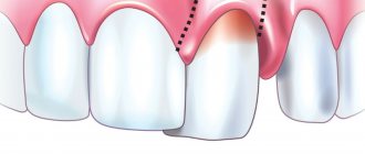
This is what varicose veins look like in stage II of the disease
Causes of varicose veins of the lower extremities
VV of the lower extremities can develop under the influence of a number of factors and circumstances, the main ones being:
- Pregnancy. This is a key risk factor for the disease. This explains the fact that varicose veins occur several times more often in women than in men. In this case, the disease develops under the influence of an increase in the volume of circulating blood and compression of the retroperitoneal veins of the pregnant uterus.
- Obesity. The connection of this condition with the development of VV has been proven by a number of studies. At the same time, a direct connection was found between increased body weight and an increased risk of developing pathology.
- A lifestyle characterized by prolonged static loads with regular heavy lifting or prolonged immobility in a standing or sitting position.
- Dishormonal conditions. Their role in the development of the disease has increased significantly over recent years. This is due to the widespread use of hormonal contraceptives, the spread of hormone replacement treatment for osteoporosis and during the premenopausal period
- Heredity. The role of this factor in the development of varicose veins on the legs has not been unambiguously confirmed today.
- A disorder of the valvular apparatus of the veins, leading to a downward flow of blood under the influence of gravity every time a person stands up. The muscles that are located around the deep veins contract as you walk. These veins are subject to emptying, causing venous pressure to increase. Blood enters the superficial veins through communicating vessels with insufficient valves. As a result, they are filled with blood, which leads to their stretching and expansion (varicose veins).
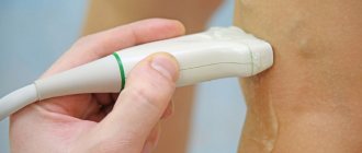
Varicose veins of the lower extremities - diagnosis
In addition to reviewing your medical history as well as a physical examination, testing for vein disease may include the following:
- Duplex scanning is a type of procedure that evaluates the blood flow and structure of the veins in the legs.
- Triplex ultrasound is a procedure similar to duplex ultrasound, which uses color to highlight the direction of blood flow.
- Magnetic resonance venography (MRV) is a diagnostic procedure that uses magnetic resonance technology and intravenous contrast material to visualize veins.
Diagnosis of varicose veins of the lower extremities is carried out with the patient standing
Diagnostics is necessary so that the doctor can prescribe a treatment method that is most suitable for the patient.
Classification and stages
Like any disease, VV has several stages, differing from each other in the degree of spread of the pathology and symptoms. Among them, the following stages are distinguished:
- Initial (or compensation).
- Second (or subcompensation).
- Third (or decompensation).
A detailed description of each stage can be found here.
It is worth noting that complications can occur at any of the above stages, but their greatest likelihood is inherent in the last two. VV can serve as an impetus for the development of diseases such as:
- Thrombophlebitis.
- Erysipelas.
- Deep vein thrombosis.
- Trophic eczema.
A visit to a specialist, made at the first signs of the disease, will help reduce the risks of worsening the situation and begin removing varicose veins. You should not ignore even minor symptoms, because this can lead to undesirable and extremely negative consequences.
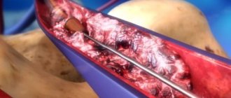
Varicose veins of the lower extremities, treatment. Operation?
Don't panic if you are diagnosed with varicose veins of the lower extremities. Sclerotherapy treatment may help. This procedure is based on the use of a special solution that is injected into varicose veins. Varicose veins of the lower extremities disappear quite quickly. Sclerotherapy treatment is suitable for treating small veins such as spider veins. Are you afraid of a knife? Varicose veins of the lower extremities can be removed (treatment without surgery). You just need to find a decent doctor.
Diagnostics
Diagnosis of varicose veins, the symptoms of which are described above, poses the following tasks:
- Determining the presence of pathology in each individual patient. It often happens that people who do not have varicose veins are confident in their presence, and vice versa. However, only an experienced phlebologist, based on an external examination and a series of comprehensive studies, can make an accurate diagnosis.
- Establishing the type characteristic of venous pathology. The doctor determines exactly which veins have undergone pathological damage, and also establishes the extent of this damage and possible or already occurring consequences.
- Prescribing the correct course of treatment. Based on the diagnosis and the characteristics of each specific organism, the attending physician makes a choice in favor of one or another treatment or a set of therapeutic measures.
- Assessment of the level of effectiveness of therapy, which is carried out by the attending physician during the elimination of the disease or after the patient’s complete recovery.
The main methods for diagnosing VV include:
- Plethysmography.
- Thermography.
- Magnetic resonance imaging.
- Ultrasound angioscanning.
- Computed tomography.
- Clinical studies: conversation with the patient, his external examination and manual examination.
- Radionuclide phlebography.
- Intravascular ultrasound.
- X-ray phlebography.
Most often, it is enough for a professional specialist to conduct a clinical examination and ultrasound angiography in order to diagnose varicose veins in the legs. At the Yuzhny clinic, you can undergo a full examination in order to make an accurate diagnosis by an experienced phlebologist. Our specialists work closely with clients, clearly explaining to each of them the specifics of the disease and possible ways to combat it.
Varicose veins. Laser treatment in Moscow
Life does not end if you have varicose veins of the lower extremities. Laser treatment will help. The doctor inserts a tiny fiber into the varicose veins through a catheter. The fiber sends out energy that destroys the affected part.
This is a tiny laser fiber that completely eliminates varicose veins
Do varicose veins of the lower extremities remain? Laser treatment, which may seem expensive, is very effective, so the answer is no. Not every patient worries if he has varicose veins of the lower extremities. However, laser treatment leaves only positive reviews. With it, varicose veins of the lower extremities will be defeated! Laser treatment (you will find out the cost at the clinic from the administrator) is short-lived and inexpensive.
Treatment methods
Modern methods of treating varicose veins are aimed at reducing the degree of disability and trauma, which contributes to a faster recovery of the patient. Main therapeutic techniques include:
- Sclerotherapy. This method involves introducing a special medication into the lumen of varicose veins of the legs, causing a chemical burn of the internal venous wall. This leads to their gluing and cessation of pathological blood flow through them. Can be used alone or in combination with other types of manipulation. It is carried out without prior anesthesia with skin punctures using a thin needle. The duration depends on the scale of the lesion.
- Foam sclerotherapy, which involves the preparation by a specialist of a special medication of foam that can use an impressive area of the internal walls of the affected venous vessels. Used to treat large diameter veins.
- Endovenous laser coagulation, which is performed using a laser device on the main trunks of the leg veins and allows you to stop the pathological flow of blood through the affected veins due to the burn of their inner walls and their subsequent gluing. Laser treatment for varicose veins is available at the Yuzhny Medical Center.
- Miniphlebectomy, aimed at eliminating subcutaneous nodes and tributaries enlarged by varicose veins through punctures of the skin. It has excellent cosmetic effects and is used alone or in combination with other therapeutic methods under local anesthesia.
- Elimination of incompetent perforating veins, performed for the prevention of venous insufficiency and treatment of trophic disorders, including ulcers.
- Combined phlebectomy, which is a combination of some methods of treating veins, based on the indications and nature of venous pathologies.
Yuzhny Medical Center offers its clients modern approaches to the treatment of varicose veins. Qualified specialists work here, ready to conduct a competent examination in order to make a diagnosis and carry out effective treatment, more about which you can read here.
Material and methods
Since the beginning of 2015, MSCT venography was performed on 121 people. Initially, 30 lower extremities were examined in individuals without signs of CVD (control group). The study group included 91 patients (52 women and 39 men aged 32 to 65 years) with CVD with clinical class C0-C6. Class C0—C1 occurred in 15 (16.5%) patients, C2—C3 — in 45 (49.5%). 31 (34%) patients had various trophic skin disorders (C4-C6).
All studies were performed on a 128-slice Philips Ingenuity CT multislice computed tomograph with the Intell Space Portal image processing software package, followed by reconstruction of a volumetric image in 3D mode.
The scanning was carried out in an automatic program mode, which implied sequential non-stop administration of a bolus of contrast agent and saline solution.
Scan mode: collimation 64×0.625; pitch 0.923; kilovoltage 100 kV; mAS 188; reconstruction parameters: axial + cranial orientation.
MSCT venography was performed according to the method we developed. In a clean dressing room or in a study room, the vein of the dorsum of the foot was catheterized using an intravenous catheter G22-G24. The patient was placed on the table on his back. One of two infusion syringes (A) was filled with 50 ml of non-ionic contrast agent (Ultravist). An isotonic sodium chloride solution was drawn into the second infusion syringe (B) at the rate of 1 ml of 0.9% saline per 1 cm of height of the subject. Both infusion syringes were inserted into the auto-injector. Using an infusion line, the injector was connected to an intravenous catheter and the infusion mode was switched on from A to B, with an injection rate of the radiopaque mixture of 4 ml/s.
Computer marking of the scanned limb was carried out, including the pelvis and foot. After preliminary scanning, the scanning area was finally set (the entire lower limb and pelvic area) with a direction from the pelvis to the foot. The scanning time parameters were entered into the program.
A pneumatic cuff was placed over the ankles, the pressure in which was raised to 60 mm Hg, and the administration of a radiopaque mixture began, which, depending on the calculated volume, lasted about 40 s. After completing the administration of the entire volume of contrast and isotonic sodium chloride solution, the pressure in the second cuff placed at the mid-thigh was raised to 60 mmHg, and the patient took a deep breath, held his breath and tensed the muscles of the anterior abdominal wall. From this moment, the first main scan began, the total duration of which was 12-15 s. After this, the patient exhaled and performed five dorsiflexion movements of the foot. After completing the test, the patient returned to the starting position. After 40 s, the second main scan began, after which the study was completed and a three-dimensional image of the limb and veins was reconstructed using automatic Intell Space Portal data processing protocols embedded in the computer.
Complications with varicose veins
It is worth understanding that improper treatment of the disease or complete refusal of it can lead to complications. The latter appear not only in cosmetic defects of the lower extremities, but also in more serious forms. Among them:
- Trophic eczema, which subsequently develops into an ulcer.
- Thrombotic lesions of the venous system, including thrombophlebitis of the superficial veins and deep vein thrombosis of the lower extremities.
Venous blood is a kind of “sewer” for the body’s tissues and is saturated with substances and products of cell metabolism that are relatively harmful to the human body. Cells of the skin and subcutaneous tissue, as well as muscles and bones, discharge products of tissue respiration and other waste material into the venous system, which carries them to the heart, lungs, kidneys and liver. In case of disturbances in the functioning of the venous system, the content of these products in the tissues of the body increases.
A vein dilated by varicose veins leads not only to an increase in the concentration of harmful products in the tissues, but also to an increase in their swelling. This disruption of the outflow of harmful products, combined with swelling observed over a long period of time, leads to the death of skin cells and subcutaneous tissue and their subsequent replacement by venous eczema, represented by a dense and dotted structure of a dark color. The death of the surface layer of the skin is the cause of trophic ulcers.
results
Analysis of the obtained 3D images in the study and control groups showed a high degree of information content of the studies, which is confirmed by a clear contrast in all cases of the main venous collectors of the lower limb. The veins of the soleus and gastrocnemius muscles were contrasted in 100% of cases when performing MSCT venography.
We considered it appropriate to make a separate analysis of the structure of the venous collectors of the gastrocnemius and soleus muscles due to their different anatomical structure and functional purpose. The soleus muscle has a long, fusiform shape and is primarily involved in flexion of the foot. The gastrocnemius muscle is a typical biceps muscle, the main purpose of which is not only and not so much in flexion of the foot, but in active and powerful flexion of the lower leg. Based on this, we can note the predominant static load on the first and dynamic on the second. It is natural that the degree of participation of the venous collectors of these muscles in the unified functioning of the muscular-venous pump of the lower leg will be different and not always unambiguous.
The veins of the soleus muscle in the study were very diverse in shape, size, location, number, location and angle of entry into the deep veins of the leg. In contrast, the veins of the gastrocnemius muscle showed a fairly pronounced uniformity.
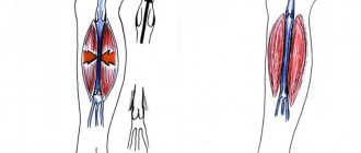
In a study of healthy individuals, it was found that in 89.9% of cases the veins of the soleus muscle had a cylindrical shape (Fig. 1)
Rice. 1. Paired intramuscular veins of the gastrocnemius (A) and soleus (B) muscles are cylindrical in shape. Main type of building. Variant of the norm. and were represented by: single (48%), paired (26%), V-shaped (28%) and Y-shaped (78%) trunks, having a mixed type of structure in 96% of cases (Fig. 2).
Rice. 2. Variants of the structure of the intramuscular veins of the leg. At the same time, the diameter of the venous trunks did not exceed 0.5 cm, the angle of entry of the intramuscular veins into the deep veins of the leg was on average 40°, valves were found in 100% of observations. The number of paired trunks of the veins of the soleus muscle ranged from 2 to 6, single trunks - from 2 to 10. Single trunks in combination with paired ones were found in 64% of observations. In these cases, single and paired vein trunks had identical shapes. A stable relationship between the shape of the muscular veins and the number of their trunks has not been established. The length of the cylindrical trunks of the veins of the soleus muscle (from the end of the indirect perforator to the place of confluence with the deep veins of the leg) was subject to significant fluctuations, averaging up to 10 cm. The localization of the veins of the soleus muscle did not have a certain strict pattern, but their bulk was concentrated in the middle third muscle mass of the lower leg, and only a small number of them were found at the border of the middle and lower thirds. The veins of the soleus muscle drained into the posterior tibial vein in 70% of cases, and into the peroneal vein in 30% of cases. The connection of the veins of the soleus muscle with the anterior tibial veins, as well as their flow directly into the popliteal vein, has not been recorded.
In the overwhelming majority of observations (93%), the veins of the gastrocnemius muscles, like the veins of the soleus muscle, were normally cylindrical in shape, represented by single and paired trunks collecting blood from each head of the gastrocnemius muscle, with clearly visible valves. Their diameter also did not exceed 0.5 cm. The usual place of confluence of the veins of the gastrocnemius muscle was the popliteal vein. However, in 18% of cases they drained into the posterior tibial vein, and in 2% into the anterior tibial vein. As a rule, in these cases, the formation of the popliteal vein trunk occurred above the joint space of the knee joint.
The classification of muscular-venous sinuses by A.N. was initially taken as the basis for systematizing the established variants of the anatomical structure of the intramuscular veins of the leg. Vedensky et al. [23]. In accordance with it, intramuscular veins were classified as reticulate (15%), main (47%) and mixed (38%) types of structure.
Our studies have shown the advisability of including in this classification, along with the described variants of the structure of intramuscular veins, the following forms: cylindrical, fusiform (spindle-shaped), local and extended.
It should be noted that, having traced the course of the main intramuscular venous lines in 3D mode, it was possible to visualize the intramuscular venous collectors along their entire length and conclude that the arches were closed in 97.3% of observations.
The main difference in the anatomical structure of the intramuscular veins of the leg in patients with CVD was the presence of fusiform forms with local (Fig. 3)
Rice. 3. Fusiform local form of intramuscular veins of the leg in a patient with varicose veins of class C2 (according to CEAP). or extended ectasia (Fig. 4).
Rice. 4. Fusiform local form of intramuscular veins of the gastrocnemius muscle (A) and fusiform extended form of intramuscular veins of the soleus muscle (B) in a patient with varicose veins of class C2-C3. Moreover, the frequency of their detection in the gastrocnemius muscle was 2 times lower than in the soleus muscle; the diameter of the veins in some observations reached 1.5 cm.
Among patients with class C0-C1, 86.7% had a cylindrical shape of intramuscular veins, combined in 13.3% of cases with slight local ectasia (without loss of parallelism of the vessel walls). Fusiform ectasia was not noted in these patients.
In class C2—C3, a fusiform local form of intramuscular veins was present in 64.4%, 24.4% had a fusiform extended extension (Fig. 5)
Rice. 5. Fusiform extended form of intramuscular veins of the gastrocnemius muscle (A) in a patient with class C3 varicose veins. and only 11.1% had a cylindrical variant.
Among patients with trophic skin disorders and leg ulcers (C4-C6), 12.9% had a cylindrical structure of intramuscular veins. The local fusiform form was found in 38.7% of patients. In 48.4% of patients, intramuscular veins had extended fusiform ectasia (Fig. 6, 7).
Rice. 7. Fusiform extended form of intramuscular veins of the soleus muscle (B) in a patient with varicose veins of class C5-C6.
Rice. 6. Fusiform extended form of intramuscular veins of the gastrocnemius (A) and soleus (B) muscles in a patient with class C4 varicose veins.
In 2 patients, we were able to establish the presence of a venous aneurysm of the calf muscles of post-traumatic origin, as follows from the anamnesis (Fig. 8).
Rice. 8. Post-traumatic venous aneurysm of the gastrocnemius muscle. Clinical case.
Prevention
Varicose veins on the legs, which are treated today using various methods, can be avoided by following preventive measures. Due to the fact that the risk of developing VV is much higher in women, it is they who need to not ignore the prevention of this disease. However, men should also not ignore preventive measures aimed at preventing the development of varicose veins in the legs. Key activities include:
- The use of local preparations (gels, ointments, creams) that help strengthen the walls of blood vessels, optimize the functioning of valves, reduce the risk of blood clots, eliminate swelling and heal wounds.
- The use of knee socks, tights, stockings and elastic bandages that have a compression effect. This is an excellent tool in the fight against varicose veins. These products can be purchased in specialized stores after consultation with a doctor, which is necessary due to the relative difficulty in independently determining the required type of compression garments.
- Specific exercises performed on a daily basis. They are able to stop even the dilation of blood vessels that has already begun. It should be borne in mind that if you have a tendency to BB, you will have to give up heavy physical activity, but in no case should you ignore an active lifestyle. For example, light jogging, swimming, yoga and skiing help maintain healthy leg veins.
- Preventive tablets for varicose veins are recognized as a more effective method of preventing VVs than the use of local drugs. However, any oral medication should be used only as directed and under the strict supervision of a competent specialist.
In order to prevent the situation from worsening, you should stop self-medicating at the first manifestations of the disease and consult a doctor. This will make it possible to make a correct diagnosis in a timely manner and prescribe adequate treatment, which will stop the progression of the disease and reduce to zero the risks of developing other pathologies.
Varicose veins. Radio frequency treatment
What to do if you have severe varicose veins of the lower extremities? RFA treatment in Moscow is the safest method, which involves heating the venous wall using radiofrequency energy. Varicose veins of the lower extremities disappear permanently. RFA treatment is very convenient.
How quickly do varicose veins of the lower extremities disappear? RFO treatment in Moscow will convince you of its rapid effect. You will remove varicose veins of the lower extremities. RFO treatment is performed at the highest level. Look at the photos and results - make sure the procedure is effective.
FAQ
Very often people are interested not only in the question of how to treat varicose veins. Many patients suffering from this disease are interested in what they can do and what they cannot do, in order not to worsen their health condition and not provoke the emergence of other health problems. Below are frequently asked questions of interest to people with VV.
Is it possible to get vaccinated against coronavirus if you have varicose veins?
The answer to the question of whether coronavirus vaccination is allowed for varicose veins is yes. This pathology is not a limitation for vaccination against COVID-19 in the absence of its exacerbation. If a person does not suffer from acute thrombophlebitis, then this refers to decompensation of varicose veins of the legs, and he is not prohibited from being vaccinated against coronavirus infection.
Is it possible to drink coffee if you have varicose veins?
Caffeine has the ability to increase blood pressure and increase heart rate, which are unfavorable factors for fragile, swollen veins damaged by varicose veins. Coffee has the following effects on blood vessels:
- Increased load on the vein walls.
- Increased blood pressure.
- Short-term venous expansion.
Therefore, with varicose veins, you can drink coffee, but not exceeding the daily norm. Completely giving up your favorite tonic drink will not lead to the restoration of veins affected by pathology, so you should not torture yourself and not drink coffee. You just shouldn't drink more than 1-2 cups a day. It is also recommended to dilute coffee with milk.
Is massage allowed?
Comprehensive treatment of varicose veins at an early stage includes massage. However, it requires proper execution.
For varicose veins, you can only do a light massage of the lower extremities. It is also indicated for patients with uncomplicated forms of varicose veins.
It is advisable to perform a professional manual massage for patients with varicose veins, but it is imperative to take into account all the features of the course of the disease. It is recommended that you consult with a specialist before you begin massaging the area where the veins are affected by varicose veins.
Is it possible to warm your feet?
When the legs are heated, the veins expand, blood circulation increases and the load on the venous walls only increases. This can worsen the already poor condition of varicose veins. This is why it is recommended to limit hot baths for patients with varicose veins. It will be better to reduce the temperature of the water from hot to warm, which will not cause vasodilation and will not lead to a worsening of the person’s condition. You should always remember that consultation with a specialist is necessary regardless of whether we are talking about hot baths or vaccination for varicose veins.
Are running and squatting allowed?
Experts recommend starting jogging at the first signs of developing VV. It is important to ensure that these activities are systematic. During running, the blood is saturated with oxygen. Therefore, it is better to give preference to jogging in the forest or park, where the air is always clean.
However, you should adequately assess your capabilities and endurance, and avoid excessive stress, which is contraindicated for varicose veins. It is important to monitor a gradual increase in loads that do not exceed values that are comfortable for the body.
A person with BB should not feel tired while jogging. Only short-distance running using compression socks is allowed. In case of thrombophlebitis, jogging should be avoided. The admissibility of running and squats with pelvic varicose veins should be discussed with your doctor.
What is the best treatment for varicose veins?
Today there is no clear answer to the question of which therapeutic method is the most effective for varicose veins. The fact is that success in treatment depends on a number of factors that must be assessed by a qualified specialist in each specific case. Only after this can they make a final decision on prescribing one or another treatment for IV.
In order to prevent the situation from worsening, you should stop self-medicating at the first manifestations of the disease and consult a doctor. This will make it possible to make a correct diagnosis in a timely manner and prescribe adequate treatment, which will stop the progression of the disease and reduce to zero the risks of developing other pathologies.
Varicose veins of the lower extremities - drug treatment
It is not always necessary, but if you are concerned about pain, ulcers or just discomfort, then, as a rule, you need to go to the hospital. But do not forget about complications, some of which lead to death, therefore, in order not to worsen the general condition, do not ignore treatment and seek medical help if you have varicose veins of the lower extremities. The doctor will explain conservative treatment and its essence.
One of the most popular venotonic drugs
How to treat varicose veins of the lower extremities with ointments
Today, modern drugs can surprise you with their effectiveness and quick results, however, you should not do anything without the advice of a doctor.
- Anti-varicose leg cream – relaxes and relieves pain. Strengthens the walls of blood vessels.
- Heparin thins the blood and helps reduce the formation of blood clots.
- Lyoton 1000 - relieves swelling, strengthens the walls of blood vessels, but does not completely remove varicose veins of the lower extremities. Treatment (the cost of Lyoton is more than that of other creams) in this way is still incomplete.
Of course, the easiest way is to go to the pharmacy and start treatment yourself, however, you don’t need to flatter yourself with hopes that the ointment or gel will provide you with reliable treatment and will not cause complications. Consult a doctor and do not deceive yourself.