Definition of microsporia
The name microsporia was given by fungi of the genus Microsporum, which provoke pathological changes. There are an average of 60-70 cases of the disease per 100 thousand people. Of the entire group of dermatophytosis (skin infectious diseases), this pathology is the most contagious (infectious). As a rule, a person becomes infected through contact with infected animals or objects with their fur on them. Transmission of microsporia in the active phase from person to person is possible, although it is observed extremely rarely - in only 2% of cases of their total number.
First actions after detection of microsporia
“In the first 3 days after identifying the patient, a dermatologist examines people who have been in contact with him. Next, observation is carried out with mandatory examination 1-2 times a week for 21 days. In an apartment where there was a sick animal (cat, dog), all furniture, floors, carpets, and animal bedding are treated with a disinfectant with an antifungal effect. After treatment, the sick person’s belongings are washed with hot water and soap. The animals are sent to a veterinary hospital for examination and treatment.”
Kuldibaeva Alina Talgatovna
expert
Pulmonologist, Allergist-immunologist, Pediatrician
Lichen is the common name for dermatophytosis, trichophytosis, microsporia in animals
Ringworm (dermatophytosis, trichophytosis, microsporia) is a fungal skin disease of cats and dogs. The disease is contagious to people and other animals! The disease is caused by fungi of the genus Microsporum, Epidermophyton, Trichophyton . Even with long-term storage in the environment, these mushrooms remain dangerous for a long time due to their spores. The spores can survive in the environment for several months to several years. The incubation period (the period from infection to the onset of skin signs) is from 4 days to 4 weeks.
Symptoms
The incubation period of microsporia is 4-7 days, that is, some time passes between the introduction of fungal spores and the appearance of the first symptoms of the disease. At first, peeling is observed at the site of contact. Then 1-2 round or oval lesions with clear boundaries with a diameter of up to 5 cm appear, as well as several small ones - their diameter usually does not exceed 1 cm. The hair inside such lesions is broken and protrudes above the skin level by an average of 3-4 mm.
On smooth skin, the lesions look like raised, swollen red spots covered with light gray scales. As the disease progresses, they become larger, and their relief increases due to the formation of a raised ridge along the edges. In most cases, vellus hair is also involved in the pathological process. With microsporia of smooth skin, the itching is mild and sometimes completely absent.
Microsporia of the scalp is characterized by the location of lesions on the back of the head, crown and near the temples. In the purulent-infiltrative form, convex lesions appear, upon pressing on which, light yellow purulent contents are released through the follicular openings.
If there is a sick person nearby
“If a person with microsporia lives in the same house with healthy people, you need to:
- disinfect the premises daily (wash clothes, towels, bedspreads, wipe surfaces with a solution of laundry soap and soda);
- examine pets for ringworm;
- provide the patient with a separate room and personal hygiene items (individual bedding, linen, towels, combs, etc.);
- do not share common household items, especially towels;
- All family members should wash their hands with soap more often during the treatment of the patient.”
Kuldibaeva Alina Talgatovna
expert
Pulmonologist, Allergist-immunologist, Pediatrician
The purulent-infiltrative form is the most severe, since in addition to the release of toxic waste products of the fungus into the blood, a secondary infection may occur. As a result, intoxication phenomena occur:
- temperature increase;
- general weakness;
- aches in joints and muscles;
- headache;
- soreness of regional lymph nodes.
The deep suppurative form most often develops due to untimely consultation with a doctor, improper treatment (self-medication), as well as against the background of other diseases that weaken the patient’s defenses.
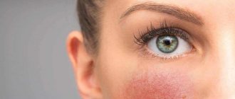
A focus of microsporia on the skin. Photo: Turkish Journal of Family Medicine & Primary Care / ResearchGate
Treatment
Treatment of microsporia in most cases is carried out on an outpatient basis, that is, the patient is not hospitalized. A hospital stay is advisable only in cases where there are concomitant pathologies that require diagnosis and treatment. Due to the high resistance of the pathogen to antifungal drugs, the course of treatment can last several weeks or even months. Treatment methods are divided into general, systemic and local.
General methods
If microsporia spreads to the scalp, once a week you should carefully shave the hair at a distance of approximately 1 cm from the lesion to ensure normal access to it. Regardless of where the source of inflammation is located (on the scalp or on smooth skin), you should adhere to the rules of hygiene, washing the skin around it 2-3 times a day.
Important!
Sports and physical activity should be temporarily limited, since sweat on the areas affected by ringworm is undesirable, as is frequent contact with water. This is why it is better to take a shower instead of a bath. It is advisable to change washcloths every 8-10 days, and towels every 2-3 days.
Systemic treatment
To eliminate the external manifestations of microsporia and avoid its further relapses, it is necessary to take antifungal drugs in tablets and capsules. The following medications have proven themselves to be the best:
- Griseofulvin. Used orally at the rate of 12 mg per 1 kg of body weight per day. It is recommended to take the drug with a teaspoon of olive or sunflower oil.
- Terbinafine. Adults are recommended to take 250 mg of the drug once a day (for children, depending on age and weight, the daily dosage varies between 60-120 mg). The average course of treatment is 10 weeks. The drug is taken after meals.
- Itraconazole For adults, the dosage is 200 mg once daily. The drug is taken after meals. Typically, the course of treatment varies between 4-6 weeks.
Prevention of microsporia in children
“You need to teach your child how to communicate properly with animals:
- The child should know not to touch sick animals. After petting a cat or dog, you need to wash your hands.
- You should not let animals, especially cats and dogs, into your bed or warm them under your shirt. After playing with them, you should immediately wash your hands with warm water and soap.
- Contact with stray animals should be avoided; domestic animals should be examined, including those that do not come into contact with wild animals.
- It is unacceptable to use other people's objects - such as a comb, handkerchief, etc. You cannot wear someone else's clothes.
- Examinations of children in organized groups (kindergartens, children's camps) should be carried out at least 2 times a year using a Wood's lamp.
- If a disease is detected, the child should be isolated from other children.”
Kuldibaeva Alina Talgatovna
expert
Pulmonologist, Allergist-immunologist, Pediatrician
Local treatment
For local treatment of microsporia, ointments, gels, alcohol tinctures and creams are used. They have an antifungal effect, cleanse the skin and promote the regeneration of damaged tissue. The following drugs are recommended:
- ciclopirox (cream, course of treatment 4-5 weeks);
- ketoconazole (cream, ointment, course of treatment 5-6 weeks);
- salicylic acid 3% + sulfur 10%;
- 2% alcohol tincture of iodine;
- 10% solution of ichthammol (course of treatment up to 3 days);
- potassium permanganate, solution 1:6000 (course of treatment 1-2 days);
- furatsilin, solution 1:5000 (course of treatment 1-2 days);
- rivanol, solution 1: 1000 (course of treatment 1-2 days).
Important!
During pregnancy and lactation, the use of systemic antifungal drugs is contraindicated. Despite their high effectiveness, during this period women are advised to limit themselves to local treatment only.
Criteria for successful treatment:
- elimination of all symptoms of microsporia;
- three consecutive negative results of a control microscopic examination;
- lack of glow under ultraviolet radiation using a Wood's lamp.
Since even with successful treatment the possibility of relapse of the disease cannot be ruled out, the patient needs clinical observation for 1-3 months. Control microscopic examinations are carried out on average once every 10 days.
Damage to the scalp by fungi of the genus Microsporum. Photo: Surg Cosmet Dermatol / ResearchGate (CC BY-NC 4.0)
A patient with microsporia should be hospitalized in the following situations:
- low efficiency of outpatient treatment;
- a large number of lesions on the body;
- deep suppurative form of the disease;
- presence of severe concomitant diseases;
- isolation from healthy people is necessary (applies to children from boarding schools and orphanages, as well as from large or dysfunctional families).
How to distinguish microsporia from other diseases
Separately, differential diagnosis is carried out with diseases such as psoriasis, trichophytosis and pityriasis rosea. The clinical picture of these diseases is in many ways similar to microsporia, but there are several important differences:
- With the superficial form of trichophytosis, small foci of peeling of a round or irregular shape are observed, while hair thinning and inflammation are weakly expressed. When conducting a differential diagnosis, the specialist pays attention to broken hair: its base is covered with muff-like layers, and peeling is observed around it. Fluorescence testing plays a key role.
- With pityriasis rosea, attention is drawn to such a characteristic feature as the absence of clear boundaries of the source of inflammation. The flaky skin resembles crumpled tissue paper, and the lesion itself has a characteristic pinkish color. During microscopic examination, the characteristic emerald glow of pathogenic microorganisms is not observed.
- Psoriasis is characterized by dry lesions and clearly defined boundaries. There are no muff-like layers on the affected hair, and the scales have a silvery tint.
To diagnose microsporia, a Wood's lamp is used. Under ultraviolet light, pathogenic fungi glow a certain color. Photo: freepik.com
MICROSPORIA Article magazine “Attending Physician” 04.01
MICROSPORIA
Article in the journal “Attending Physician” 04.01
V.M.Rukavishnikova, KMN
Modern features of the clinic and treatment of MICROSPORIA
What are the sources and routes of transmission of microsporia? What is the clinical picture of typical microsporia and its atypical forms? What is the rational therapy for microsporia?
Zooanthroponotic microsporia is the most common highly contagious disease of the skin and hair, common to humans and animals.
It is characterized by damage to coarse hair of the head, upper lip, chin, eyebrows, eyelashes, pubis, labia, as well as vellus hair of smooth skin. The causative agents of microsporia are zoophilic, anthropophilic and geophilic fungi of the genus Microsporum. Of the more than 20 species of this fungus, the most important in human pathology is the zoophilic fungus - fluffy microsporum. This is a kind of cosmopolitan fungus, practically the only causative agent of microsporia in the world, with the exception of African countries.
Microsporia predominates in European countries, especially in the Mediterranean, the USA and South America, Japan, Israel, Kuwait, Qatar, and the United Arab Emirates [1, 3, 5, 7, 9].
Today, microsporia has become most widespread even in regions with a traditionally high incidence of trichophytosis. Thus, in Dagestan, Uzbekistan, Tajikistan, Turkmenistan, Bashkortostan, Kazakhstan, Armenia, where isolated cases of microsporia were previously observed, today it accounts for up to 83–99.7% of all fungal hair diseases [3, 5].
In Russia, the incidence of microsporia is about 71.6 per 100 thousand population. In Moscow and the Moscow region it accounts for 96.2% of all dermafitia with hair damage [3].
Microsporia mainly (up to 65%) affects children, including newborns.
The main source of infection (80.5%) are cats, mainly stray ones. Cats living in medical institutions pose a particular danger, as they infect sick patients. However, cats sold at the Bird Market, near Durov’s Corner, as well as in pet stores and special clubs can also be dangerous, although they are equipped with special certificates, and some of them are even vaccinated. We witnessed how an expensive, elite breed of cat, distinguished by its particularly beautiful shape and color, became a source of infection for three generations in a family. Given to her granddaughter for her birthday, it infected her grandparents, the hero of the occasion, as well as her parents.
In most cats, which serve as a source of infection, foci of microsporia are determined clinically in the form of areas of baldness on the skin of the face, around the mouth and nose, on the outer surfaces of the ears, front and hind legs, and on the tail. The skin in areas of baldness appears flaky with the presence of unevenly broken hair, and sometimes new hair growth is observed in the center of such areas. Under a Wood's lamp, the green glow of the affected hair, characteristic of microsporia, is determined.
In another group of cats, lesions may not be visible to the eye, but are detected during fluorescent examination. Finally, in approximately 2–2.4% of cats, the lesions are not visible to the eye and are not detected under a Wood's lamp, but when combed from their hair, a culture of fluffy microsporum can be obtained.
Although one of the synonyms for fluffy microsporum is microsporum canis (canine), dogs appear as a source of infection in only 4% of patients with microsporia.
Rare animals that suffer from microsporia and can become a source of infection for people include monkeys, tigers, lions, wild and domestic pigs (especially piglets), horses, sheep, silver-black foxes, rabbits, rats, mice, hamsters, guinea pigs and other small rodents, as well as birds - pigeons, crows, chickens, which are hunted by sick cats. Animals become infected from the cats themselves or from their fur falling on plants, straw, or grain. In addition, fluffy microsporum can be carried on their legs by domestic insects, in particular cockroaches.
In 5.5% of patients with microsporia, the sources of infection are people - relatives, friends, neighbors - if basic sanitary and hygienic rules are not observed, as well as sexual partners when foci of mycosis are localized on the external genitalia, pubis, abdomen, upper thighs.
Homeless people and beggars, in addition to lice and scabies, can become carriers of fungal diseases.
Household items - a stroller left overnight in the entrance and favored by cats, toys, combs, underwear, etc. become a source of infection in 2-2.5% of patients with microsporia.
| Figure 1. Microsporia of smooth skin |
The incidence of microsporia varies throughout the year and largely depends on the appearance in cats, which are the main source of infection, of kittens that are more susceptible to infection and microsporia. Although pregnancy in cats lasts seven weeks and offspring appear several times a year, two outbreaks of microsporia disease in humans can be identified. The first occurs in May-June and is associated not only with the birth of kittens that are attractive to children, but also with greater freedom for children in the summer, their greater contact with the animal world when moving to the village, to the country, to health, sports and labor camps. Another increase in incidence is observed in September - November, when children return to the city and are carefully examined not only by their parents, but also by health workers when they enter schools and kindergartens. In this case, both fresh and erased, previously unrecognized forms of microsporia are identified [3].
| Figure 2. Disseminated foci of microsporia |
The incubation period for microsporia is usually five to seven days. After it, on smooth skin, mainly on open areas of the face, neck, chest, upper and lower extremities, single (from one to three) round-oval erythematosquamous spots 1–4 cm in diameter, clearly outlined by a peripheral ridge, appear (Fig. 1 ). If the kitten is warmed under a shirt, taken to bed, and the primary foci of mycosis are rubbed while washing with a washcloth, multifocal, disseminated variants of microsporia arise (Fig. 2). The spread and fusion of mycosis foci is also facilitated by irrational treatment, in particular lubrication with corticosteroid creams.
| Figure 3. Foci of microsporia with “stumps” of hair wrapped in sheaths |
Typical foci of microsporia on the head are usually located on the crown, in the parietal and temporal regions. They look like round-oval “bald patches” up to 3–5 cm in diameter with clear boundaries and “screenings” near them. The hair in the lesions is dull, all broken off at the same level, at a height of 4–6 mm, as if trimmed. Apparently, this is why microsporia is called “ringworm” in everyday life. The surface of the focus of mycosis appears rough, shagreen due to protruding “stumps” of hair, shrouded in grayish or whitish sheaths (Fig. 3). Under a Wood's lamp, the affected hair glows bright green, resembling a freshly mown meadow.
Microsporia is traditionally considered a disease of childhood. However, nowadays adults are also often affected by this mycosis. Apparently, unfavorable social and environmental conditions, an increase in neuroendocrine diseases and immunodeficiency conditions have an effect. If in 1932 A. M. Arievich observed microsporia in 6 adults out of 6000 patients, now adults account for up to 35% of cases of the disease. Women get sick four times more often than men.
In children, microsporia, as a rule, is diagnosed in a timely manner; atypical forms and errors in diagnosis are observed in 5% of cases; in adults, this figure increases almost four times and amounts to 19% of cases [3].
Of the atypical forms of microsporia, erased, trichophytoid varieties deserve special attention. They are not so rare: in 8.5% of patients, and in adults 2.5 times more often. Such microsporia occurs unnoticed, hardly bothers patients and does not force them to immediately consult a doctor. It is often confused with seboreids, seborrheic dermatitis, psoriasis and other diseases, and incorrectly selected treatment does not bring results. In this regard, such variants of microsporia become chronic, becoming the cause of further dissemination of mycosis in the sick person and its spread in the environment. The duration of trichophytoid microsporia has been recorded from seven months to two years.
Such forms of microsporia are usually characteristic of aggravated patients (tuberculosis, Sjögren's disease, pellagroid dermatitis, etc.). Clinically, they are manifested by diffuse or focal peeling, sparse hair, or the formation of areas of focal alopecia. The hair in the lesions is dull, without covers, broken off in different ways - at the level of the skin and at a height of 10–15 mm. Areas of alopecia are either very small, about the size of a pinhead, or gigantic, polycyclic in shape when merging.
On smooth skin, foci of microsporia appear as low-inflammatory, slightly flaky or depigmented round-oval spots. When they merge, polycyclic foci of mycosis may appear, blurred, indistinct, without clear boundaries, slightly itchy. In one of our patients, a 69-year-old cat lover, trichophytoid microsporia of the scalp was not diagnosed for two years. In the past, she suffered from tuberculous lymphadenitis and mesodenitis and took maintenance doses of prednisolone for a long time (for 10 years) for Shagren's syndrome. Microsporia was manifested in her by peeling of the scalp, hair loss, mainly in the occipital region, and itching. All phenomena intensified in the warm season and subsided in the cold season. The disease was treated for a long time and unsuccessfully with drugs used for seborrheic dermatitis or psoriasis. The diagnosis of microsporia was helped by the appearance of fresh typical foci of mycosis on the skin of the face and neck. When the patient was shaved, it turned out that almost the entire scalp was involved in the pathological process. An identical culture of fluffy microsporum was obtained from all foci of mycosis on the head, face, neck, as well as from squamous-keratotic rashes on the skin of the palms.
Sometimes an unusual source of infection also misleads the doctor, and microsporia may not be diagnosed even in children, which leads to the trichophytoid form. Thus, an eight-year-old girl fell ill after a week of contact with a sick pigeon, which she and her father had captured from a cat apparently suffering from this mycosis. While nursing the pigeon, the girl took it in her arms, swaddled it, and fed it. A clearly defined spot appeared on the top of the child’s head, covered with silvery scales. The hair in the lesion was not broken off, there were no subjective sensations.
The girl was diagnosed with psoriasis and was treated with diprosalic and ditrastic. During this time, the skin process spread to the entire scalp and became complicated by folliculitis and perifolliculitis. The changes were regarded as a complication of psoriasis by secondary pyococcal flora, and therapy was supplemented with antibiotics - geoxizone and fucarcin. The child's condition kept getting worse. Hair fell out in large quantities, and the girl experienced pain when combing it. With a presumptive diagnosis of discoid lupus erythematosus, the patient was referred to the Central Clinical Hospital.
A detailed examination revealed diffuse peeling of the entire scalp, sparse hair, atrophic bald patches, which were especially numerous in the parietal-occipital region. In some places the peeling was insignificant, but areas of scar atrophy of various sizes and shapes were clearly defined, without clear boundaries, almost without inflammatory phenomena. Near the scarred skin, the hair fell out easily, was dull, thinned, curled, and refracted light unevenly. The length of the hair was maintained or it broke off either at skin level or at a height of 10–15 mm. Scraping the skin in the lesions was painful. After removing the scaly crusts, the skin appeared moist and inflamed. After shaving and washing, it turned out that the entire surface of the head was covered with multiple lesions (more than 60), some of them were the size of a lentil grain, others were 2-3 cm in diameter, there were also giant areas of 8x12 cm. Enlarged and painful posterior cervical and parotid lymph nodes. Microscopically, clusters of small spores were detected on the surface of the affected hair. Under Wood's lamp, a not entirely characteristic grayish-whitish glow was detected. However, a typical culture of fluffy microsporum was obtained on a nutrient medium. Thus, late diagnosis and inadequate treatment led to the fact that the focus of superficial microsporia, which usually passes without any consequences, was complicated by the dissemination of the fungal process throughout the entire scalp, the formation of areas of cicatricial atrophy and focal alopecia, and lymphadenitis.
Perhaps, deep - infiltrative-suppurative, kerion-like or granulomatous variants of microsporia with pronounced symptoms deserve even more attention and alertness. They are recorded in 4.5–6.5% of patients with this mycosis. They are characterized by severe disease, pain, fever and other symptoms of intoxication. The course of the disease is complicated by lymphadenitis and allergic rashes, with a rapid outcome in cicatricial atrophy and irreversible focal alopecia.
As a rule, deep forms of microsporia occur in weakened children or adults, mainly in women with endocrine or immune pathologies (dysfunction of the reproductive and thyroid glands, pituitary dwarfism, lymphogranulomatosis, blood diseases). Sometimes superficial variants of microsporia are transformed into infiltrative-suppurative ones under the influence of inadequate treatment, as well as repeated injuries, including with a washcloth during washing and going to steam rooms. This is facilitated by frequent sea bathing, and most importantly, constant stay in a damp, tight-fitting bathing suit.
In adults, foci of deep microsporia are usually detected on the skin of the legs or pubis and labia, in children - on the scalp.
Kerion-like foci of mycosis appear to be large, confluent, occupying large areas of the scalp or the entire area of the pubis and outer labia. This is usually a conglomerate of merged and suppurating painful folliculitis, perifolliculitis and abscess elements. Due to edema, infiltration and impetiginization, microsporia foci rise above the skin level. Their surface is covered with rough purulent crusts with loose and partially melted hair stuck together in them. After removing the crusts and hair from the gaping openings of the hair follicles, a creamy pus is released like honey from a honeycomb (kerion-honeycomb). Regional lymph nodes are enlarged and painful. Such patients are characterized by severe symptoms of intoxication, allergic rashes up to erythema nodosum of the legs. Often, such forms of microsporia are mistaken for ulcerative-vegetative pyoderma, infiltrative-suppurative trichophytosis, psoriasis complicated by pyoderma.
In young girls with hypertrichosis, deep microsporia of the legs may occur, usually mistaken for vasculitis, Majocchi granuloma, reticulosis. In these cases, relatively small (2-3 cm in diameter), deep, single, follicular-nodular lesions are located on the lower leg in the form of a ring.
We present a fairly typical story of the formation of infiltrative-suppurative microsporia in a seven-year-old boy. It all started with the appearance, usual for this mycosis, of a small round patch of baldness in the left parietal region. The hair in it was characteristically broken off. However, the parents associated the appearance of this lesion not with contact with the kitten, but with a fall from a bicycle, a superficial abrasion and its contamination, especially since both events coincided in time. The lesion was therefore considered pyodermic. Treatment with corticosteroid creams and antibacterial agents provoked the spread of the fungal process to the entire left half of the head. Due to swelling, infiltration, suppuration, enlargement of the cervical and preauricular lymph nodes, the head and face seemed asymmetrical. The surface of the skin in the focus of mycosis was covered with rough purulent and purulent-bloody crusts with sparse hair stuck together in them. At the slightest movement of the head and neck, the child experienced severe pain. The boy was lethargic, apathetic, pale, and there were periodic rises in temperature. Along with the atypical focus of mycosis described above, characteristic small foci of mycosis appeared on the head in the area of the left eyebrow, on the forehead and temple. Fluorescent examination revealed a typical green glow in them. The diagnosis was confirmed microscopically and by isolating an identical culture of fluffy microsporum from all foci of mycosis.
Infiltrative-suppurative microsporia of the skin of the pubis and labia can be classified as atypical in localization and course. This form of microsporia often (according to our data, in two out of five cases) leads to infection of sexual partners. Due to the atypical localization and course, this form of microsporia is not immediately diagnosed. Erroneous and inadequate treatment makes clinical manifestations even more uncharacteristic.
Currently, the localization of microsporia foci on the skin of the pubis and labia has increased more than tenfold compared to 1976. The peculiarities of the anatomical structure of this area, replete with blood vessels and nerve endings, contribute to the rapid formation of deep invasive, extremely painful infiltrates, the appearance of complications in the form of lymphadenitis and allergic rashes, and the appearance of symptoms of intoxication due to the absorption of lysed tissues, bacteria, fungi and their metabolic products.
We present in more detail the medical histories of two women, one of whom infected her sexual partner, the other herself was infected during sexual contact.
An 18-year-old woman had no diagnosis of microsporia for four months. She became infected from her nephew, who had microsporia, by using a washcloth to wash him. A superficial focus of microsporia on the skin of the inguinal fold resolved after a week of lubrication with a 2% iodine solution. The patient considered the episode to be over. Therefore, when, after a month and a half, superficial flaky lesions appeared on the skin of the pubis, she in no way connected them with untreated microsporia. Application of corticosteroid creams (suspicion of allergic rashes), frequent sea bathing (the patient was at sea at that time) and almost constant stay in a tight-fitting bikini led to the spread of the pathological process both on the surface and in depth. Folliculitis, perifolliculitis, and abscess elements formed, merging into a single conglomerate rising above the surface of the skin. “Dropouts” appeared on the anterior surface of both thighs and the skin of the lower abdomen (Fig. 4). The surface of the foci of mycosis was covered with rough purulent and purulent-bloody crusts. The inguinal lymph nodes became enlarged and painful. Painful subcutaneous nodes appeared on symmetrical areas of both legs. The disease was regarded as deep ulcerative-vegetative pyoderma, complicated by erythema nodosum of the legs. The patient was unsuccessfully treated with oxacillin, kefzol, vibramycin in combination with short courses of prednisolone. The pain and symptoms of intoxication only increased. The diagnosis of microsporia was helped by the appearance of a typical focus of mycosis on the skin of the abdomen of the patient’s sexual partner. After a detailed examination of the woman, as the presumed source of microsporia, her infiltrative-suppurative form was diagnosed. The clinical diagnosis was confirmed microscopically, fluorescently and culturally.
A 25-year-old patient who became infected with microsporia from a patient had a similar history of the disease. A week after sexual contact, a bright itchy spot appeared above the pubis, quickly becoming covered with scaly crusts. The patient was being treated for ureoplasmosis at that time, and the spot was regarded as allergic. Under the influence of short therapy with Triderm cream, the spot faded and became almost invisible. A month after this episode, while staying at sea, superficial scaly rashes appeared on the pubic skin, merging into a large area of curly outlines. Deciding that these were allergic rashes, the patient used Lorinden cream and 2% salicylic alcohol. For ease of lubrication, the patient shaved her pubic hair. This provoked the process to spread into depth. An extremely painful infiltrate of deep follicular nodular elements formed. Deep pyoderma was diagnosed. Tsifran and vilprofen were used orally, fucarcin and lincomycin paste were used externally. However, pain, infiltration and impetiginization only increased. To clarify the diagnosis, after two months from the onset of the disease, the patient was sent to the Central Clinical Hospital.
On the pubis, occupying its entire central part, passing onto the skin of the outer labia and twisting into cords in the upper and lateral parts, there was an extensive infiltrate, rising 1-2 cm above the skin level. Its contours were emphasized by a bright peripheral ridge. The skin over the focus of mycosis was bluish-brown, tense, stretched, with a bumpy surface (Fig. 5). Touching the hearth turned out to be unusually painful. The pubic hair completely retained its length; the cap usual for microsporia was not visible. The central part of the lesion was practically devoid of hair. The remaining hair was difficult to epilate, and epilation was accompanied by severe pain. The inguinal lymph nodes were enlarged and painful. Papulovesicular microsporidae were present on the dorsum of both hands. A luminescent examination of the focus of mycosis on the pubis and labia revealed a characteristic green glow. Microscopic examination revealed small spores located on and inside the hair. A typical culture of fluffy microsporum was obtained on a nutrient medium.
Treatment of patients with microsporia seems to be an important, socially significant problem.
This disease usually affects not only children, but also their parents. Due to the high contagiousness of mycosis, children are prohibited from attending kindergartens and schools. They miss school classes and find themselves separated from the children's group for a long time. Parents are forced to interrupt work and take sick leave for care. Thus, the family suffers significant moral and material damage.
A speedy recovery of patients with microsporia is achieved with timely recognition of mycosis and adequate treatment. It includes the combined use of systemic and external antifungal agents along with pathogenetic drugs. The latter increase the effectiveness of treatment and reduce the frequency and severity of adverse reactions and complications.
In patients with microsporia, two systemic antimycotics are used: the antibiotic griseofulvin and lamisil. Moreover, antimycotics are used in high doses in patients with microsporia. The fact is that the causative agent of mycosporia is characterized by the highest resistance compared to other dermatophyte fungi. For example, the daily dose of griseofulvin, equal to 16 mg/kg in patients with rubrophytosis and 18 mg/kg in trichophytosis, should be increased to 22 mg/kg for mycosporia. The high stability of fluffy microsporum spores is due to the presence of a very dense six-layer shell, reinforced by longitudinal and circular costal projections [11].
Griseofulvin is a chlorine-containing antibiotic, a product of biosynthesis of mold fungi of the genus Penicillium. Therefore, when taking griseofulvin and simultaneous treatment with penicillin antibiotics, mucocutaneous allergic complications very often occur. The daily dose of griseofulvin, which is selected at the rate of 22 mg per kilogram of weight, is divided into three to four equal parts and taken with one teaspoon of vegetable oil. The oil stimulates the evacuation of bile and promotes the dissolution of griseofulvin. In addition, due to the presence of vitamin E (a-tocopherol), the metabolism of griseofulvin slows down and the duration of action of the drug increases. Griseofulvin is better absorbed in an acidic environment, so it is advisable to drink it with some sour juice (lingonberry, cranberry, lemon, apple, etc.).
To reduce the hepatotoxicity of griseofulvin, it is recommended to take it with hepatoprotectors - liver preparations, Liv-52, Karsil, Silibor, etc.
Given the poorer absorption of griseofulvin in the presence of helminths, deworming is indicated before or during griseofulvin therapy [8]. For this, various anthelmintic drugs are prescribed, for example, decaris (levamisole), which eliminates helminths and at the same time has immunomodulatory properties. Griseofulvin somewhat enhances immunodeficiency conditions, against which, as a rule, microsporia occurs. Levamisole neutralizes the immunosuppressive effect of griseofulvin. During griseofulvinotherapy, foci of focal infection usually worsen, which become sources of superinfection of foci of mycosis, therefore their sanitation is indicated.
The course of continuous treatment with griseofulvin lasts 1.5-2 months. Initially, it is used daily until two negative fungal tests are obtained seven days apart; then the antibiotic is used for two weeks every other day and another two weeks twice a week. At the end of therapy, a control clinical and laboratory examination is carried out, and in case of negative results, the patient is considered cured. Although griseofulvin is a fairly gentle drug, it still has hepato-, nephro- and neurotoxicity, photosensitizing, embryotoxic and cocarcinogenic properties.
Griseofulvin is contraindicated in hepatitis that was suffered no more than a year ago and is manifested by subjective sensations and/or elevated levels of bilirubin and liver enzymes; peptic ulcer of the stomach and duodenum; kidney diseases; neuritis, especially of the optic and auditory nerves; malignant and fast-growing benign tumors; blood diseases; photodermatoses and related conditions; cerebrovascular accident; uterine and other bleeding.
It should be taken into account that an adequate dose of griseofulvin in patients with microsporia should not exceed the maximum permissible - 1.0 g (8 tablets). Otherwise, the toxic-allergic properties of the antibiotic appear. In adults and large children weighing more than 60 kg, prescribing an adequate dose of the drug is practically impossible.
A worthy alternative to griseofulvin is terbinafine (trade name Lamisil).
The drug has high activity against various types of fungi. It is especially active against dermatophyte fungi, the causative agents of the most common dermatophytosis: rubrophytosis in adults and microsporia in children.
Lamisil suppresses squalene epoxidase, which is extremely sensitive in fungi (10 thousand times more sensitive than in humans). As a result, at the very early stages (at the level of the squalene epoxidase cycle), the formation of ergosterol, the main component of the cytoplasmic membranes of the fungal cell, is blocked. Without receiving this building material, the fungal cell becomes defective, cannot grow and develop, but only survives. This is how the fungistatic effect of Lamisil manifests itself. However, the predominant fungicidal effect of this antimycotic is. It directly depends on the accumulation of squalene in the cell, excluded from the cycle of further transformations into ergosterol. These active high molecular weight hydrocarbons accumulate in the fungal cell. Moreover, the volume of squalene granules gradually increases, since they actively extract lipids from cell membranes. Ultimately, a cell membrane with a damaged structure cannot withstand it and ruptures, leading to cell death [6, 7].
Lamisil does not require any specific conditions for administration and is quickly absorbed from the gastrointestinal tract. Its blood level is stable. The drug practically does not interact with various medications, as well as enzyme systems, including cytochrome P-450. Its action is selective and directed mainly at the fungal cell. Lamisil enters the skin and its appendages by simple diffusion, as well as excretion by the sebaceous glands.
The peculiarities of the distribution of Lamisil due to its lipophilicity and connection with chylomicrons include its lymphatic transport. Through the lymphatic vessels, Lamisil directly reaches infiltrative-suppurative and abscessing foci of mycosis with lymphadenitis and lymphangitis. In this regard, the greatest activity of Lamisil is observed in patients with severe complicated infiltrative-suppurative forms of dermatophytosis with hair damage. According to M. Tsoi and M. D. Alaeva (1996), with superficial trichophytosis, lamisil accelerated the recovery process by 3.3 days, with infiltrative trichophytosis - by 6.3 days, with suppurative trichophytosis - by 8.2 days compared with griseofulvin therapy [10].
The rapid resolution of complicated forms of dermatophytosis during therapy with Lamisil is thus associated with its high antifungal activity, lymphatic transport, pronounced antibacterial properties comparable to the action of gentamicin, as well as anti-inflammatory effect. The latter is due to the suppression of peroxidase activity of hydroxyl radicals of polyunsaturated fatty acids.
Above, we have already noted the high resistance of fluffy microsporum to antimycotics. In this regard, N. S. Potekaev et al. (1997) observed the cure of children with microsporia with lesions of long and vellus hair, who were prescribed Lamisil 94 mg per day with a body weight of up to 20 kg and 186 mg per day with a body weight of 20 to 40 kg, which is 50% higher than the dose, proposed in the annotation for the drug [4].
In adults with complicated forms of microsporia, we successfully carried out short but intense courses of therapy with Lamisil. The drug was prescribed at a daily dose of 7 mg/kg or two tablets (500 mg) per day. In the next three to four weeks, we observed complete resolution of microsporia foci in all patients without exception. Healthy hair began to grow where scar atrophy had not developed, microsporidae, including foci of erythema nodosum of the legs, simultaneously resolved, regional lymph nodes became painless and decreased in size. In no case were any side effects or deviations in the biochemical status noted [6].
I would also like to note the better tolerability of Lamisil compared to its analogue, Exifin. One of our patients began, on her own initiative, to take the cheaper exifin instead of Lamisil. After four to five days of taking exifin at a daily dose of 500 mg, itchy papulovesicular allergens appeared on the inner surface of both forearms. They resolved after the use of adsorbents and antihistamines. After resuming therapy with lamisil at a daily dose of 500 mg, allergens did not appear.
And one more thing: in the family of one of our patients, everyone except her had respiratory infections. We associated its stability with an increase in the body's defenses as a result of taking Lamisil. It is known that Lamisil does not have a negative effect on the metabolic activity of leukocytes, chemotaxis, and phagocytosis. Moreover, under the influence of lamisil therapy, indicators of cell-mediated immunity improve, which is associated with the release of large amounts of antigenic material as a result of the death of fungal cells [12].
The effectiveness of treatment of patients with microsporia increases with the simultaneous use of systemic antimycotics and external agents. Along with epilation and weekly shaving of hair, a 2% tincture of iodine is used, which is used to lubricate the foci of mycosis in the morning. In the evenings, domestic antifungal ointments are rubbed into them: sulfur-salicylic, sulfuric, sulfur-tar, bifosin. This iodine-ointment treatment is highly effective and cheap and accessible.
For relatively calm foci of microsporia, it is advisable to lubricate them once a day with 10% salicylic-quinosol dimexide, which quickly leads to the resolution of foci of mycosis on the scalp.
For infiltrative-suppurative microsporia, to free foci of mycosis from crusts and pus, compress bandages with 10-20% solutions of ichthyol, 5% solution of mumiyo in dimexide, 50% solution of licorice root, as well as lotions with 0.5 -1% solutions of chlorhexidine bigluconate (Ghibitan) or ultrasonic irrigation with a 0.5% solution of this drug using the Rossa apparatus [2, 5].
Effective foreign azole and allylamine compounds can be used as external antimycotics. Among them, we prefer Lamisil 1% cream, Mycosporus 1% cream and Travogen 1% cream. These drugs are usually well tolerated because they have anti-inflammatory properties. In case of acute inflammatory phenomena, their combination with corticosteroids is not required. At the same time, they have pronounced antifungal activity, and lamisil cream also has an antibacterial effect, which is important for the rehabilitation of microsporia foci complicated by secondary pyococcal flora.
The inclusion of external antifungal agents in the complex of treatment measures for patients with microsporia allows one to achieve rapid recovery with the smallest course dose of expensive systemic antimycotics that are not indifferent to the body.
Diagnosis of microsporia
The clinical picture and history of contact with animals provide the doctor with grounds for making a preliminary diagnosis. To confirm it, the following diagnostic studies are prescribed:
- Inspection using a fluorescent filter. This method is based on the ability of fungi of the genus Microsporum to fluoresce when using ultraviolet radiation. For this purpose, a device known in medicine as a Wood's lamp is used. This lamp emits ultraviolet light, which, when passed through a darkened filter, helps identify pathogenic fungi: when examined, they glow pale yellow or greenish.
- Histological examination. An area of skin is scraped for microscopic examination, which makes it possible to identify the fact of infection and the degree of inflammation. However, microscopy does not determine the type of pathogen.
- Cultural research. To determine the species of the pathogen and its sensitivity to antifungal drugs, the fungus is sown and grown on a nutrient medium. The disadvantage of the cultural method is that you have to wait at least a week to get a reliable result.
- Additionally, clinical tests of blood and urine are performed. If the results deviate from the norm, the tests are repeated once every 10 days. A biochemical study of blood serum is done before the start of the therapeutic course and 3 weeks after its completion.
Sources and routes of infection
The most common source of infection is the fungi Microsporum canis, which parasitizes mainly dogs and cats. To a lesser extent, rabbits, sheep, rodents, poultry, pigs and large wild animals are susceptible to microsporia.
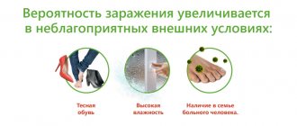
Infection can occur in any environment. However, the combination of heat and high humidity is most favorable for the fungus, so outbreaks of microsporia are characterized by seasonality - most often they occur in late spring and early autumn. Microsporum spores can live in soil for up to several months, so frequent contact with the soil may pose a certain risk. An additional predisposing factor may be increased sweating and disruption of the secretion of the sebaceous glands. Timely washing of hands with soap reduces the risk of infection to a minimum, even if fungal spores have already reached the skin.
Important!
For infection, the so-called entry gate of infection is necessary, which can be abrasions, scratches, calluses and other microtraumas of the skin. If the integrity of the skin is not compromised, the fungus has virtually no chance.
When and how can a child become infected?
Microsporia is a highly contagious (highly contagious) disease caused by dermatophyte fungi (literally, growing on the skin) of the genus Microsporum.
There are several types of this fungus that cause the development of three forms of the disease.
Anthropophilic and zoophilic forms of microsporia are widespread in Russia.
The disease occurs throughout the year. However, zoophilia is characterized by seasonality: the largest number of cases are recorded in late summer and autumn.
Reasons for the development of microsporia
Microsporia develops after fungi of the genus Microsporum enter the skin. Contact of the fungus with the skin itself does not always lead to infection: susceptibility to infection develops as a result of insufficient hygiene rules and weak immunity, so children under 12 years of age are most susceptible to microsporia. Single infections often develop into outbreaks in children's groups. Another predisposing factor is insufficiently high density of keratin in the skin and hair in children. For this reason, lesions of the scalp are much less common in adults.
Adults are not immune from microsporia, but they experience it much less frequently than children. This is due to a more advanced immune system, established hormonal levels and compliance with hygiene rules. Among adults, microsporia most often affects young women, which is explained by the presence of fungistatic organic acids in their skin.
Fungi of the genus Microsporum are very stable in the external environment: even in conditions unfavorable for most pathogenic microorganisms, they are able to maintain their infectious properties for several months (sometimes even several years).
The cause of microsporia in humans is contact with a sick animal. Photo: Todestoast / Wikipedia (CC BY-SA 4.0)
Diagnostic measures
Diagnosis and treatment of ringworm are carried out by a dermatologist. The doctor examines the patient and identifies typical manifestations of microsporia. Examination of skin scrapings under a microscope reveals fungal mycelium and changes in the structure of hair and skin. Differential diagnosis makes it possible to exclude trichophytosis from the patient’s history, which has similar manifestations upon microscopy of the patient’s biomaterials.
Microflora culture appears to be a more informative diagnostic technique. Laboratory staff determine the type and genus of fungi. Based on the laboratory report, the dermatologist selects drugs that will cure the patient.
Luminescent examination makes it possible to identify pathological lesions on the skin of the patient and those living with him. This diagnostic method is based on the green glow of the fungal mycelium under the influence of a gas-discharge light source.
Prevention
The prevention of microsporia is based on compliance with the rules of personal hygiene: regular, thorough hand washing reduces the likelihood of infection to a minimum. You should avoid contact with sick animals and regularly conduct preventive examinations of your pets. If you suspect ringworm in an animal, you should immediately visit a veterinarian and carry out the course of treatment prescribed by him.
Important!
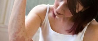
A sick animal can be easily recognized by the presence of foci of thinning hair with visible pink skin, which is often covered with light gray scales.
Remember that an apparently healthy animal can be a carrier of fungal spores, so when in contact with any animals on the street (farm, nursery, etc.), you must make sure that there are no abrasions, scratches or other damage to the skin of your hands, and do not forget to wash your hands . It is important to explain these rules to children, who are especially vulnerable to infection by fungi of the genus Microsporum.
When identifying patients with microsporia in a team (including in a family), it is necessary to organize preventive and focal disinfection. In any public places, sanitary, hygienic and disinfection measures must be regularly carried out (primarily this applies to saunas, baths, hairdressers, swimming pools, gyms and hotels).
If microsporia is detected in groups, the child is not allowed to enter kindergarten/school, and adults are not allowed to work/educate.
If you suspect contact with a person or animal with microsporia, it is recommended to use special antifungal shampoos based on selenium sulfide. Such shampoos should be used for 3-4 weeks, even for purely preventive purposes.
Since weakened immunity is one of the main factors predisposing to the disease, it is necessary to follow a few simple rules that will help maintain and strengthen the body’s defenses:
- ensure sufficient amounts of protein and vitamins in the diet;
- minimize bad habits;
- avoid nervous stress;
- maintain an adequate level of physical activity;
- sleep at least 8 hours a day.
Classification of microsporia
Microsporia is usually classified according to two main criteria: the pathogen and the depth of the lesion.
Classification by pathogen:
- anthropophilic fungi Microsporum audouinii and Microsporum ferrugineum;
- zoophilic fungi Microsporum canis and Microsporum distortum;
- geophilic fungi Microsporum Gypseum and Microsporum nanum.
Classification by depth of damage:
- superficial with damage to the scalp;
- superficial with damage to the skin;
- deep suppurative.