Causes of obliterating atherosclerosis of the arteries
The disease causes severe circulatory failure in the legs, dooms patients to excruciating suffering and makes them unable to work. The process is localized mainly in large vessels (aorta, iliac arteries) or medium-sized arteries (femoral, popliteal arteries).
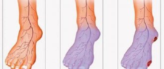
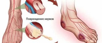
The most striking sign of ischemia of the lower extremities is intermittent claudication, characterized by the appearance of pain, a feeling of numbness and compression in the leg muscles when walking. This sensation forces the patient to stop, after which the pain and compression gradually disappear, but when the load is resumed, the pain returns. The affected leg is usually paler than the opposite leg and cold to the touch. Even minor injuries (scratches, bruises, abrasions) heal poorly and can cause ulcers. The feeling of numbness and pain at rest is often also caused by ischemia of the nerve trunks (ischemic neuritis). Prolonged course of the disease leads to the development of gangrene and inevitable amputation.
The degree of damage to the vascular bed is based on the walking distance. The pain forces the patient to stop, waiting for it to disappear. Without treatment, the disease continues to progress, which leads to a decrease in walking distance, limitation of the patient’s physical activity, and the inability to lead a normal lifestyle. Unfortunately, most often patients associate these pain sensations simply with muscle fatigue due to age, or with venous problems, defining the pain as a cramp, thus delaying seeking medical help and deepening the degree of damage to the arterial bed. In severe cases, the patient cannot walk 10 meters without stopping, but then it gets even worse: pain appears at rest, initially passing in an upright position, but after a short period of time the pain becomes constant, taking painkillers becomes ineffective. Gradually, the lumen of the artery narrows, leading to complete closure. Doctors are forced to admit that patients seek medical attention. help is late when the damage to the limbs is irreversible.
Risk factors:
- smoking;
- regular increase in blood pressure;
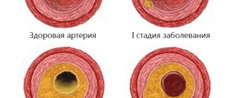
- high concentration of total cholesterol and its components in the blood;
- overweight (obesity);
- sedentary lifestyle (hypodynamia);
- diabetes;
- cardiovascular diseases in close relatives.
Damage to the vascular system of the lower extremities is a life-threatening condition, and ranks third in the structure of mortality from cardiovascular diseases. This figure is due to the development of such a severe complication as gangrene of the limb, which often leads to the need for amputation, and with high amputation, the mortality rate is 25%. Along with this, it should be noted that 50% of patients die within a year without amputation after the diagnosis of “critical ischemia”.
Symptoms of obliterating atherosclerosis of the arteries of the lower extremities
Pay attention to the following symptoms in yourself or your loved ones:
- fatigue in the calf muscles or thigh muscles when walking 500 meters or less,
- a feeling of leaden heaviness in the legs or muscle pain that makes you stop when walking,
- changes in the skin on the legs in the form of peeling, thinning, hair loss.
Important!
These are signs of atherosclerotic damage to the arteries of the lower extremities. An examination by a vascular surgeon is required in the near future. Do not be surprised if, when diagnosing lesions of the arteries of the lower extremities, your carotid arteries are checked and a cardiological examination is prescribed. Atherosclerosis is a systemic disease, and, as a rule, different groups of blood vessels are affected.
If sharp pain in the foot or lower leg occurs simultaneously with paleness and coldness of the skin, you must immediately call an ambulance, otherwise you may not have time to save the limb.
Discussion
Intraoperative iatrogenic vascular injury is a serious complication. These types of iatrogenic injuries occur during traditional (open) surgical interventions, endovascular procedures (angioplasty, stent placement, etc.) and, of course, laparoscopy. If not treated promptly, such injuries can cause serious harm to health, and in extreme cases, death. Vascular injury during laparoscopic surgery is fortunately rare. The results of studies conducted in individual countries showed an incidence of 0.1 cases per thousand procedures [23, 24]. This is lower than the incidence of damage to other organs (intestines, urethra or bladder) [25–27]. Although angiosurgeons rarely encounter such iatrogenic injuries, it is important to be aware of this threat. There are also descriptions of unsuccessful cases of vascular restoration complicated by thrombosis and the development of acute ischemia [16, 20, 21, 28].
According to the literature, the terminal aorta and iliac vessels are damaged more often than other vessels. Simultaneous artery and vein injury, noted in one case in our practice, is reported to occur in 10% of all vascular injuries during laparoscopic manipulation [16, 29, 30].
As a rule, damage to a large artery or vein, accompanied by severe bleeding, is diagnosed fairly quickly. However, the possibility of later detection of bleeding cannot be excluded. This may be due to pneumoperitoneum and the patient being in the Trendelenburg position, or a retroperitoneal hematoma, so bleeding may appear some time after laparoscopy [20, 23, 30]. There are also reports in the literature of vascular damage that was not immediately noticed and was diagnosed several months later in the form of a false aneurysm or arteriovenous anastomosis [20]. In our practice, such cases have not been observed.
Damage to large vessels (aorta, iliac arteries) requires immediate laparotomy to stop bleeding and restore the integrity of the vessel. The most preferred is a midline laparotomy. However, in one of our observations, an extraperitoneal pararectal access was performed when urologists damaged the left external iliac artery.
Restoring the integrity of the vessel should be carried out exclusively by an experienced angiosurgeon. General surgeons (gynecologist, urologist) should stop the bleeding and call an angiosurgeon or transfer the patient to a multidisciplinary specialized hospital with round-the-clock work as quickly as possible. According to Chapron et al. [16], a vascular surgeon is not always called for consultation - only in 71.4% of cases.
The ideal is complete restoration of damaged vessels. Ligation of the iliac arteries or veins can be performed only in exceptional cases to stop bleeding, followed by prompt transfer to a specialized hospital. It is usually possible to apply a vascular suture to the damaged artery and thus restore its integrity [20, 21, 29]. If this is impossible, as it will lead to narrowing of the vessel, then plasty using a patch (synthetic or autovenous) is indicated [20, 31]. Sometimes it is possible to resect the edges of the damaged artery and perform an end-to-end anastomosis [20, 31]. In case of significant damage to the artery along its length or intimal detachment, prosthetics of the damaged portion of the vessel should be performed [20]. In our observation, 3 patients underwent iliac artery replacement. Also, in 1 case, in addition to prosthetics, it was necessary to perform thrombectomy from the femoral and popliteal arteries and fasciotomy of the anterior group of leg muscles due to the development of severe post-ischemic edema of the leg.
With concomitant damage to the vein, it is preferable to suturing the damaged vessel rather than ligating it, since subsequently, even with thrombosis, the patency of the vein will be restored and the risk of developing venous insufficiency is reduced.
The main methods for diagnosing obliterating atherosclerosis of the arteries of the lower extremities:
- measurement of the ankle-brachial index (the ankle-brachial index is a parameter that allows you to assess the adequacy of arterial blood flow in the lower extremities);
- duplex scanning of the arteries of the lower extremities is the “gold standard” for screening patients (detection and follow-up);
- MSCT angiography of the aorta and arteries of the lower extremities is the “gold standard” of preoperative examination and in cases where information from ultrasound scanning is insufficient;
- X-ray contrast angiography.
How is CT angiography of the iliac arteries done?
Multislice CT of the cardiovascular system is recommended to be performed on tomographs that scan at least 64 sections per revolution of the X-ray tube. Such devices reduce examination time and guarantee high image quality. Semi-open tomographs increase the comfort of the procedure.
The device for MSCT is a device equipped with a wide annular part. X-ray tubes are fixed in it, which make circular movements during the examination. The emitters illuminate the abdominal area, and the tomograph transmits the data, converting them into layer-by-layer photos of the iliac artery, to a computer monitor. The scanning is carried out in an axial projection, but if necessary, it is possible to reconstruct the examined area in any other plane or in the form of a three-dimensional image.
MSCT angiography of the iliac arteries, 3D reconstruction
The table with the patient moves inside the annular part of the tomograph, providing gradual illumination of the vessel. The patient lies horizontally, the procedure takes about 30 minutes. It is important to remain still during the scan to improve image quality.
The examination results are recorded on electronic media, which are given to the patient free of charge on the same day. The disk contains more than 200 photos, a 3D model (if necessary), their description and a doctor’s report.
Angiography of the iliac artery is carried out with the obligatory introduction of a contrast agent into the lumen of the vessel, which significantly increases the information content of the method. This method of examination requires preliminary preparation of the patient.
Treatment methods for obliterating atherosclerosis of the arteries of the lower extremities:
Conservative therapy
In the early stages of obliterating atherosclerosis of the arteries of the lower extremities, conservative treatment is indicated, which must be comprehensive, and all unfavorable factors causing vasospasm must be excluded. A necessary condition for successful treatment is quitting smoking(!). Physical activity is of paramount importance. Patients with intermittent claudication should walk daily for 30-45 minutes - this promotes the development of small arteries, leads to an increase in muscle strength and an increase in the distance traveled without pain.
If pain and compression appear in the leg muscles, the patient should stop, and after these sensations disappear, continue walking. Often, cycling or swimming are much better tolerated than walking (however, they do not replace it). Correction of increased blood pressure, normalization of blood cholesterol levels, blood glucose levels in patients with diabetes mellitus. Drugs are used that reduce the tone of small vessels, increase the flexibility of red blood cells and prevent the formation of blood clots in the vessels. Physiotherapeutic and balneological procedures, hyperbaric oxygenation are also used.
Surgical treatment of arteriosclerosis of the arteries of the lower arteries, vessels
Reconstructive operations:
- X-ray endovascular treatment methods.
Under the control of X-rays using special long thin instruments, through a small puncture in the femoral artery (less often other arteries), we can reach the affected vessel (area of blood vessels) of the lower extremities. Modern technical capabilities make it possible to expand a section of the vessel from the inside with a special balloon and, if necessary, install a thin metal frame (stent) that prevents re-narrowing.
- Open surgical interventions.
For patients with blockage of a vessel lasting no more than 7-9 cm, the internal modified layer of the artery with atherosclerotic plaque and blood clots is removed (endarterectomy). With a more significant spread of the occlusive process, pronounced calcium deposition in the artery wall, a bypass of the blood flow (bypass) or resection of a section of the artery and its replacement with a synthetic prosthesis or biomaterial (prosthetics) is indicated.
Amputation
If, despite the treatment, ischemia of the affected limb increases and gangrene progresses, amputation is indicated: its level must be strictly individual and carried out taking into account the blood supply to the limb.
Preparing for the study
Before contrast CT angiography of the iliac arteries, the patient takes a blood test for creatinine, an elevated level of which indicates renal failure. If you wish, you can undergo rapid testing on site, at the diagnostic ]"Magnit"[/anchor].
Registration for the procedure occurs during a personal visit to the clinic, by phone or on the website. The patient is offered a free consultation with a radiologist who will talk about the features of the study and help prepare for CT angiography of the iliac artery.
If you are allergic to iodine preparations, you should notify your doctor to avoid undesirable consequences. A preliminary consultation with a specialist will also be required if there are contraindications to computed tomography.
Symptomatic interventions:
- Sympathectomy
(intersection of the nerve plexuses responsible for spasm (narrowing) of the arteries) is performed for repeated blockages of the arteries and in addition to reconstructive operations. This operation improves blood circulation in the extremities by dilating small arteries. - Revascularization osteotomy
is also an adjuvant technique that improves blood circulation by stimulating the formation of new small vessels in the lower extremities after bone injury. - Arterialization of the venous bed
is rarely used at this time, since its implementation is associated with various technical difficulties, and long-term results are worse than the methods described above.
conclusions
1. When diagnosing iatrogenic damage to a large vessel during laparoscopic manipulation, one should switch to open surgery, perform either a median laparotomy, or, if the external iliac artery is damaged, an extraperitoneal approach.
2. In case of damage to the iliac vessels or aorta, close cooperation between the general surgeon (gynecologist, urologist) and the angiosurgeon is necessary, which significantly increases the chances of a favorable outcome. You should not attempt to restore the integrity of the vessel without the appropriate skills. It is necessary to stop the bleeding and call a mobile angiosurgical team or transfer the patient to the vascular department as quickly as possible.
3. Each operating room should have 2-3 vascular clamps to be able to stop bleeding if a major vessel is damaged.
Indications and contraindications for CT angiography
Iliac artery occlusion is a dangerous pathological condition and has serious consequences for the body. The clinical picture is characterized by symptoms, the presence of which is an indication for CT angiography:
- pain in the legs, numbness, coldness of the extremities;
- convulsions, fatigue when walking (intermittent claudication);
- dryness and flaking of the skin in the lower extremities;
- formation of ulcers, cracks, foci of necrosis on the legs;
- thickening of the nail plates on the toes;
- signs of impotence in men.
Such conditions indicate a violation of blood flow in the iliac arteries and require immediate diagnosis and treatment. The cause may be stenosis, embolism, thrombosis of the vessel, or blockage of the lumen by atherosclerotic plaques.
Abdominal injuries accompanied by symptoms of internal bleeding are also an indication for immediate evaluation. In this case, a CT scan is prescribed to localize the damage and determine the nature of the operation.
The radiation exposure that is present with this method and the use of contrast cause the following contraindications to CT angiography:
- pregnancy at any stage;
- iodine intolerance, allergic reactions;
- diseases of the thyroid gland with increased hormone levels;
- diabetes mellitus during treatment with Metformin and its analogues;
- acute and chronic renal failure;
- conditions accompanied by radiation intolerance.
For patients with diabetes mellitus, Metformin is prescribed, which is a contraindication to CT with contrast. At a preliminary consultation, it is necessary to discuss the advisability of stopping the drug for several days.
Children under 5 years of age and the lactation period are relative contraindications. Breastfeeding mothers should express milk at least twice after the examination. For children, the procedure is performed if indicated in specialized clinics after preliminary consultation with a pediatrician.
CT angiography of the iliac artery with contrast
The use of a contrast solution helps to assess the characteristics of blood flow, the condition of the vascular wall, and visualizes the presence of tumors. Getting into the lumen of the canal, the iodine-containing substance reflects X-rays. In the photographs, the circulatory network acquires a clear pattern; the slightest changes in the state of the vascular system are visible.
Angiography of the iliac artery using CT consists of two stages. First, the doctor conducts an examination without the use of contrast, taking a series of pictures. Then a special solution is injected into the lumen of the vessel, either by injection or using a catheter. The procedure continues, the tomograph transmits photographs of thin sections of tissue in the area of the iliac artery to the computer monitor.
The resulting images are compared and the radiologist gives an opinion on the condition of the area under study, conducting a differential diagnosis of pathological changes in the vessel.
The use of contrast does not have a toxic effect and is safe for humans; the drug is eliminated from the body within 1-2 days with the help of filtration organs.
MSCT angiography of the iliac arteries with contrast injection