Kidney cancer is a chronic disease in which a malignant tumor forms on one or both kidneys, growing in most cases from the cells of the epithelial layer. Recently, there has been a trend towards an increase in kidney oncology; approximately 250 thousand people are diagnosed with this diagnosis every year. The disease is prone to relapse and metastasis, but the prognosis when a kidney tumor is detected in the early stages is considered relatively favorable.
According to statistics, kidney cancer accounts for 2-3% of detected malignant tumors. Of all kidney tumors, 90% are malignant. Neoplasms in the kidney occur most often in men in the age group of 55-75 years. In adult urology, kidney cancer is in third place in terms of incidence, followed by prostate and bladder cancer.
Our clinic in Moscow specializes in the early detection of kidney cancer and the use of modern methods in treatment, which has a beneficial effect on the health of patients.
With the help of innovative equipment, modern consumables and instruments, our oncologists achieve impressive positive results. The cost of examination and treatment is calculated individually for each patient.
Kidney cancer treatment stages 1, 2, 3. Symptoms, signs, metastases, prognosis.
Content:
- What types of kidney tumors are there?
- How common is kidney cancer?
- What factors predispose to the development of kidney cancer?
- Symptoms of kidney cancer
- Diagnosis of kidney cancer
- Treatment methods for kidney cancer Surgical treatment
- Radiation therapy
- Chemotherapy (drug therapy)
- Immunotherapy
- Targeted therapy
- Hyperthermia and hyperglycemia
Cost of services
- Consultation with an oncologist— RUB 5,100.
- Consultation with a chemotherapist— RUB 6,900.
- Kidney resection - 141,100 rubles.
- Nephroureterectomy - 84,400 rubles.
- Kidney stenting under X-ray television control (without the cost of the stent) - 34,300 rubles.
- Chemotherapy (infusion, without the cost of drugs) - 19,000 rubles.
- Immunotherapy (without the cost of medications) - 19,000 rubles.
Book a consultation 24 hours a day
+7+7+78
For foreign citizens we offer a preliminary free consultation
Leading specialists of the Center will answer your questions.
To get a consultation
Find out the price
The kidneys are paired organs located on either side of the spine in the space between the abdominal organs and the back muscles. Each kidney is surrounded by fatty tissue, and the adrenal gland is located on top of the kidney.
The kidneys are organs of the urinary system. They produce urine, which removes excess water and salts from the body, as well as human waste products. Urine collects in the renal pelvis and is discharged through the ureter into the bladder.
Diagnostic Imaging Techniques
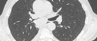
Computed tomography (MSCT or CT). A CT scan provides images of internal organs using x-rays taken from different angles. A computer combines these images into a detailed 3D image that shows any abnormalities or tumors. Sometimes, to obtain a more informative image, a contrast agent is used - a special dye (injected into the patient's vein) that provides better detail in the images obtained using CT. For example, kidney cysts do not take up the contrast agent, but kidney tumors do and become clearly visible. If the patient has severe chronic kidney disease or kidney failure, it is not safe to use a contrast agent. Computed tomography of the urinary tract is called CT urography. Please note that PET-CT is not a useful test for renal cell carcinoma.
X-ray. An x-ray is a method of creating images of the internal structures of the body using x-rays.
Magnetic resonance imaging (MRI). Uses magnetic fields to produce detailed images of the body. When scanning, a special dye, gadolinium, may be used to create a clearer image (injected into the patient’s vein).
Cystoscopy and nephroureteroscopy. Sometimes used for cancer of the ureter or renal pelvis, rarely for renal cell carcinoma (if preliminary imaging studies show a tumor or stone in the bladder). During these procedures, the patient is sedated and a thin tube with a video camera and light is inserted through the ureter into the bladder and up into the kidney. The sedative effect of the medication allows the patient to relax, calm down, or fall asleep. This procedure can be used to take a biopsy and obtain tumor cells for examination under a microscope, and sometimes to destroy small tumors completely.
After conducting diagnostic tests, the oncologist at the Rassvet Clinic will study the results with you. If the diagnosis of cancer is confirmed, the research results will allow us to establish the stage of the disease and choose the necessary treatment tactics.
What types of kidney tumors are there?
There are two main types of malignant tumors in the kidney - tumors that develop from the tissue of the kidney itself - renal cell carcinoma and tumors that develop from the renal pelvis.
Of the benign tumors, the most common are angiomyolipomas (tumors consisting of fatty, muscle tissue and blood vessels). These tumors have characteristic signs that can be detected with a special x-ray examination (computed tomography). If they are large, angiomyolipomas must be removed because they can be easily damaged even by minor injuries and lead to dangerous internal bleeding.
Classification
Oncologists distinguish several types of kidney cancer. Each of them differs in the type of cells and growth characteristics.
The most common type when diagnosed with kidney cancer is renal cell carcinoma, accounting for about 85% of diagnoses. It develops in the proximal renal tubules, which make up the renal filtration system.
Urothelial carcinoma is also called transitional cell carcinoma. On average, it accounts for 5% to 10% of cases diagnosed in adults. Urothelial carcinoma begins in the renal pelvis, the part of the kidney where urine accumulates before moving to the bladder. This type of lesion is similar to bladder cancer; both types of tumor originate in the same cells.
Kidney sarcoma is diagnosed less frequently than others. This type of neoplasia originates in the connective tissue that forms the framework of the kidney, the soft tissue that surrounds the organ (capsule), or the fat deposits around it. Kidney sarcoma is usually treated with surgery, but often returns to the kidney area or metastasizes. After removal, chemotherapy is required; a second surgical intervention may be required.
Wilms' tumor is typical for children, but it is treated in a completely different way than cancer in adults. This tumor accounts for up to 1% of all tumors; treatment includes radiation and chemotherapy, then removal of the kidney.
Lymphoma can affect both kidneys, but is more often associated with enlarged lymph nodes, called lymphadenopathy, in other parts of the body, including the neck, chest, and abdomen. Sometimes lymphoma can appear as a single tumor in the kidney, only then affecting regional lymph nodes. If lymphoma is a possibility, your doctor will do a biopsy and may recommend chemotherapy instead of surgery.
How common is kidney cancer?
Kidney cancer accounts for 4% of all detected malignant neoplasms. Kidney tumors occur primarily in people aged 50-70 years, although the disease can occur in teenagers and even children. Kidney tumors are slightly more common in men than in women, and more common in urban populations than in rural areas.
In the Republic of Belarus in 2010, 1833 new cases of this disease were identified. The incidence of malignant kidney tumors in the Republic of Belarus is constantly increasing and in 2010 amounted to 19 per 100,000 inhabitants.
Most common types of kidney cancer cells
Today, more than 30 different types of kidney cancer cells are known.
Clear cell carcinoma. This type of cancer accounts for about 70-80% of cases. Clear cell carcinoma cells range from slow-growing (grade 1) to fast-growing (grade 4). Particularly effective treatments for clear cell kidney cancer are targeted therapy and immunotherapy.
Papillary cancer. Papillary kidney cancer occurs in 10-15% of patients. This type of cancer is divided into 2 subtypes: type 1 has a relatively low grade and a favorable prognosis; type 2 is characterized by a high degree of malignancy and a pronounced tendency to metastasize.
Localized papillary kidney cancer is often treated with surgery. Locally advanced metastatic papillary kidney cancer - targeted drugs (inhibitors of blood vessel growth). The use of immunotherapy to treat metastatic papillary cancer is still under investigation.
Chromophobic type. Occurs in 4-5% of cases. This is another unusual and different type of cancer. It may have indolent (slow) tumor growth. The tumor has aggressive growth and a low tendency to metastasize. Clinical trials are currently underway to find the most effective treatments for this type of cancer.
Oncocytic type. Occurs in 2-5% of cases. This is a slow-growing type of kidney cancer that rarely (or does not) metastasize.
Ductal type. Ductal carcinoma occurs in 1-2% of cases. This type of cancer is more often diagnosed in people between 20 and 30 years of age. It begins in the collecting ducts of the kidney and, due to its connection with the pyelocaliceal system, is closely associated with transitional cell carcinoma. Ductal carcinoma is difficult to treat, even with a combination of systemic chemotherapy and surgery.
Sarcomatoid type. Each tumor subtype of kidney cancer (clear cell, papillary, chromophobe, etc.) may have highly disorganized features when examined microscopically. Pathologists often describe these types as “sarcomatoid.” This is not a separate tumor subtype, but the identification of sarcomatoid elements indicates a very aggressive form of kidney cancer. There is promising research into the effectiveness of immunotherapy treatments for people with sarcomatoid types of kidney tumors.
Medullary type. This is a rare and very aggressive form of kidney cancer. It most often occurs in people of the Black race and is associated with sickle cell anemia (hereditary hemoglobinopathy). Combinations of chemotherapy with blood vessel growth inhibitors are considered effective treatment options based on some scientific evidence. Clinical trials are currently underway to determine the best treatment.
Angiomyolipoma. A benign tumor that has a unique appearance on CT scans and when viewed under a microscope. Treatment usually involves surgery or (if the tumor is small) active surveillance. Significant bleeding is rare and is more common in pregnant and premenopausal women. An aggressive form of angiomyolipoma, epithelioid, can in rare cases grow into the renal vein, inferior vena cava, and spread to nearby lymph nodes or organs (for example, the liver).
The oncology department of the Rassvet Clinic conducts high-precision histological, immunohistochemical and molecular genetic studies. Accurate determination of the cell type of a kidney tumor allows for proper treatment planning.
What factors predispose to the development of kidney cancer?
- Smoking is one of the most significant risk factors for kidney cancer. Smokers are approximately twice as likely to develop kidney cancer as non-smokers;
- Obesity, especially in women;
- Overuse of painkillers may also contribute to the development of this disease;
- Professional factors. The risk of this disease is increased among workers in the metallurgical industry, tanning industry and when working with asbestos and cadmium. It must be emphasized that the influence of the latter factors is not very large and is not recognized by all experts;
- Genetic factors present in cases of familial kidney cancer (eg, Von Hippel-Lindau syndrome). These cases are characterized by the development of the disease at a young age, the frequent development of tumors in both kidneys and the occurrence of multiple foci of cancer in the kidney.
Stages of the disease
How kidney cancer manifests itself is determined by the stage of the disease. In addition, staging is necessary to select the most effective therapy and determine the prognosis and rehabilitation plan. The stage allows us to clarify how far the tumor has spread from its original site, whether it has damaged neighboring tissues, nearby lymph nodes, and whether it has metastasized. All this data helps determine the volume of tumor tissue in the body, determine the severity of the condition and develop individual therapy. It is equally important to determine the preliminary prognosis and survival duration based on the stage data.
Stages of kidney cancer range from I (first) to IV (fourth). Typically, the lower the number, the less the cancer has spread. Within a stage, an earlier letter (or lower number) indicates a lower stage. Although each person faces different cancer dynamics, cancers with similar stages tend to have similar management approaches and are often treated in the same way.
Kidney cancer is usually assigned a clinical stage based on the results of a physical examination, biopsy, and imaging tests performed. If surgery was done, pathological stage (also called surgical stage) is determined by examining the tissue removed during surgery Source: Molecular Epidemiology of Kidney Cancer. Zaridze D.G., Mukeria A.F., Shangina O.V., Matveev V.B. Oncourology, 2022. p. 107-119.
Stage I : This is usually referred to as T1N0M0. The tumor measures up to 70 mm, located only in the kidney itself - this is T1. Nearby lymph nodes are not affected - this is N0, there are no distant metastases - this is M0.
Stage II : This is usually referred to as T2N0M0. The tumor is more than 70 mm in size, located only in the kidney itself - this is T2. Nearby lymph nodes are not affected - this is N0, there are no distant metastases - this is M0.
Stage III : may be referred to as T3N0M0. In this case, the tumor may damage one of the main veins - the vena cava or the renal vein, or has spread to the perinephric tissues, but has not affected the adrenal gland or has not gone beyond the boundaries of the fascia - this is T3. Nearby lymph nodes are not affected - this is N0, there are no distant metastases - this is M0.
There may be a variant T1-T3N1M0: with it, the tumor has any size, can extend beyond the borders of the kidney itself, but not further than the borders of the fascia. If the cancer affects the closest lymph nodes - this is N1, but there are no metastases to other nodes and tissues - this is M0.
Stage IV : May be described as T4N0-1M0. The tumor itself affected the kidney, but did not go beyond the fascia, but could damage the adrenal gland - this is T4. The nearby lymph nodes may or may not be affected - there will be any N. There are no metastases to other tissues - this is M0.
Possible option T1-4, N0-1 M1. This is an initial tumor of any size, whether or not there are lesions in nearby lymph nodes, but metastases to distant lymph nodes or organs are determined - this is M1.
When assessing what kidney cancer looks like using these criteria, it is necessary to take into account additional criteria - age, severity of the general condition and associated complications.
Symptoms of kidney cancer
Since the kidneys are located deep in the human body, tumors often grow for a very long time and reach large sizes before signs appear that allow them to be suspected.
Before the advent of sophisticated examination methods in medicine, the diagnosis of kidney cancer could be suspected based on three symptoms:
- lower back pain;
- the presence of blood in the urine;
- palpable tumor.
Now these symptoms indicate an advanced stage of kidney cancer and are rare. Most tumors are now discovered incidentally during ultrasound examinations performed for other diseases or symptoms:
- One of the most common symptoms of kidney cancer is blood in the urine. Often this is followed by acute pain in the kidney area, caused by blockage of the ureter with blood clots, which disappear after the clots pass in the urine;
- Low back pain is the second most common symptom of kidney cancer. The pain may be dull, which is associated with stretching of the fibrous capsule of the kidney or compression of the nerve plexuses by the tumor. Acute lower back pain is usually associated with bleeding and the formation of clots that obstruct the flow of urine;
- The presence of a tumor in the kidney area, which can be palpated, is characteristic of a widespread tumor process.
- Sometimes with kidney tumors, dilation of the veins near the testicle is observed. If such enlargement occurs suddenly or after 40 years of age, you should consult a doctor, as this may be a symptom of a serious pathology.
Kidney cancer is characterized by a high frequency of development of various manifestations associated with the tumor's production of various biologically active substances, which has given rise to calling kidney cancer a “therapeutic tumor.”
Kidney tumors can produce a variety of protein substances that can lead to an increase in body temperature, an increase in the level of calcium in the blood, a decrease or increase in the number of red blood cells, an increase in blood pressure, an acceleration of ESR, and a blood clotting disorder.
All these conditions disappear after complete removal of the tumor. The return of these symptoms usually indicates a relapse of the disease or the development of distant metastases.
Postoperative period
After laparoscopic surgery, recovery is much faster and the patient feels almost no pain. Small incisions are left on the skin, which heal quickly. You will be able to get up and eat after the first day, this will speed up your recovery. And you will be discharged from the hospital within 2-3 days. Rehabilitation takes 1-2 months.
After open surgery, this will not be easy due to cut muscles or even removed ribs. On average, it takes from 1 week to 1 month before you are allowed to go home. But rehabilitation will last about 1.5 years. You will be prescribed painkillers, recommended moderate exercise to restore blood circulation, and prescribed a special diet. After the operation, you will need to regularly visit a urologist for monitoring purposes, and also undergo a CT examination on average every six months.
How long you live after surgery for kidney cancer depends on the type and stage of cancer, as well as how you undergo the surgery.
Immunotherapy
Kidney cancer after surgery can go into remission or metastasize. For such patients, immunotherapy is relevant - maintaining the body's resources in order to encourage them to fight the remaining disease. For treatment, tumor cells from a removed tumor are used: a vaccine is made from it, which stimulates the immune system to fight cancer and recognize cancer cells as hostile. This technique reduces the likelihood of relapse. It is important that such treatment is chosen by a very experienced doctor, so as not to aggravate the condition or encourage unwanted cells to grow.
Diagnosis of kidney cancer
If a kidney tumor is suspected or in the presence of nonspecific lower back pain, ultrasound examination of the kidneys is now widely used.
The advantages of this study are its low cost, accessibility, safety, and the absence of harmful effects of X-rays. Ultrasound can clearly distinguish a regular kidney cyst from a tumor or suspicious formation that requires a computed tomography scan.
The disadvantages of ultrasound are that the results are not entirely reliable in obese patients; sometimes the tumor is difficult to detect due to screening of the ultrasound by the ribs or gas in the intestine. In addition, the results of the study depend on the experience and qualifications of the doctor performing the ultrasound.
Metastasis
Metastases are secondary tumor foci that are formed as a result of the spread of tumor cells throughout the body from the primary formation. Clear cell kidney cancer can metastasize through hematogenous and lymphogenous routes. In the first case, tumor cells spread through the bloodstream into the internal organs. The lungs, brain, and bones are most often affected. Cancer metastases can also spread through the lymphatic vessels and form secondary lesions in the lymph nodes. The sentinel nodes are the first to be affected - the first ones on the path of lymph outflow.
Treatment methods for kidney cancer
The main factor influencing the choice of treatment for kidney cancer is the extent of the tumor, mainly the presence or absence of tumor spread to other organs - metastases.
Surgery
Most often, with this pathology, an operation is performed to remove the kidney with the tumor and surrounding tissues, including the fatty tissue surrounding the kidney, lymph nodes and sometimes the adrenal gland (radical nephrectomy).
In some cases, complete removal of a tumor from the body requires removal of other organs adjacent to the tumor.
In some patients with small tumor sizes, laparoscopic radical nephrectomy is performed. This operation is performed without a large incision through several small punctures. The removed kidney containing the tumor is usually removed through a small incision in the lower abdomen.
The advantage of this intervention is faster recovery of the body after surgery.
In some patients with kidney cancer, the tumor can grow into the venous system of the kidney and even extend into the largest vein of the human body - the inferior vena cava. In these cases, the operation becomes much more complicated and may be accompanied by large blood loss.
Currently, the so-called organ-preserving treatment of kidney tumors has increased significantly. If the tumor size is small and its localization is favorable, it is possible to completely remove the tumor and the adjacent area of kidney tissue without removing the entire kidney (partial nephrectomy).
The final decision to perform a particular operation is made by the surgeon after analyzing the computed tomography data. For small tumors in very elderly patients with contraindications to surgery, observation may be acceptable. With this approach, surgery is performed only when the tumor increases in size, which occurs only in a small number of such patients.
An alternative treatment for small kidney tumors is radiofrequency ablation - destruction of the tumor using a special probe that generates high temperature, inserted into the tumor under the guidance of ultrasound, computed tomography or laparoscopy.
The advantage of this treatment is that it is less traumatic and well tolerated by elderly or seriously ill patients. In a number of patients with kidney cancer, distant metastases are already determined upon initial treatment.
Despite the disappointing prognosis for such a common disease, some interventions can prolong the life of patients or improve its quality. So, although removal of a kidney for metastatic kidney cancer cannot cure the patient, it has been found that this operation makes it possible to more effectively treat this disease with the help of medications, primarily immunotherapy.
Radiation therapy
Kidney cancer is poorly sensitive to radiation. Currently, radiation therapy for kidney cancer is used exclusively for pain relief in patients with bone metastases or brain damage.
Chemotherapy
To date, the results of the use of traditional antitumor chemotherapy drugs in patients with kidney cancer remain disappointing. Almost all existing chemotherapy drugs have been used for kidney cancer. Unfortunately, none of the chemotherapy drugs have been shown to be effective against kidney cancer.
Immunotherapy
Currently, immunotherapy for metastatic kidney cancer is one of the effective treatment methods. Basically, two immunological drugs are used for kidney cancer - interleukin-2 and interferon-alpha. Given the similar effectiveness of both drugs, fewer side effects develop when treated with interferon, so interferon is predominantly used in clinical practice.
Targeted therapy
In recent years, a number of new drugs have been developed with “targeted” action on various molecular mechanisms of tumor development. The use of a number of drugs for advanced kidney cancer has proven to be more effective than traditional immunotherapy.
However, these drugs are currently characterized by extremely high costs, which prevents their widespread introduction into clinical practice. Studying the possibilities of using targeted therapy drugs is currently ongoing.
Hyperthermia and hyperglycemia
Dissatisfaction with the results of treatment of patients with common forms of malignant neoplasms was the impetus for the search for fundamentally new methods that enhance the capabilities of chemoradiotherapy. There is currently evidence of the effectiveness of general hyperthermia in the treatment of advanced kidney cancer. Elevated temperature, in addition to its direct destructive effect on the tumor, can enhance the effect of antitumor drugs.
Advantages of contacting MEDSI
- Experienced doctors.
We employ not only urologists, oncologists, but also other doctors who have all the necessary knowledge and skills to identify kidney cancer based on the main objective signs and symptoms and treat the pathology not only in the early but also in the late stages of development - Highly accurate diagnostics.
MEDSI can perform: multislice computed tomography with dynamic contrast and 3D image reconstruction to detect the smallest tumors, magnetic resonance imaging on a Philips Ingenia device with an increased tunnel diameter, ultrasound with a Doppler attachment, navigational biopsy under ultrasound control, etc. - Personal selection of treatment methods for kidney cancer at various stages with positive prognosis.
We understand that every case is unique. In order for all measures taken to be successful, doctors take into account the location of the tumor, its type, the patient’s health condition and other factors. - Modern technologies.
We use the da Vinci Si robot and perform laparoscopic and endoscopic interventions. This allows you to increase their accuracy and reduce recovery time, eliminating the risks of certain complications. - Fast and comfortable rehabilitation.
Our patients with the first stage of the disease are discharged on the third day after surgery, and a week later they go to work and return to their previous lifestyle. In serious cases, a longer recovery process is required: on an outpatient basis or at MEDSI sanatoriums. However, in any case, rehabilitation is carried out as completely, comprehensively and effectively as possible. - Monitoring patients.
After the course of treatment, the doctor continues to supervise the patient. This is important for the final restoration of health, maintaining quality of life, and preventing relapses of the disease. The patient visits his doctor once every 3-6 months. If necessary, additional tests and studies are prescribed. Particular attention is paid to helping prevent relapses of the disease. Experienced doctors are ready to eliminate risk factors, stabilize blood pressure, create a balanced nutrition program, etc.
To clarify the specifics of kidney cancer treatment or make an appointment with a doctor, just call +7 (495) 7-800-500. Our specialist will answer all questions. Recording is also possible through the SmartMed application.
What kind of observation is required after treatment?
After completion of treatment, it is necessary to undergo periodic examinations to timely detect relapse or progression of the disease. For citizens of the Republic of Belarus with cancer, observation is carried out in oncology clinics at their place of residence.
How often you should be examined, as well as the amount of necessary research, will be determined by your doctor, depending on the stage of the disease and the treatment provided.
The standard scope of examination includes:
- Ultrasound of the abdominal organs;
- chest x-ray;
- general and biochemical blood test;
- general urine analysis.
Some useful aspects of normal renal anatomy.
Normally, each person has two functional kidneys that produce urine. The kidneys are located in the retroperitoneal space, below the chest, their projection falls on the lumbar region. At the back, the kidneys are protected by powerful back muscles. The kidneys are surrounded by a membrane (Gerota's fascia) and fatty tissue. The bud itself is covered with a thin membrane (bud capsule), similar to the thin skin of a red apple. Blood drains from the kidney through the renal vein, which drains into the inferior vena cava, which carries blood to the heart.
Adrenal glands - located on top of the kidneys, but are not part of them. The adrenal glands regulate water and electrolyte balance and the level of sex hormones in the body. In addition, in response to stress, they produce a special hormone - adrenaline.
Kidneys – regulate water and electrolyte balance (potassium, sodium, calcium, magnesium). They produce urine and regulate blood pressure. The kidneys also produce a hormone called erythropoietin, which stimulates the production of red blood cells (erythrocytes).
After removal of a kidney or diseases that affect the kidney tissue, the kidney functions listed above are affected. The kidneys have a good compensatory mechanism and, with minor disturbances, are able to maintain normal homeostasis (the internal state of the body) due to compensatory hypertrophy.