Proper preparation
Before performing endoscopy of the bronchi and lungs, the patient must remember the basic rules of preparation for the procedure:
- The examination is carried out on an empty stomach. Otherwise, food or a drink will accidentally enter the respiratory tract due to an accidental cough or vomiting.
- If a course of medication is prescribed, it is recommended to talk with your doctor; you may need a break for one day.
Before performing a pulmonary endoscopy, it is necessary to plan so that the last meal takes place no later than 21 hours before the start of the procedure.
Bronchoscopy
Bronchoscopy is a method of examining the mucous membranes of the trachea and bronchi using a special device - a bronchoscope. A tube equipped with lighting equipment and a video camera is inserted into the respiratory tract through the larynx. This modern equipment provides research accuracy of over 97%, which makes it indispensable in diagnosing various pathologies: chronic bronchitis, recurrent pneumonia, lung cancer.
Preparation for bronchoscopy
Before bronchoscopy, it is necessary to undergo a number of diagnostic tests:
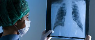
- radiography of the lungs,
- ECG (electrocardiogram),
- blood tests (general, for HIV, hepatitis, syphilis),
- coagulogram (blood for clotting)
- and others according to indications.
Dinner should be no less than 8 hours before the procedure.
On the day of the study, smoking is prohibited (a factor that increases the risk of complications).
Bronchoscopy is performed strictly on an empty stomach.
In the morning, it is necessary to do a cleansing enema (prevention of involuntary bowel movements due to increased intra-abdominal pressure).
It is recommended to empty your bladder immediately before the procedure.
If necessary, the doctor will prescribe mild sedatives on the day of the procedure. Patients with bronchial asthma must have an inhaler with them.
For people suffering from cardiovascular pathology, preparation for bronchoscopy is carried out according to an individually developed program.
Methodology
Immediately before the procedure, the doctor will ask you to sign a consent form for the manipulation. Be sure to discuss with your doctor the likelihood of consequences, as well as the risks of the examination!
The study is carried out in a specially equipped endoscopy room, where the same sterility conditions are observed as in the operating room. The procedure is performed by a doctor who has undergone special training in bronchial examination.
The duration of bronchoscopy is 30-40 minutes.
By spraying the patient, bronchodilators and painkillers are administered to facilitate the advancement of the tube and eliminate discomfort.
Patient's body position: sitting or lying on his back.
It is not recommended to move your head or move. To suppress the urge to vomit, you need to breathe often and not deeply.
The bronchoscope is inserted through the oral cavity or nasal passage.
In the process of moving to the lower sections, the doctor examines the internal surfaces of the trachea, glottis and bronchi.
After the examination and the necessary manipulations, the bronchoscope is carefully removed, and the patient is sent to the hospital for some time under the supervision of medical staff (to avoid complications after the procedure).
Feelings after bronchoscopy
Feelings of numbness, a lump in the throat and nasal congestion will persist for up to 30 minutes. During this time and after another hour, it is not recommended to smoke or eat solid food. Also, doctors do not recommend driving a car on this day, since the administered sedatives can impair concentration.
Possible complications
The risk of negative consequences, although minimal, is possible. Therefore, you should immediately consult a doctor if you notice the following symptoms:
- hemoptysis for a long time;
- pain in the chest;
- audible wheezing;
- feeling of suffocation;
- nausea and vomiting;
- rise in body temperature.
How is tracheobronchoscopy performed?
- On the eve of the procedure, the patient undergoes an examination by a doctor. If necessary, it is possible to prescribe a sedative at night, this will reduce the level of anxiety and allow you to get a good night's sleep.
- Sedative medications can be taken immediately before the start of endoscopy, but the decision remains with the doctor.
- Dentures must be removed; the flexible tube of the endoscope can displace them towards the respiratory tract.
- The neck should be free, so choose clothes that are loose at the top.
- During the preliminary examination, it is necessary to inform if there is a history of allergic reactions to medications or products.
- To ensure that the endoscope passes painlessly and a cough reflex does not occur, local anesthesia is applied to the mucous membrane of the nasal cavity and oropharynx using a sprayer.
- The position during the procedure is determined by the doctor. There are two options: lying on your back or sitting on a chair.
- At the end of the endoscope there is a camera that will allow the doctor to insert the instrument under visual control while simultaneously examining the lungs on both sides.
- Most often, the device is inserted through the nose, but sometimes through the oral cavity.
- Special forceps are used for biopsy. The procedure for taking a sample from the mucous membrane is painless and extends the time by only 1-2 minutes.
Diagnostic and therapeutic capabilities of modern bronchoscopy
Today, more than a hundred years after the “father of bronchoscopy” Gustav Killian first inserted an endoscope into the trachea and removed an aspirated meat bone from a patient, bronchoscopy is one of the leading methods for diagnosing and treating respiratory diseases. Most lung diseases are in one way or another associated with bronchial pathology. The airways provide access to any areas of the lung, allow one or another instrument to be passed to them and obtain a variety of information about the state of the respiratory organs, and also represent an additional route for administering medications to pathologically altered areas of the lung.
History of development
The development of bronchoscopy can be divided into three stages. At the first, which began at the end of the 19th century. and continued until the end of the 50s of the twentieth century, bronchoscopy was performed under local anesthesia, usually using rigid bronchoesophagoscopes
, which had a dual purpose - examination of the tracheobronchial tree and esophagus. The progress of bronchoscopy during this period was greatly facilitated by the work of Ch. Jackson, J. Lemoine, A. Soulas, A. Olsen, N. Andersen, and in our country - A. Delens, V. Voyachek, V. Trutnev, A. Likhachev and Chalky. Bronchoscopy during this period was performed mainly for foreign bodies in the respiratory tract and was carried out mainly by otolaryngologists. The procedure was very traumatic, and the patients had a hard time bearing it.
With the advent and improvement of general anesthesia, pulmonary surgery began to actively develop and indications for bronchoscopy expanded significantly. This was facilitated by the creation in the late 50s - early 60s of respiratory bronchoscopes (N. Friedel; R. Hollinger; G.I. Lukomsky), which made it possible to perform bronchoscopy under anesthesia with myoplegia and injection ventilation of the lungs, which significantly alleviated the suffering of patients and made the study safer. The progress of bronchoscopy at this second stage of its development was facilitated by the appearance of lens telescopes with direct, lateral and retrograde optics, various instruments for biopsy, extractors, scissors, and electrocoagulators. At this stage, bronchoscopy passed into the hands of thoracic surgeons.
A true revolution in bronchology and the beginning of the third, modern stage in the development of bronchoscopy was the creation in 1968 of a flexible bronchofiberscope
[1], with the help of which it became possible to examine the lobar, segmental and subsegmental bronchi of all parts of the lung, perform a visually controlled biopsy, and administer medicinal solutions. The fiberoptic bronchoscope has significantly changed the technique of bronchoscopy. It was again performed under local anesthesia, causing almost no discomfort to the patients. Bronchofibroscopy began to be successfully performed on an outpatient basis, in pulmonology hospitals and offices, and in intensive care units. It seemed that the need for rigid bronchoscopes had disappeared forever. However, the creation of high-energy medical lasers determined a new direction in bronchology - surgical endoscopy, and rigid endoscopes were again required for it. Therefore, modern bronchoscopy is equipped with both flexible and rigid endoscopes and instruments that allow performing a wide range of diagnostic and therapeutic manipulations in the trachea and bronchi both under local anesthesia and under general anesthesia.
Indications for bronchoscopy
The main indications for bronchoscopy are shown in Table. 1. They should be divided into diagnostic and therapeutic.
Diagnostic bronchoscopy
Tumors of the bronchi and lungs
Tumors of the bronchi and lungs
Tumors of the bronchi and lungs are one of the main indications for bronchoscopic examination. Currently, verification of centrally located endobronchial cancer reaches almost 100% and is carried out by visually controlled biopsy with wire cutters. Diagnosis of the so-called “early cancer”, which also includes tumors in situ, is more difficult. Fluorescent chromobronchoscopy helps to identify such neoplasms, almost invisible to the naked eye.
with the introduction of special drugs - photosensitizers [2, 3].
Diagnosis of peripherally located tumors, especially small ones, can also be quite difficult, since getting to such tumors through the bronchi is very difficult.
Their biopsy is performed under the control of an X-ray television screen and, in addition to cutters, scarifying brushes and controlled curettes are used. However, even in experienced hands, verification of peripheral lung tumors rarely reaches 60-70% and in complex cases should be combined with percutaneous puncture biopsy under computed tomography (CT) control.
Diagnosis of peribronchial cancer also requires great skill, especially in the early stages. The presence of such a tumor can be suspected based on X-ray and CT data, and to verify it, a puncture biopsy
the bronchial wall in a suspicious place using a special needle. Thus, all darkening or cavitary formations in the lungs, hilar or located on the periphery, suspicious for the oncological process, are direct indications for bronchoscopy and various methods of bronchoscopic biopsy, the choice of which is determined by the doctor performing the study.
Mediastinal neoplasms and lymphadenopathy
Mediastinal neoplasms and lymphadenopathy may also be indications for bronchoscopy. With enlarged paratracheal and bifurcation lymph nodes and mediastinal tumors located in close proximity to the trachea, material for cytological examination can be obtained using transtracheal puncture biopsy
. However, the not very reliable results of such a study have now ceased to meet the requirements of practice and bronchoscopic methods have been replaced by more invasive, but much more informative methods: mediastinoscopy, pleuromediastinoscopy [4, 5] and video thoracoscopy [6]. They should be resorted to in cases where bronchoscopic biopsy methods are ineffective.
Diffuse lung diseases
The same trend can, to a certain extent, be attributed to the diagnosis of diseases accompanied by diffuse changes in the pulmonary pattern (the so-called diffuse lung diseases - DLD), which require morphological research for their verification. Since the early 70s, after the work of N. Andersen, transbronchial pulmonary biopsy
, performed using a bronchoscope, has become the leading method for diagnosing DLD [5]. Over time, however, it became clear that with transbronchial lung biopsy it is not always possible to obtain enough lung tissue to successfully carry out differential diagnosis in a number of DLD, especially those accompanied by fibrosing processes in the pulmonary parenchyma. And although the relatively less invasiveness still allows transbronchial lung biopsy to remain the method of primary endoscopic diagnosis of DLD, it is increasingly complemented by thoracoscopic biopsy, performed using wire cutters or endostapplers, hermetically stitching the lung parenchyma while simultaneously cutting off a section of lung tissue of the required size [6].
Important diagnostic information for many lung diseases, and primarily for DLD, can be obtained by studying material obtained using bronchoalveolar lavage (BAL)
.
The latter today is an almost obligatory study in the diagnosis and treatment of DLD such as cryptogenic fibrosing alveolitis and sarcoidosis. BAL performed repeatedly during the treatment of these diseases makes it possible to monitor the effectiveness of therapy and determine its prognosis. Bronchoscopy and BAL are also indicated for suspected fungal diseases of the bronchi
(bronchomycosis) and some
parasitic diseases of the lungs
(for example, with Pneumocystis pneumonia).
Inflammatory processes in the lungs
A bronchoscope allows you to look deeply into the airways. This makes it possible for patients with descending tracheobronchitis to determine the distal border of the lesion of the bronchial tree and the intensity of inflammation in it. Bronchoscopy is effective in searching for a draining bronchus in acute lung abscesses, as well as in the differential diagnosis of bacterial suppuration and disintegrating cancer in the presence of a cavity in the lung. The difficulties of absolute sterilization of bronchofibroscopes somewhat complicate the microbiological diagnosis of inflammation and require the use of special catheters that protect the material collected in the bronchi from contamination with the contents of the oral and nasal cavities. Our experience in the use of bronchoscopy in patients with acute and chronic inflammatory lung diseases allows us to give preference to the use of a rigid bronchoscope, amenable to thermal sterilization methods, if microbiological diagnosis of pulmonary suppuration is necessary.
Pulmonary hemorrhage and hemoptysis
If we strictly follow the terminological logic, hemoptysis is a manifestation, a symptom of pulmonary hemorrhage. However, in practice, pulmonary hemorrhage (or hemoptoea) refers to the release of pure blood or intensely bloody sputum when coughing, and hemoptysis (hemophthisis) refers to the coughing up of sputum tinted with blood or containing streaks of blood. Thus, there is a quantitative difference between hemoptoea and hemophthisis [4, 5]. Both pulmonary hemorrhage and hemoptysis are direct indications for diagnostic bronchoscopy, since this is the only way to determine the source of bleeding or at least its approximate location.
The causes of pulmonary hemorrhage and hemoptysis are extremely diverse. In addition to the pathology of the tracheobronchial tree and lung parenchyma, among them are diseases of the blood and circulatory system, hemorrhagic diathesis and capillary toxicosis, pulmonary embolism, pulmonary endometriosis, etc. The relative frequency of these causes has changed over time. Thus, in the 30s and 40s, destructive pulmonary tuberculosis was in first place among all causes of pulmonary hemorrhage. Currently, the most common cause of hemoptysis in the pulmonology clinic is chronic bronchitis accompanying bronchiectasis or focal pneumosclerosis, in which in foci of chronic inflammation against the background of decreased blood flow along the branches of the pulmonary artery, excessive vascularization develops due to dilation of the bronchial arteries and multiple anastomoses arise between the large and small circles of blood circulation.
Due to the shunting of blood from the bronchial arteries into the branches of the pulmonary artery, hypertension occurs in the microvasculature of the lungs, which the fragile walls of small vessels cannot withstand, and the blood enters the respiratory tract. Similar mechanisms are observed in the area of foci of destruction of lung tissue of specific and nonspecific etiology. During bronchoscopy in these cases, the source of bleeding, as a rule, cannot be seen, but it is quite possible to determine at least its approximate location, especially if the study is performed at the height of hemoptysis. This is very important for determining treatment tactics for each individual patient.
The causes of hemophthisis and hemoptoea diagnosed in patients in the thoracic surgical department are presented in Table. 2. Undoubtedly, the most serious cause of pulmonary hemorrhage and hemoptysis was and is bronchial tumors and, above all, cancer, which can only be verified using bronchoscopy. This allows us to conclude that in all cases of pulmonary hemorrhage and hemoptysis, bronchoscopy is a mandatory study, the main purpose of which is to identify or exclude a malignant neoplasm of the lungs
.
Chronic cough
Among the indications for diagnostic bronchoscopy, the so-called treatment-resistant cough should also be mentioned, i.e. cough that does not respond to intensive treatment for at least 1 month, the cause of which remains unclear. And although lung tumors, according to R. Irwin et al. [7], are rarely accompanied by an isolated cough syndrome (without any radiological manifestations), our experience in examining persistently coughing patients [4, 5] gives us reason to assert that bronchoscopy is one of the most important studies in the complex diagnosis of the causes of chronic cough.
Broncho-obstructive syndrome
Bronchoscopy plays an important role in the differential diagnosis of chronic obstructive pulmonary diseases and obstruction of the trachea and bronchi, accompanied by broncho-obstructive (asthmoid) syndrome. This primarily applies to tumors, foreign bodies (including those of endogenous origin - broncholitis) and cicatricial strictures of the trachea and large bronchi, in which radiological symptoms may be completely absent, and the clinical picture is very similar to an attack of bronchial asthma [8, 9].
Therefore, in cases where patients have signs of difficulty breathing that are not relieved by modern drug therapy, a bronchoscopic examination is indicated, which often reveals one or another organic pathology in the large respiratory tract.
Therapeutic bronchoscopy
Removal of aspirated foreign bodies
Removal of aspirated foreign bodies
The therapeutic capabilities of bronchoscopy have long been limited to the extraction of aspirated foreign bodies, and even now this is the only bloodless method of removing them from the bronchi.
The development of flexible extractors and the considerable experience accumulated to date suggests that most aspirated foreign bodies in adults can be removed using a bronchofiberscope under local anesthesia and even on an outpatient basis [5]. However, foreign bodies in the respiratory tract sometimes present the bronchologist with the most unpleasant surprises, forcing the use of general anesthesia and hard instruments and requiring maximum concentration of strength and skill, and sometimes inspiration.
Drainage of intrapulmonary purulent foci
The therapeutic effect of bronchoscopy as a method of draining intrapulmonary purulent foci, be it bronchiectasis or lung abscesses, is undeniable. Therapeutic catheterization of the bronchi during bronchoscopy makes it possible to unblock a significant part of the intrapulmonary abscess cavities [5], and long-term transnasal drainage [10] ensures the constant introduction of antibacterial drugs into the cavity and frees patients from repeated bronchoscopy and catheterization. A technique for immunoreplacement therapy has been developed in the form of intracavitary administration of a suspension of autologous macrophages [11], making bronchoscopic treatment even more effective.
Chronic obstructive bronchitis
The therapeutic role of bronchoscopy in chronic obstructive bronchitis (COB) has traditionally been limited to restoring airway patency with stimulation or imitation of impaired bronchial drainage function and local use of antibacterial and secretolytic agents. After the first publications by A. Soulas and P. Mounier-Kuhn, who described the method of treating patients with chronic nonspecific lung diseases using a bronchoscope, many different methods of bronchoscopic treatment of COB were proposed. Some of them were abandoned as having not been tested by practice, others took a strong place in the arsenal of therapeutic agents for patients with diseases of the bronchopulmonary system [5, 12].
Currently, sanitation bronchofibroscopies
, carried out under local anesthesia in a course method with a frequency of 1 time every 2-3 days.
The duration of the course depends on the severity of the pathological process and the effectiveness of treatment and ranges from 3 to 20 sessions. If the sputum is purulent and there is a significant amount of it, 10 ml of a 0.5-1% solution of potassium furagin, warmed to body temperature, is instilled through the channel of the bronchofibroscope into the bronchi with the addition of 1-2 ml of a mucolytic ( ambroxol, acetylcysteine
).
Before removing the bronchofibroscope, antibiotics are injected into the lumen of the bronchi in a daily dose (in accordance with the sensitivity of the bronchial microflora to them). In the presence of purulent sputum with an ichorous odor, instillation of a 1% dioxidine solution in an amount of 5-10 ml is used. At the end of the procedure, the patient is placed alternately on each side for 5-7 minutes, after which he is asked to actively cough.
The emergence of new technical devices is also reflected in the endobronchial therapy of inflammatory lung diseases. The publications of E. Klimanskaya, S. Ovcharenko, V. Sosyura and others describe the use of low-frequency ultrasound and radiation from ultraviolet and helium-neon lasers
during therapeutic bronchoscopy in patients with chronic bronchitis and pulmonary suppuration, including children. The authors have obtained good results from the use of these methods, which, in their opinion, contribute to better sputum production, increased concentration of antibiotics in the bronchi and improved local immune defense of the respiratory tract.
NOT. Chernekhovskaya and I.V. Yaremaya [13] obtained a positive effect from the intrabronchial use of the immunomodulator T-activin, which, according to the authors, contributes to the restoration of the immune reactivity of the bronchial mucosa. In patients with COB, the drug was injected during bronchoscopy using a needle into the mucous membrane of the spurs of the lobar and segmental bronchi in the places of the most visually pronounced inflammation. For severe inflammation in the bronchi, the authors recommended the use of intrabronchial immunotherapy in combination with endolymphatic administration of antibiotics into interbronchial spurs.
In conclusion, we consider it our duty to remind you that sanitary bronchoscopy is a rather crude and traumatic method of treatment and should be performed in patients with COB if there are appropriate indications, which primarily include purulent complications and a pronounced obstructive component of the disease. It is not necessary to expand the indications for therapeutic bronchoscopy in patients with serous forms of endobronchitis without severe bronchial obstruction, where it is quite possible to achieve good results using inhalation, injection or oral methods of administering therapeutic drugs. Bronchoscopy is a “cannon” method of treatment and is hardly worth using when “shooting at sparrows.”
Severe bronchial asthma
If there is a significant accumulation of thick, viscous sputum in the distal parts of the bronchi in cases of ineffective expectoration, which is often observed in severe bronchial asthma, therapeutic bronchial lavage
. For the first time, massive lavage of the bronchi through an endotracheal tube was described by H. Thompson and W. Pryor in patients with alveolar proteinosis and bronchial asthma. By modifying this method, we developed a technique for therapeutic bronchial lavage through a rigid bronchoscope under conditions of pulmonary injection ventilation [5, 12]. Therapeutic bronchial lavage in patients with severe respiratory failure requires highly qualified anesthesiological care and post-anesthesia monitoring in an intensive care unit or intensive care unit. When performed correctly, this procedure effectively helps remove sputum from medium- and small-caliber bronchi that are inaccessible to other methods of endobronchial aspiration. It is important to point out the dangers of using this technique in patients with purulent forms of endobronchitis, since the absorption of liquefied and not completely removed purulent sputum can lead to increased intoxication and worsening of the patients’ condition.
In several particularly severe patients with status asthmaticus and hypoxic coma, we performed bronchial lavage under conditions of extraorgan oxygenation. The experience of using such a resuscitation aid is relatively small, but it deserves attention and can be used in specialized intensive care units.
Early postoperative period
Bronchofibroscopy has proven itself to be an effective treatment procedure for impaired bronchial obstruction in patients in the early postoperative period and, especially, in patients requiring long-term artificial pulmonary ventilation (ALV). A flexible bronchofiberscope can be easily inserted into the patient’s respiratory tract through an endotracheal or tracheostomy tube, which makes it possible to perform sanitary bronchoscopy in patients on mechanical ventilation daily, and, if necessary, several times a day [5].
In addition to the fairly ordinary situations listed above that require the use of bronchoscopy, there are a number of more rarely occurring pathological conditions in which bronchoscopy can also have therapeutic value. These include isolated cases of destructive pneumonia complicated by pyopneumothorax
. In some patients with this disease, wide or multiple bronchopleural fistulas not only do not allow the lung to expand after drainage of the pleural cavity, but also do not allow successful sanitization of the pleural cavity due to the penetration of lavage fluid into the respiratory tract. In such a situation, it is possible to insert an obturator made of foam rubber or collagen sponge through a bronchoscope into the corresponding segmental or lobar bronchus and temporarily block it [5]. This seals the lung and stops the drainage of air. This creates conditions for effective lavage of the pleural cavity and reexpansion of the lung. Such a blockade of the bronchi is possible for a period of several days to 2 weeks. During this time, the pleural moorings manage to fix the lung in an expanded state, and small fistulas can close. Temporary bronchial occlusion is also successfully used for large solitary lung abscesses, helping to reduce and obliterate their cavity [14].
In patients with severe dystonia of the membranous wall of the trachea
, manifested by the clinical picture of expiratory stenosis,
transtracheal sclerotherapy
performed during bronchoscopy can help reduce its symptoms. According to the method proposed by A.T. Alimov and M.I. Perelman [15], using a flexible bronchoscopic needle-injector, a mixture of glucose and blood plasma is injected into the tissue between the walls of the esophagus and trachea through the membranous wall of the latter, which causes the development of retrotracheal sclerosis and fixes the excessively mobile tracheal membrane. In patients, the difficulties of exhalation and expectoration are reduced and the annoying and ineffective cough that torments them is alleviated.
Endotracheal and endobronchial surgical interventions
A description of the therapeutic capabilities of bronchoscopy will be incomplete without mentioning endotracheal and endobronchial surgical interventions. At first, they were performed using high-frequency current, but recently high-energy YAG lasers—neodymium and holmium—have been predominantly used. benign tumors of the trachea and large bronchi are successfully removed during bronchoscopy
, perform recanalization of the trachea with its
tumor, granulation and cicatricial stenoses
[16, 17]. The latter occur quite often, complicating prolonged tracheal intubation or tracheostomy in patients in intensive care units. To prevent re-stenosis of the trachea after its recanalization with a laser, for peribronchial tumors compressing the lumen of the trachea or main bronchi, as well as for collapse of the tracheal walls as a result of tracheomalacia, silicone stents of various designs are used - self-fixing with the help of protrusions, T-shaped or Y-shaped , bifurcation [17].
Such spacer stents can remain in the lumen of the trachea and main bronchi for a long time and provide free patency of large airways, in some cases making it possible to do without tracheostomy.
Contraindications to bronchoscopy
Contraindications to bronchoscopy are usually relative.
These include severe respiratory failure, cardiac arrhythmias, a tendency to bronchospasm, blood clotting disorders, and severe intoxication. In these cases, we are talking mainly about diagnostic studies. Where bronchoscopy is performed for therapeutic purposes, these contraindications often fade into the background and, according to vital indications, bronchoscopy can be justified in the most severe patients, being part of the resuscitation manual.
Complications of bronchoscopy
With the increase in the number and invasiveness of bronchoscopic techniques and the expansion of indications for them, the risk of the procedure has also increased, which, despite the increased level of anesthesia, is still occasionally accompanied by quite serious complications (Table 3).
Their prevention and treatment constitute a separate and very extensive problem that cannot be covered within the limited scope of this review. Our analysis of complications of bronchofibroscopy and the so-called rigid or rigid bronchoscopy in homogeneous groups of patients [5] showed that “flexible” bronchoscopy, performed for diagnostic purposes, is generally accompanied by a significantly smaller number of severe complications, in particular those caused by diagnostic manipulations, because it is associated with less trauma to the bronchi and biopsy objects. This allows us to speak about the comparatively greater safety of diagnostic bronchofibroscopy under local anesthesia, which is especially important in outpatient practice. It is impossible to compare the safety of therapeutic bronchoscopic procedures performed using rigid and flexible endoscopes, since the indications for their use, and therefore the severity of the patients’ condition, differ significantly. It should only be emphasized that bronchofibroscopy, as well as “rigid” bronchoscopy, cannot be considered an absolutely safe method of research and treatment. This procedure requires the endoscopist to not only perform it in different ways and understand endobronchial and pulmonary pathology, but also to be prepared for the development of various, sometimes severe complications, and requires certain knowledge and skills of a resuscitation, therapeutic and surgical nature. The room in which bronchoscopy is performed, whether it is a special room or an intensive care ward, must be appropriately equipped and equipped with all the devices for successful resuscitation or immediate treatment of any complication that is potentially possible during the introduction of a bronchoscope and endobronchial manipulation with its help. Literature
1. Ikeda Sh. Flexible Bronchofiberscope. Ann.Otol., 1970; 79 (5): 916–23.
2. Chissov V.I., Sokolov V.V., Filonenko E.V. and others. Modern possibilities and prospects of endoscopic surgery and photodynamic therapy of malignant tumors. Ross. oncological magazine 1998; 4: 4-12.
3. Lam S., MacAulay C., Palcic B. Detection and Localization of Early Lung. Cancer by Imaging Techniques. Chest, 1993; 103: 1 (Suppl.): 12S—14S.
4. Lukomsky G.I., Shulutko M.L., Winner M.G., Ovchinnikov A.A. Bronchopulmonology. M., Medicine. 1982; 399.
5. Lukomsky G.I., Ovchinnikov A.A. Endoscopy in pulmonology. In the book: Guide to clinical endoscopy. Ed. V.S. Savelyev, V.M. Buyanov and G.I. Lukomsky. M., Medicine. 1985; 348-468.
6. Porkhanov V.A. Thoracoscopic and video-controlled surgery of the lungs, pleura and mediastinum. Abstract of dissertation. Doctor of Medical Sciences M., 1996; 33.
7. Irwin R., Rosen M., Braman S. Cough: A comprehensive review. Arch.Intern.Med., 1977; 137 (9): 1186–91.
8. Danilyak I.G. Bronchoobstructive syndrome. M. Newdiamed. 1996; 34.
9. Perelman M.I., Koroleva N.S. Asthmatic syndrome in diseases of the trachea. Ter. archive 1978; 3:31-5.
10. Ovchinnikov A.A., Filippov M.V., Gerasimova V.D. and others. The use of long-term transnasal catheterization in the treatment of patients with lung abscesses. Gr.hir. 1986; 4:45-9.
11. Chuchalin A.G., Ovchinnikov A.A., Belevsky A.S. et al. The use of a suspension of autologous macrophages in the treatment of lung abscesses. Klin.med. 1985; 2: 85-8.
12. Ovchinnikov A.A. Endoscopic diagnosis and therapy of chronic obstructive bronchitis. In the book: Chronic obstructive pulmonary diseases. Ed. A.G. Chuchalina. Publishing house BINOM, 1998; 423-35.
13. Chernekhovskaya N.E., Yarema I.V. Chronic obstructive pulmonary diseases. M. RMAPO, 1998; 148.
14. Ivanova T.B. Prolonged temporary bronchial occlusion in the complex treatment of acute suppurative diseases of the lungs and pleura. Author's abstract. diss. ...candidate of medical sciences. M., 1987. 22.
15. Alimov A.T., Perelman M.I. Sclerosing endoscopic therapy for expiratory stenosis of the trachea and main bronchi. Gr.hir. 1989; 1:40-3.
16. Rusakov M.A. Endoscopic surgery of tumors and cicatricial stenoses of the trachea and bronchi. M., Russian Research Center of Chemistry, Russian Academy of Medical Sciences. 1999; 92.
17. Dumon J., Meric B. Handbook of endobronchial YAG laser surgery. Hopital Salvator, Marseille, France. 1983; 97.
| Applications to the article |
| All dark spots or cavitary formations in the lungs suspicious for an oncological process are direct indications for bronchoscopy with biopsy |
| The most common cause of hemoptysis in the pulmonology clinic is chronic bronchitis |
| Sanitation bronchoscopy is a rather traumatic method of treatment and in patients with COB should be performed according to indications |
How does the patient feel?
- Bronchial endoscopy is accompanied by local anesthesia, which causes a sensation as if the nose is stuffy. First, the tongue and palate become numb, and a feeling of “lump” appears in the throat. It is difficult to swallow saliva for some time.
- Modern techniques allow the procedure to be performed without pain.
- The diameter of the device is much smaller than the lumen of the lungs, so endoscopy is a completely safe procedure; you should not be afraid that you will suffocate during the procedure; a specialist is responsible for the patient’s health.
Spontaneous breathing is not impaired in any way.
Videobronchoscopy
Videobronchoscopy is an endoscopic examination of the tracheobronchial tree. An apparatus for examining the respiratory tract (bronchoscope) is inserted through the nose and larynx into the lumen of the trachea and bronchi. The method allows you to assess the condition of the lumen, walls and mucous membrane of the respiratory tract, detect neoplasms, and perform painless collection of tissue samples (biopsy) and bronchial lavages for further study in the laboratory.
- Bronchoscopy is a serious manipulation performed as prescribed by a doctor after a preliminary basic examination.
- Before it is carried out, in accordance with the order of the Ministry of Health of the Russian Federation, an X-ray examination of the chest and sputum analysis for VK are required.
- To avoid complications, the study is carried out strictly on an empty stomach.
In what cases is bronchoscopy prescribed?
Bronchoscopy is indicated in the following situations:
- In order to clarify the diagnosis in the presence of changes identified during X-ray examination.
- To identify tumors of the tracheobronchial tree when central lung cancer is suspected.
- To find out the causes of difficulty breathing, bleeding from the respiratory tract, and long-term cough.
- To take pieces of bronchial tissue for further laboratory study (biopsy).
- For collecting mucus from the bronchi for the same purpose.
- To remove foreign bodies from the respiratory tract.
- For the administration of medicinal substances and sanitation (“washing”) during therapeutic bronchoscopy.
How to prepare for bronchoscopy?
- Since accidental entry of food or liquid into the respiratory tract during vomiting and coughing can cause severe inflammation of the lungs, and in some cases, respiratory arrest and cardiac dysfunction, bronchoscopy is performed strictly on an empty stomach. The last meal should be 12 hours before the test.
- On the day of the examination, several small sips of water are allowed at least 5-6 hours before the procedure.
- If you need to take medications, consult with your doctor about your dosage schedule on the day of your bronchoscopy.
- After the examination, it is recommended not to drink or eat for 1 hour.
- After bronchoscopy, for about half an hour the patient still has a feeling of numbness in the throat and a hoarse voice - there is no need to be alarmed, this is a consequence of anesthesia of the vocal folds, it goes away along with the action of Lidocaine (like the feeling of numbness in the jaw after dental treatment with anesthesia).
Features of the study
Your consent to undergo bronchoscopy implies that you understand the need for this study and understand its essence. If you have anxiety and anxiety, do not be afraid to ask your doctor questions. Please tell your doctor or nurse if you have any allergies or drug intolerances; whether you have had a bronchoscopy before, whether you have asthma, and any changes in your health recently.
During the examination, anesthesia with Lidocaine will first be carried out step by step. The procedure may provoke a cough, but does not interfere with breathing or cause pain. Typically, the examination lasts about 20 minutes, but since at least half of the entire examination time is occupied by anesthesia of the respiratory tract, due to the individual characteristics of the body, it may proceed faster or longer. When the examination is completed, the bronchoscope is removed from the bronchi easily and quickly.
After research
If you experience weakness and drowsiness, stay for a while in the endoscopy department under the supervision of our staff. Because you were given anesthesia in your larynx before the test, it is not safe to take food or liquid right away to avoid possible inhalation. Your swallowing reflex will be fully restored in about 1 hour. Since the device is inserted into the bronchial tree through the nose, and the nasal mucosa is thin and vulnerable, after the examination there may be minor nosebleeds, and if a biopsy was performed, streaks of blood may sometimes be found in the sputum. This goes away within 24 hours and should not cause you concern. Rarely occurring after the study, sore throat or hoarseness of the voice will also decrease within 24 hours. If you go straight home on the day of the test, it is better for someone to accompany you. At home, try to devote the rest of the day to rest.
The anesthesia may last longer than expected, so you should not:
- drive,
- maintain machinery and technical equipment,
- drinking alcohol.
When will I know the result of the bronchoscopy?
The doctor will inform you of the results of the study immediately. However, if to clarify the diagnosis it was necessary to take tissue or contents of the bronchi for analysis, it will take from 1 to 7 days before the final result becomes known. Details of the necessary treatment should be discussed with the doctor who recommended the study and referred you for bronchoscopy.
What can you do after bronchoscopy?
It will take about 20-30 minutes for the numbness in the throat to go away, then the patient is allowed to eat. Usually a person understands this himself when the feeling of a foreign body and cold disappears. If the endoscopy was accompanied by the collection of biological material for additional laboratory testing, then the time of meal intake is determined by the doctor.
By following the basic rules and recommendations of the doctor, the patient will not feel discomfort and will soon receive the results of the diagnostic procedure performed.
Recommendations for nursing staff on preparing a patient for endoscopic examinations.
Algorithm for preparing for bronchoscopy .
Introduction The psychological state of the patient is a fundamental condition, along with quality preparation, for safe and effective endoscopy. Thanks to the achieved positive psychological result in the examination, the feeling of anxiety before the examination and during the endoscopic examination is leveled. Communication between the medical professional and the patient before these types of examinations is of no small importance. First of all, from an emotional point of view, it is important for patients to know: what kind of doctor will examine them, his experience, qualifications, and how the examination will take place. It should be noted that modern technology for conducting endoscopic examination requires the utmost concentration of attention from the medical worker to monitor the general condition of the patient being examined and has the ability to provide him with psychological assistance in a timely manner. A medical worker at the stage of preparation for an endoscopic examination, through adequate psychological contact, has the opportunity to minimize the patient’s feeling of anxiety. The department's medical worker explains to the patient the essence of the study and the corresponding rules of behavior.
Preparation for the procedure: 1. Inform the patient that:
- 3-4 days before the study, it is necessary to exclude alcohol intake (as it sharply worsens the tolerability of the study and distorts the picture of the mucous membrane of the organs in question due to its irritating effect, and also strengthens the cough and gag reflexes)
- on the eve of the study, the last meal at 19:00 is a light dinner (tea, broth, fermented milk products, juice, bread);
- You can take medications prescribed by your doctor
- the evening before the examination, you should stop smoking (nicotine increases the gag reflex and salivation, which will make breathing difficult during FEBS, and it also reduces the potency of local anesthetics used during bronchoscopy)
- if you are worried before the study, then the night before going to bed you can take sedatives prescribed by your doctor or weak sedatives (valerian tablets, novopassit, etc.)
- on the day of the examination - fasting, it is necessary to avoid drinking any liquids, if absolutely necessary - the last drink of water 1 hour before the examination (boiled water - no more than 100 ml)
- Taking medications by mouth on the day of the examination is possible 1 hour before the examination, wash them down with a small amount of water
- Less than an hour before the test, you are allowed to take medications sublingually and use an inhaler.
- Patients with diabetes who regularly use insulin should skip the morning injection.
- Patients suffering from epilepsy or seizures should start taking anticonvulsant medications prescribed by their doctor 2-3 days before the study. 3-4 hours before the test, you must take this drug in crushed form with a small amount of water (up to 100 ml). It is necessary to warn your doctor about the possibility of developing a seizure.
- at the FBS you need to take: a towel, an outpatient card or medical history, protocols of previous studies, a referral, radiographs of the lungs, medications that you regularly use for heart pain, choking (nitroglycerin, inhaler, etc.)
- Before the examination, be sure to warn the doctor performing the examination that you have serious diseases or intolerance to anesthesia drugs
- Before the examination, you must unbutton your shirt collar, which may make breathing difficult during the examination.
- Before the examination, as prescribed by the doctor, an intramuscular or intravenous injection of the drug is performed, after which you may feel drowsiness, a slight increase in heart rate, and dry mouth.
Figure 1. Performing bronchoscopy
Algorithm of action during the procedure: 1. Explain to the patient the purpose and progress of the study, the rules of behavior during the study and obtain his consent. 1. Provide psychological preparation to the patient. 2. Inform the patient that the test is performed in the morning on an empty stomach. He should exclude food and water the night before, and not smoke. 2. Make sure that the patient removes removable dentures before the examination. 3. Show the patient to the endoscopy room at the appointed time with a towel, medical history and a referral. 4. Irrigate the mucous membrane with 10% lidocaine spray. 5. Follow SOPs for conducting the study. 6. Take the patient to the room and warn him not to eat or smoke for two hours after the examination. 7. Inform the patient that: - after the study, do not take water or food for 30 minutes (until the feeling of a “lump in the throat” disappears), then do not consume spicy, rough food and alcohol - after the biopsy for 12- Do not consume hot food or drink for 18 hours. - after the examination, hoarseness is possible, which will disappear within a few hours.
Fiberoptic bronchoscopy
Fiberoptic bronchoscopy - what is it?
Fiberoptic bronchoscopy is a procedure for examining the bronchi and trachea. A fiberoptic bronchoscope is inserted into their lumen.
A fiberoptic bronchoscope is a thin, flexible probe containing a fiber-optic fiber. The device broadcasts on the screen a view of the internal organs being examined.
This type of research is needed for:
- Viewing the anatomical features of the tracheobronchial tree;
- Assessment of mucous membranes by condition;
- Biopsies of required organs;
- Obtaining information for histology and cytology;
- Bacteriological research;
- Removing viscous sputum;
- Administration of medications;
- Other medical enterprises;
- Detection of bronchotracheal tumors;
- Confirmation of lung disease;
- Identifying the causes of coughing up blood.
The procedure should not be performed on a patient if:
- There is bronchial type asthma;
- The patient cannot tolerate local painkillers;
- There is severe pulmonary insufficiency;
- There is severe cardiovascular insufficiency;
- There are mental problems.
Before fiberoptic bronchoscopy is performed, you need to take an x-ray of the chest organs, an electrocardiogram and a computed tomography.
Before the study, you do not need to eat anything in the evening. You must take a towel with you.
A fiberoptic bronchoscope is inserted through the patient's nose. Local anesthesia is performed with a two percent solution of lidocaine in the reflex zones.
There should be no pain during the procedure; there may be some discomfort and a reflex cough.
The procedure takes one hour.
The indication for fibrobronchoscopy is the treatment of lung collapse of a segmental or lobar nature. Fiberoptic bronchoscopy is done for patients with whom it is impossible to contact, patients who have chronic atelectasis of lung tissue, and if respiratory therapy is ineffective.
Fiberoptic bronchoscopy will show whether there are foreign bodies in the organs being tested, aspirated stomach contents, or a malignant neoplasm that caused the collapse.
If difficulties arise with the airway, the fiberoptic bronchoscope is passed next to or through the endotracheal tube. The cause of its obstruction may be overinflation of the cuff or dried secretion. To examine the subcuff space, pull the endotracheal tube back a certain number of centimeters.
If secretions have accumulated in the trachea, the vocal cords are swollen or paralyzed, then complete tracheal obstruction is possible. This disorder is detected by a fiberoptic bronchoscope.
Fiberglass bronchoscopy also helps with complex intubation. The fiberoptic bronchoscope occupies most of the cross-section of the endotracheal tube. The fiberoptic bronchoscope can be passed through an endotracheal tube with an internal diameter of at least eight millimeters. It is possible to insert a fiberoptic bronchoscope into the trachea nearby. If the patient is intubated, then he needs to undergo manual or mechanical ventilation with a breathing bag.
The cause of coughing up blood can be injury to the mucous membrane of the ETT or catheter when removing secretions. If coughing up blood is severe, then fibrobonchoscopy is performed using a rigid bronchoscope to intensively remove blood and ensure the passage of oxygen.
Osteoscintigraphy
If infiltration and other formations are detected in the lungs, fiberoptic bronchoscopy is also performed.
If a brush or conventional biopsy of the respiratory tract is needed, fiberoptic bronchoscopy is performed to obtain samples. A bronchoscope is inserted into the distal bronchus and an isotonic solution is installed. Pneumocystis carinii, Mycobacterium tuberculosis and other harmful microorganisms may be detected in samples.
If there is an infiltrate that interferes with breathing, a lung biopsy is performed during fiberoptic bronchoscopy.
During the procedure, especially during artificial ventilation, there is a danger:
- Depressurization;
- Air leak around the fiberscope;
- Excessive suction;
- Exceeding the upper pressure limit;
- Hypoventilation;
- Bartotrauma;
- Bleeding;
- Fever.
To avoid these dangers, you need to constantly monitor oxygen, be prepared to stop arrhythmia, high blood pressure (connection to a cardiac monitor), be ready to administer bronchodilators to the patient, and administer platelet and plasma infusions if the patient has coagulopathy.
Onco.Rehab Clinic recommends partners who will perform modern diagnostic procedures necessary for your examination and treatment.
