What types of pneumonia are there?
If a person gets pneumonia outside the hospital, then the pneumonia is called community-acquired.
Nosocomial pneumonia, on the contrary, develops in hospitals and nursing homes. This also includes pneumonia associated with mechanical ventilation.
Dividing the disease according to its origin helps to select the necessary antibiotics in the first hours of the disease.
The more areas of the lungs are affected by inflammation, the more dramatic the development of pneumonia. Depending on the location of the zones of inflammation, there are unilateral, bilateral, polysegmental, and lobar.
“Atypical” and “typical” pneumonia
Any pneumonia is dangerous if it is not detected in time and the correct treatment is not prescribed. The word “atypical” stuck after the emergence of SARS in 2003. This pneumonia required completely different treatment. In atypical pneumonia, complaints and symptoms may differ from classic pneumonia - not a high temperature, the symptoms are more similar to ARVI.
Typical pneumonia is caused by “classical” pathogens. These include Streptococcus pneumonia (pneumococcus), as well as Haemophilus influenza (hemophilus influenzae).
Non-classical microorganisms cause atypical pneumonia. For example, Legionella, Mycoplasma pneumoniae, Chlamydia pneumoniae, Chlamydia psittaci, Coxiella burnetiid.
Pneumonia caused by respiratory viruses are grouped separately - Influenza A, Influenza B, Rhinoviruses, Parainfluenza, Adenovirus, Respiratory syncytial virus, Metapneumovirus, Coronaviruses (SARS Cov-1, SARS Cov-2, MERS).
The differences between “atypical” and “typical” are relative. If you take an x-ray of the lungs, it is possible to assume an atypical pathogen based on the characteristics of the obtained x-ray.
Prevention of pneumonia
To avoid developing pneumonia, you should regularly follow these recommendations:
- observe the rules of personal hygiene - do not eat or touch your face and mouth with unwashed hands;
- Perform light physical activity every day, do exercises, or walk a lot in the open air;
- eat food enriched with fiber, vitamins and minerals (especially during an epidemic);
- rest regularly, avoid stress if possible, maintain a daily routine;
- ventilate the room several times a day;
- harden the body (just not during any illness);
- get rid of bad habits, especially smoking, as this bad habit increases the risk of developing pneumonia;
- during seasonal epidemics, it is necessary to avoid public places with large crowds of people;
- do not overcool or overheat the body;
- Get vaccinated if possible.
Timely contact with a competent specialist guarantees the absence of secondary diseases in the future. Pulmonologists at the Yusupov Hospital will ensure a quick recovery for the patient, protecting him as much as possible from the development of complications. You can see a doctor by first scheduling a consultation on our clinic’s website or by calling.
What determines the severity of pneumonia?
- Pneumonia may be limited to fever, cough with sputum without breathing problems. This is a mild form description.
- Severe course is manifested by respiratory disorders, multiple organ failure, and sepsis.
- A person's immune response determines the extent of lung damage. The more massive the response, the more severe the disease.
- Obesity, chronic heart and lung diseases, and diabetes worsen the prognosis of the disease.
Who gets pneumonia more often?
- The older a person is, the higher the risk of disease.
- Patients suffering from COPD, bronchiectasis, asthma, chronic heart disease (heart failure), stroke, diabetes.
- A previous viral infection (ARVI) provokes bacterial or fungal pneumonia.
- Smoking and excessive alcohol consumption contribute to the disease
- Other lifestyle factors - such as prisons, homeless shelters, exposure to environmental toxins (such as solvents, paints, or gasoline)
Complications of pneumonia
Complications of pneumonia can affect both the lungs themselves and other organs.
Direct pulmonary complications
The most common complications are those affecting the lungs themselves:
- Pulmonary fibrosis . One of the most dangerous complications. Lung tissue is replaced by connective tissue, the respiratory area is significantly reduced, and respiratory failure occurs. At first it is noticeable only when moving, and then at rest. The more the connective tissue grows, the more progressive the pneumonia itself.
- Acute respiratory failure – complete hypoxia. It begins with a feeling of lack of air; in the absence of medical care, a hypoxic coma occurs.
- Pleurisy – deposits of fibrin on the pleura, accumulation of fluid in the pleural cavity, formation of adhesions. As a result, the mobility of the diaphragmatic dome is significantly limited. As a result, chronic respiratory failure develops. If not only fluid, but also pus accumulates in the cavity, purulent pleurisy develops, which is the cause of death.
- Destruction is a response to purulent-necrotic infection. Accompanied by a sharp increase, pain in the sternum, and the release of foul-smelling sputum. If measures are not taken in a timely manner, the probability of death is 2-5%; with intensive medical therapy under the supervision of pulmonologists, the prognosis is favorable.
Heart complications
Pneumonia is quite difficult for those who have heart problems.
A significant problem is the risk of complications to the heart itself:
- Myocarditis is inflammation of the heart muscle. At the same time, conductivity is also impaired. As a result, the patient is faced with the problem of palpitations, shortness of breath, and swelling of the veins.
- Pericarditis is inflammation of the pericardial layers. The consequences are severe shortness of breath, especially when moving, and general weakness.
- Endocarditis is the formation of vegetations on the heart valves. The person begins to experience shortness of breath and severe sweating.
Symptoms and signs of pneumonia
Complaints with pneumonia are sudden. Fever, chills, fatigue, chest pain combined with cough (with or without phlegm), shortness of breath, difficulty breathing, increased breathing occur and increase over several hours.
Blood tests will help diagnose the disease: leukocytosis or leukopenia is the result of the body's inflammatory response. Inflammatory markers such as ESR, C-reactive protein and procalcitonin may increase, although the latter is largely specific to bacterial infections.
A mandatory test for diagnosing pneumonia is chest x-ray.
Causes of pneumonia
Both infection and other irritants can lead to the development of pneumonia.
Infectious pneumonia can be caused by:
- pneumococci (a type of streptococcus),
- staphylococci,
- coronaviruses,
- influenza and parainfluenza viruses,
- mushrooms (genus Candida and yeasts),
- mycoplasma,
- adenoviruses,
- klepsiella
- herpes,
- Pseudomonas aeruginosa,
- legionella,
- enteroviruses (Coxsackie, ECHO),
- rhinovirus.
Bacterial pneumonia is the most common. Pneumonia caused by viruses and fungi is less common
If pneumonia is non-infectious, then the reasons may be the following:
- prolonged exposure to chemical vapors;
- injuries (road accidents, unsuccessful surgeries)
- burns.
- irradiation in the treatment of cancer.
Pneumonia due to COVID 19 infection.
The course of the disease does not go beyond the general concept of pneumonia. Classic symptoms are present: fever, chills, muscle pain, cough.
80% suffer from pneumonia without breathing problems and at home.
20% have severe manifestations of the disease: breathing is difficult, the person begins to breathe quickly, and there is a need to use additional oxygen. If it worsens, failure of important organs such as the heart and kidneys may occur. The longer the patient is in the hospital, the greater the likelihood of hospital infections and fungi.
Is hospitalization necessary for COVID 19 pneumonia?
No, hospital treatment is not always required.
Outpatient treatment is possible for patients with mild pneumonia. Patients who are initially healthy, with normal breathing, and without concomitant diseases are treated at home.
Hospitalization is necessary for patients whose oxygen saturation is less than 94% and who are breathing rapidly.
I feel like I'm having trouble breathing. Am I having respiratory failure?
The easiest way to understand that you are developing respiratory failure is to count your “inhalation and exhalation” per minute. If more than 21, then you should call a doctor. Another way is to measure the oxygen in your blood. Many people already have a pulse oximeter at home. With this device you can monitor saturation - if it is below 94%, then regard this situation as a deterioration and seek help from doctors.
The first signs of pneumonia in adults
There are the following symptoms, the presence of which indicates the development of pneumonia:
- fever (up to 40 degrees);
- chills (muscle tremors);
- squeezing pain in the chest;
- cough of various types;
- headaches, dizziness;
- pale skin;
- decreased appetite;
- cyanosis (blue nose);
- wheezing when breathing;
- convulsions;
- nausea, vomiting;
- diarrhea;
- shortness of breath;
- shortness of breath;
- increased sweating;
- general weakness and decreased ability to work.
If you discover several of the above symptoms, you must immediately consult a doctor, explaining to him your complaints, including the signs of inflammation in the lungs. In adults, pneumonia develops very rapidly, and if treated improperly, it can lead to even more serious pathologies, sometimes even death. In this case, it is very important to find an experienced and competent doctor who can make the correct diagnosis and prescribe treatment that suits the specific clinical picture.
The use of CT in the diagnosis of viral pneumonia.
Computed tomography has high sensitivity compared to radiography and detects changes in the lungs at the initial stages of the disease earlier than the results of laboratory tests.
- The advantage of the method is that it detects changes in the lungs even in people infected with COVID-19, but who have no symptoms of infection.
- Despite its high sensitivity, CT cannot give an accurate answer about the causes of pneumonia (bacteria, viruses, fungi).
What is “ground glass” in a CT scan?
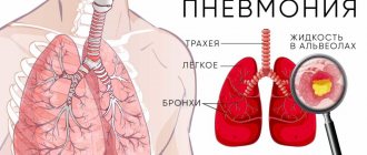
Densification of the lung tissue “frosted glass” is the initial phase of pneumonia. Occurs when the alveoli gradually fill with fluid.
When the bronchi and alveoli fill with fluid, areas of compaction will appear, which are called consolidation. Consolidation of lung tissue occurs during a prolonged inflammatory process.
I have COVID 19 and CT-2 What does this mean?
It is important to understand how much lung tissue is affected. The more the lung tissue is involved in inflammation, the more difficult it is to obtain oxygen from the inhaled air. Without oxygen, a person quickly dies. For example, with pneumonia caused by COVID 19, it takes several hours for the lungs to worsen. The sooner a decision is made about hospitalization, oxygen therapy and treatment, the greater the chance of recovery.
To assess changes, the percentage of lungs damaged by pneumonia is calculated. The measurement is carried out “by eye”. The obtained result is compared with the scale of prevalence of changes:
- CT-0 – no signs of pneumonia
- KT-1 – up to 25%
- KT-2 – from 25 to 50%
- KT-3 – from 50 to 75%
- KT-4 – over 75%
The percentage score sorts patients who urgently require hospital treatment from those who can be treated at home.
If your CT scan is 2, this corresponds to mild pneumonia. Such changes are not accompanied by difficulty breathing and do not require hospitalization. But CT 2 can, if treated incorrectly, turn into CT 3 and 4. Therefore, supervision by a doctor is mandatory!
If the doctor does not hear wheezing in the lungs when listening, does that mean I do not have pneumonia?
This is not true. Diagnosis requires demonstration of changes in lung tissue on radiography (CT) and clinical manifestations (eg, fever, shortness of breath, cough and sputum production), changes in blood tests.
Main types
Understanding what pneumonia is, you need to know what types it comes in. This is important for treatment choice and prognosis.
According to provoking factors, pneumonia is classified into the following types:
- Bacterial. It is caused by pneumococci when it enters the respiratory tract. Most often, the disease develops as a complication of acute respiratory viral infection or other chronic diseases that lead to weakened immunity.
- Viral. The disease develops when infected with viruses of various types. The peculiarity of the disease is its rapid development.
- Fungal. The pathology occurs against the background of the presence of a chronic fungal infection in the human body, in particular the Candida fungus. The disease is characterized by the absence of pronounced symptoms.
Symptoms of different types of pneumonia may vary. The disease in each specific case differs in the nature of its course. If a certain provoking factor is confirmed, it will be necessary to purchase various medications. That is why, if symptoms of pneumonia appear in an adult, it is necessary to urgently undergo examination. Self-medication is strictly forbidden.
With strong immunity, the risks of developing pneumonia in adults, even against the background of provoking influences, are significantly reduced. The following factors worsen the natural defense reactions of the human body:
- Hypothermia.
- Unbalanced diet.
- Tobacco smoking.
- Stressful situations.
- Alcohol abuse.
- Chronic diseases.
The risk group includes people over 65 years of age, as well as those who have various chronic diseases. Working in hazardous industries where you have to inhale harmful substances increases the likelihood of pneumonia.
Treatment of pneumonia
To treat pneumonia, you need to know the name of the infection. In patients, the name of the pathogen is unknown at the very beginning of the disease, so empirical antibiotic therapy is used - treatment aimed at the likely pathogen. For all patients with CAP, treatment regimens have been developed aimed at destroying S.pneumoniae and atypical pathogens.
For most patients, combination therapy with beta-lactam antibiotics or macrolides is used. Alternative regimens include fluoroquinolone monotherapy.
Treatment of SARS CoV-2
- The principles of treating pneumonia are general. Prescription of antiviral and antibacterial therapy.
- In severe cases, in a hospital setting, high-flow oxygen therapy, monoclonal antibodies, anticoagulants, and pulmonary ventilation are prescribed.
- Antibiotics do not work against the COVID 19 virus.
Treatment
Treatment is based on stopping the cause of the disease, relieving symptoms and maintaining the body’s reserves:
- Anti-infective drug therapy. Drugs are selected strictly based on what is the causative agent. Therefore, therapy can be antibacterial, antiviral, antifungal.
Among antibacterial drugs, amoxicillin, macrolides, cephalosporins, cephalosporins, fluoroquinolones, carbapenems, and aminoglycosides have a good effect. If pneumonia is of a mixed nature or the exact pathogen has not been identified, sometimes several drugs are combined. If the pneumonia is of a viral nature, anciclovir or cidofovir is prescribed. Foscarnet and other drugs. It all depends on the type of virus. For fungal pneumonia, antimycotic drugs are prescribed. If the pathogen is unknown, a combination antibiotic therapy of 2-3 drugs is prescribed. The course of treatment can last from 7-10 to 14 days, it is possible to change the antibiotic.
- Symptomatic therapy. For symptoms, patients with pneumonia are prescribed expectorants, antipyretics, and antihistamines (antiallergic) drugs.
- Physiotherapy. This type of treatment is relevant only at the stage when there is no fever. Effective options for physiotherapy are UHF, massage, electrophoresis.
Rehabilitation after pneumonia
In addition to drug therapy aimed at bacteria, virus and inflammation, it is necessary to restore lung function to its original healthy level.
After an illness, changes form in the lungs. This includes “frosted glass” and fibrosis of the lung tissue, pneumofibrosis. In these areas, gas exchange is difficult, the nutrition of the alveoli and bronchi is disrupted, and the protection of the bronchi is reduced. Infection and pneumonia affect a person at the same time, depriving him of strength, confidence, and reducing the quality of life. Efforts must be made to prevent fibrotic changes in the lungs. The problem is solved by a rehabilitation program consisting of a set of medical procedures, inhalations, breathing simulators, and physical therapy exercises.
More about rehabilitation
Pathogenesis of pneumonia
The mechanism of origin - the pathogenesis of pneumonia is complex. It all depends on how the infection entered the body.
The ways in which infections and toxins enter the body are different:
- Through the bronchi ( bronchogenic pathogenesis). Bacteria and viruses enter the lungs by airborne droplets - directly with inhaled air or indirectly (from the nose), upon contact of mucous membranes (nose, eyes) with a surface on which there are viruses and bacteria.
- Through the blood (hematogenous pathogenesis). Such pneumonia most often develops against the background of sepsis - purulent-septic blood damage.
- Through lymph . From the lymph, microorganisms penetrate the epithelial cells of the bronchopulmonary system and adhere to them.
- From the inside (endogenous pathogenesis). In this case, the mechanism of the onset of the disease begins with a disturbed microflora (for example, the synthesis of mucins (glycoproteins) is disrupted in a person). Because of this, problems arise with mucociliary clearance, the task of which is to directly remove rhinobronchial secretions.
After bacteria, toxins, and viruses enter the lungs, cytokine proteins (peptide information molecules) come into play. They are activated by the movement of neutrophils (parts of leukocytes), macrophages taking part in the inflammatory process. It is cytokines that regulate the development of local protective reactions.
But if bronchopulmonary protection is impaired, the immune system is weak, autoalergic and allergic reactions occur in the body, and pneumonia occurs.
If fungi are to blame for the disease, then a characteristic feature of the disease development mechanism is the active proliferation of fungal cultures, the migration of leukocytes and the accumulation of serous fluid in the bronchioles and alveoli.
Our specialists
Chikina Svetlana Yurievna
Candidate of Medical Sciences, pulmonologist of the highest category. Official doctor, expert at Russian congresses on pulmonology.
30 years of experience
Kuleshov Andrey Vladimirovich
Chief physician, candidate of medical sciences, pulmonologist, somnologist, member of the European Respiratory Society (ERS).
Experience 26 years
Meshcheryakova Natalya Nikolaevna
Candidate of Medical Sciences, pulmonologist of the highest category, associate professor of the Department of Pulmonology named after. N.I. Pirogov.
Experience 26 years
Nikitina Natalia Vladimirovna
Deputy chief physician, pulmonologist, allergist of the highest category. Full member of the European Academy of Allergy and Immunology.
Experience 15 years


