Paget's disease is a dangerous pathology in which the restoration of bone tissue slows down, and an abnormal formation appears in its place. The disease can progress on any bones of the human skeleton and usually affects 3 or more bones. In the absence of timely diagnosis and prompt access to qualified medical help, treatment will be extremely difficult and will require a very long period of time.
Causes of the disease and risk factors
Paget cells, from which malignant tumors develop, are glandular cells derived from the epithelium of the milk ducts. The main theories of Paget's cancer development are epidermotropic and in situ transforming theories.
The epidermotropic theory, to which oncologists are more often inclined, is based on conclusions demonstrating that in the vast majority of cases Paget's disease occurs against the background of other forms of breast cancer (BC). According to this theory, cancer cells move along the milk ducts from the primary tumor to the nipple, where they attach and divide.
The transformative theory explains the development of Paget's cancer by spontaneous malignancy of epithelial cells of the nipple and areola. This is confirmed by the fact that in a certain percentage of patients with Paget's disease, no other breast tumors were found.
Risk factors:
- hereditary predisposition (often typical for cases of the disease in men);
- age over 60 years;
- excess weight;
- alcohol abuse;
- injuries and damage to the nipple;
- hormonal imbalances - early puberty, late menopause, infertility;
- concomitant diseases – scleroderma, cervical cancer;
- taking hormonal medications;
- contact with carcinogens (chemicals, radiation).
Pathogenesis of Paget's disease
Normally, bone structures are constantly renewed thanks to the coordinated work of osteoclasts (cells that destroy bone tissue) and osteoblasts (cells that form new bone tissue). This is necessary to maintain strength. With deforming osteodystrophy, the coordinated operation of these mechanisms is disrupted; in some areas of the skeleton, osteoclasts and osteoblasts become overactive, which causes severe deformation of the bones, a violation of their density due to the rapid destruction of old bone tissue and the incorrect chaotic growth of new one.
The affected area is prone to fractures, and musculoskeletal function is impaired.
Statistics
The disease occurs in 3% of the population. The first symptoms appear no earlier than 40-50 years of age. Men get sick twice as often. Paget's disease is usually diagnosed accidentally during a routine examination. The most common form of the disease is polyostotic, in which several paired bones are involved in the process: femur, pelvic, tibia. The bones of the skull and spine are often affected. Causes The exact causes of Paget's disease are still unknown. A connection between the development of the disease and viral infections, in particular the measles virus, has been identified. In the course of many years of research, it has been proven that close relatives of the patient are susceptible to osteodystrophy more often than others, i.e. Genomic abnormalities play a significant role in the development of the disease. Relatives are advised to regularly undergo bone x-rays and blood tests for alkaline phosphatase levels.
Symptoms of the disease
In the first stages of Paget's cancer, symptoms appear that are similar to skin diseases and manifestations of allergies, to which patients often do not attach much importance. This:
- changes in pigmentation of the nipple and areola;
- slight roughening and/or flaking of the skin, itching;
- discomfort when touched, increased sensitivity, slight pain in the affected area.
Often patients use ointments or compresses to relieve symptoms and do not consult a doctor, thereby worsening the prognosis of the disease. It is very important when the first, even nonspecific signs appear, to visit a doctor and undergo the necessary examination to make a diagnosis. Early diagnosis gives a chance for successful treatment and absence of relapses.
As the disease progresses, the following symptoms appear:
- swelling, increase in the size and shape of the nipples;
- increased pain, inflammation;
- discharge from the nipples is purulent, possibly containing blood;
- the formation of ulcers, erosions, crusts, when removed, a weeping surface is exposed;
- enlarged axillary lymph nodes;
- feeling unwell, weakness, fever.
In the later stages, retraction of the nipple is noted, but most often it flattens (the result of the growth of the underlying tumor) Source: Pogodina E.M. Paget's breast cancer / E.M. Pogodina [and others] // 2006. - No. 1. - pp. 65-70..
Causes of thrombosis
In order for a blood clot to form in a vein, a number of conditions or factors are necessary. In the classical version, there are three factors that contribute to thrombus formation (Virchow’s triad).
- hypercoagulable state (increased coagulability). There are many reasons for increased blood clotting - surgery on any part of the body, pregnancy, childbirth, diabetes, excess dietary fat, oral contraceptives, dehydration, genetic factors (rare), etc.
- injury to the inner wall of the vessel (endothelium). The vascular wall can be injured during the installation of a venous catheter, injection into a vein (due to this, thrombosis is very common in drug addicts who inject into the veins of the legs, in the groin), injuries, during radiation therapy, chemotherapy, etc.
- slowing down blood flow. It can be observed in various conditions - varicose veins, pregnancy, obesity, immobilization of a limb (for example, when wearing a cast after fractures), heart failure, forced sedentary position of the body (for example, during long air travel), compression of veins by tumors, etc.
Thrombosis can occur due to the action of one factor or a combination of them. For example, in case of a fracture of the leg bones, all three factors can be involved - increased coagulability in the case of extensive hemorrhages in the area of injury, damage to the vascular wall as a result of a mechanical shock, and a slowdown in blood flow as a result of wearing a cast.
Most often, thrombosis occurs in the veins of the lower extremities. This is due to the fact that stagnation most often occurs in these veins (obesity, varicose veins, edema, etc.).
Paget's classification of cancer
The disease is classified according to its forms, stages, location, and occurrence simultaneously with other types of breast cancer.
Forms of the disease:
- psoriatic – there are small pink papules covered with dry whitish scales;
- ulcerative - the presence of ulcerations in the form of a crater;
- tumor – the neoplasm is located in the thickness of the mammary gland;
- acute eczematoid - characterized by a weeping, fine-grained rash, ulcerations on the nipple;
- chronic eczematoid - crusts form, when they are separated, a weeping area is exposed;
- Paget's pigmented carcinoma.
Sometimes Paget's disease develops extramammary - outside the mammary gland, in areas of the body that have apocrine sweat glands (perineum and vulva in women, scrotum and penis in men).
Stages of the disease according to TNM classification:
| 0 | the tumor is localized within the tissue from which it originated and does not grow into adjacent tissues |
| I | tumor size up to 2 cm, no metastases to nearby lymph nodes and distant organs |
| II | tumor from 2 to 5 cm, metastases are possible in the lymph nodes on the affected side, which are not fused to each other and the underlying tissues |
| IIIA | tumor more than 5 cm, metastases in lymph nodes fused to each other and to the underlying tissues |
| IIIB | the primary tumor invades surrounding tissue |
| IV | there are distant metastases, the primary tumor can be of any size |
In half of the cases, Paget's cancer is located within the nipple and areola. In 40% of patients, to the manifestations in this area is added a tumor palpated in the parapapillary zone. In 10%, the disease is detected by cytological analysis of a smear of discharge from the mammary gland before the appearance of visual symptoms.
Clinical recommendations of the Ministry of Health of the Russian Federation contain the following Paget classification of cancer:
- isolated;
- in combination with intraductal breast cancer;
- in combination with infiltrating ductal carcinoma.
In the majority (67-100%) of cases of breast cancer, the nipple is combined with intraductal carcinoma of the mammary gland. Isolated nipple damage is less common (7-8%) Source: Nikitina E.A. Paget's cancer of the breast (literature review) / E.A. Nikitina [et al.] // Tumors of the female reproductive system. - 2016. - No. 4. - pp. 37-46..
Forecast
The prognosis for life of patients with Paget's cancer is moderately favorable. The average life expectancy is approximately 5 – 8 years. If infiltration and metastasis of the tumor process is detected, the period is reduced to 2-3 years. At the stage of distant metastases, the patient receives only palliative care and drug therapy to relieve symptoms of target organ dysfunction.
Paget's cancer of the breast before the appearance of obvious symptoms is almost impossible to prevent, since, like many cancerous tumors, it occurs without a visible clinic in the early stages. However, its occurrence can be predicted if all preventive measures are followed.
Diagnosis of Paget's cancer
The first signs of the disease are a reason to visit a mammologist or oncologist. The doctor will conduct a thorough collection of the patient’s life and illness history, a general examination, palpation of the breast, and prescribe the necessary studies. To make a diagnosis of Paget's disease, the following are prescribed:
- mammography;
- breast ultrasound;
- CT scan;
- tumor biopsy followed by cytological analysis of the contents;
- cytological examination of a smear.
To detect metastases in the lymph nodes and distant metastases, ultrasound of the lymph nodes, biopsy of sentinel lymph nodes, radiography, computed tomography of bones and various organs, and scintigraphy are performed.
Since Paget's disease has similar symptoms to other diseases, a careful differential diagnosis is necessary. Oncopathology should be differentiated from the following conditions:
- dermatitis and eczema of the nipples;
- psoriasis;
- herpes;
- breast tuberculosis;
- syphilis;
- mycosis fungoides;
- melanoma;
- superficial basalioma.
Rehabilitation and palliative care
All patients require rehabilitation measures to restore the function of the musculoskeletal system. Rehabilitation centers in Israel and Germany, as well as other foreign countries, operate exclusively in accordance with modern standards, so the duration of the rehabilitation period is reduced by one and a half to two times. After achieving positive results, patients need to be examined regularly: monitor the level of alkaline phosphatase once every 3 months, perform x-rays once a year. After treatment, patients maintain contact with specialists even when they are already at home.
Treatment of Paget's cancer
The main method of treatment is removal of the mammary gland followed by remote gammatherapy, chemotherapy or hormone therapy. Sometimes radiation is given before surgery to reduce the size of the tumor and reduce the area of intervention.
In the early stages, removal of the nipple-areola complex and the neoplasm with part of the affected gland is indicated. For non-invasive cancer at later stages, a simple mastectomy is performed - removal of the mammary gland with a section of the pectoralis minor muscle. Invasive Paget's cancer is an indication for extended mastectomy: removal of the breast with muscles, subcutaneous fat and lymph nodes.
Subsequently, reconstructive mammoplasty is recommended for patients.
Treatment of Paget's disease abroad in Israel, Germany and other countries
Treatment of deforming osteodystrophy is aimed at suppressing the activity of cells that destroy bone tissue, eliminating pain, preventing complications and malignant degeneration.
Symptomatic therapy
Aimed at eliminating pain and eliminating the local inflammatory reaction. For this purpose, NSAIDs (paracetamol, ibuprofen, baralgin, sodium diclofenac, etc.), hormones, and blockades are used.
Pathogenetic therapy
To suppress the activity of the process, Israeli specialists prescribe medications that prevent the breakdown of bone tissue and promote full bone formation:
- Nitrogen-containing bisphosphonates
- Calcitonin
Surgical correction
The level of development of Israeli orthopedics allows not only to perform osteotomy for therapeutic purposes, but also to perform orthopedic plastic surgery for severe deformities to restore joint function or for cosmetic purposes. Performed daily:
- Osteotomy to correct deformity
- Surgical correction of fractures with fixation of bone parts with plates or pins
- Joint replacement
For minor deformities, surgery is performed endoscopically, which is much easier for the patient to tolerate and reduces recovery time.
If the spinal cord structures are compressed, neurosurgery may be required.
Disease prognosis and prevention
The prognosis of Paget's cancer depends on timely diagnosis and treatment. The success of therapy is influenced by the stage at which the tumor is detected, the presence of affected lymph nodes and their number, morphological signs of the tumor, its aggressiveness, and the patient’s age. Often the disease is detected in late stages due to the nonspecificity of symptoms, and this type of cancer is quite aggressive, so there is a risk of relapses.
The average life expectancy for Paget's disease is three years; in the presence of metastases and an infiltrative component, it decreases to 1 year.
There is no specific prevention of the disease. To detect cancer early, you should periodically perform breast self-examination and consult a doctor if even minor symptoms appear.
Sources:
- Vavilov A.M. Clinical and morphological diagnosis of Paget’s disease / A.M. Vavilov, O.R. Katunina // Almanac of Clinical Medicine. — 2006.
- Kolobukhov A.E. Paget's cancer of the breast: treatment results / A.E. Kolobukhov [et al.] // International reviews: clinical practice and health. - 2022. - No. 2. — P. 42-47.
- Nikitina E.A. Paget's cancer of the breast (literature review) / E.A. Nikitina [et al.] // Tumors of the female reproductive system. - 2016. - No. 4. — P. 37-46.
- Pogodina E.M. Paget's breast cancer / E.M. Pogodina [and others] // 2006. - No. 1. — P. 65-70.
- Pogodina E.M. Paget's cancer of the breast: clinical picture, diagnosis, treatment / E.M. Pogodin // Bulletin of the Russian Cancer Research Center named after. N.N. Blokhin RAMS. — 1995.
- Fetisova E.Yu. Surgical approaches to the treatment of patients with Paget's cancer / Fetisova E.Yu. [and others] // Tumors of the female reproductive system. - 2015. - No. 2. — P. 35-39.
The information in this article is provided for reference purposes and does not replace advice from a qualified professional. Don't self-medicate! At the first signs of illness, you should consult a doctor.
Osteitis deformans: 100 years after J. Paget
IN
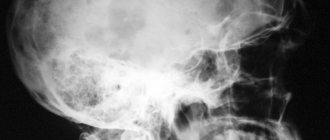
1999 marked 100 years since the death of the English surgeon and pathologist James Paget (born in 1814), whose name in medicine is associated with 3 diseases:
osteitis deformans
; other names: Paget's bone disease, deforming ostosis, deforming osteopathy), as well as
Paget's cancer
(eczema-like breast cancer) and
Paget-Schrötter syndrome
(acute subclavian vein thrombosis). In 1876 (20 years before the discovery of X-rays), J. Paget reported (published an article in 1877) about 5 patients with widespread skeletal lesions, mainly the skull and long bones of the lower extremities, accompanied by their thickening, softening and deformation, and He called this disease deforming osteitis, suggesting that it is based on inflammation of bone tissue. By 1882, he had already observed 23 cases of the disease and described in detail not only the clinical picture, but also sectional data.
The inflammatory nature of the disease was rejected in the 20s of our century, after which Paget’s bone disease (PDB) was for some time classified as a type of fibrous osteodystrophy, which also included fibrous osteitis, a systemic lesion of bone tissue described by the German pathologist F. Recklinghausen in 1891. , and fibrous bone dysplasia. But already in the 30s, each of these diseases was recognized as independent. With fibrous osteitis, hyperfunction of the parathyroid glands was established (A.V. Rusakov was one of the first to identify this connection in 1925), and fibrous bone dysplasia was classified as a congenital anomaly (the assumption of a defect in the development of osteoblasts in this disease was first made by V. R. Braitsev). KBP was isolated after the classical works of G. Schmorl (1930–1932). In our country, the pathomorphology of KBP was first described in detail by A.I. Abrikosov (1926).
Paget's disease of bone is an acquired chronic disease caused by local disorders of bone tissue remodeling.
(see diagram). It is believed that the pathological process is initiated by increased resorption carried out by osteoclasts, which leads to a compensatory increase in the formation of new bone tissue. In this case, an excess amount of collagen fibers is synthesized, located in different directions, not in an orderly manner, which leads to the formation not of lamellar, as is normal, but of coarse-fibrous, excessively vascularized bone tissue, which naturally forms in the embryonic period, and in adulthood is observed only in places of healing of fractures and with hyperparathyroidism. Repeated processes of resorption and neoplasm lead to disorganization of the architectonics of bone tissue. The structure of the bone in the lesions of KBP resembles a mosaic, where zones of abnormal, newly formed fibrous bone tissue, foci of lamellar bone and areas of resorption are randomly located. A mosaic structure is often observed over a significant extent of the affected bone.
A characteristic feature of this disease is the locality of skeletal damage:
changes are noted in any one bone (monostotic form), in several bones (usually asymmetrical) or in many parts of the skeleton. The predominant localizations of “Paget's lesions” are (in descending order of frequency): the spine (usually the lumbar region), the skull (almost always the brain), the pelvis and the long tubular bones of the extremities (most often the femur, tibia and humerus). Less commonly, changes can be detected in the bones of the upper extremities, clavicles, shoulder blades, ribs, facial skull, hands and feet, and sometimes even in the sesamoid bones. Within one bone there may be several affected areas. Their slow growth is typical (no more than a few millimeters per year), but there are also cases of more intense progression. The number of lesions, by all accounts, does not increase over time. The pathological process, as a rule, does not extend to the joints. The exception is the sacroiliac joints, which can be completely destroyed by a layer of newly formed structureless bone tissue; There are also cases of transition of changes from the vertebral body to the intervertebral disc.
Prevalence
With targeted pathological and radiological examination, KBP is often detected, and in patients over 55–60 years of age, the chance of detecting this pathology clearly increases. In a non-selective X-ray examination of the skeleton in 4603 deceased, G. Schmorl established changes typical for KBP in 138 (3%) cases, and DH Collins noted that the frequency of osteitis deformans in old age (after 80 years) reached 10%. There are many descriptions of individual cases of KPD in middle and even young age, but early onset of the disease is still considered atypical.
Geographical differences in the prevalence of the disease are known. The incidence of CKD is highest in England (4.6%), Australia and New Zealand (3–4%), France (2.4%), slightly lower in Ireland (0.7–1.7%), Spain and Germany (1.3%) and significantly lower in Italy, Greece (0.5%) and especially in Norway and Sweden (0.3%). KBP is extremely rare in Central Africa (0.01-0.02%) and almost unknown in Asian countries.
In the UK, when analyzing a large array of skeletal X-rays in 1993–1995. There was a more than 2-fold decrease in the incidence of KBP compared to a similar study conducted in 1974.
In our country, the prevalence of KBP has not been studied, but it is known that at one time individual radiologists had a significant number of observations (600 cases or more).
Etiology
In 1974, during an ultrastructural study of osteoclasts obtained from the zones of “Paget's foci,” virus-like inclusions similar to the nucleocapsid of paramyxoviruses were identified in the nuclei and cytoplasm, and later it was established that they contained antigens of the nucleocapsid proteins of measles viruses and respiratory syncytial virus. In the 1980s, measles virus mRNA sequences were isolated from osteoclasts and other bone cells in patients with osteitis deformans. Measles virus transcripts have been shown to be present not only in Paget bone osteoclasts, but also in hematopoietic stem cells, more differentiated osteoclast precursor cells, and even in circulating peripheral mononuclear cells. A number of other studies have found evidence of canine distemper virus, also a member of the paramyxovirus class, and have found that the risk of developing CDV increases with mongrel dogs kept at home, especially if the animals have not been vaccinated against canine distemper virus.
It should be noted that attempts to identify signs of viral infection in patients with osteitis deformans were not always successful, and viral inclusions reminiscent of those found in KPD are observed in bone tissue cells with giant cell tumor, pycnodysostosis, osteopetrosis and primary oxalosis. It has not yet been possible to isolate the virus from KBP.
Hereditary predisposition
The possibility of the existence of CBP in close relatives has long been known. Using isotope scanning of the skeleton, it was shown that asymptomatic changes in at least one of the first-degree relatives are detected very often
, in approximately 40% of those examined. Family genetic studies have revealed that the risk of developing CBP in first-degree relatives is increased 7-fold.
The search for susceptibility genes was initially limited to the study of the histocompatibility system. In general, these attempts were not successful, although there are reports of a slight increase in the detection of certain HLA system antigens (DQw1, DR1, DR2, Drw6, DPw4) in KPD. In recent years, attention has been drawn to one of the loci (q21–22), located on chromosome 18. The reason for this was the establishment of a connection between this locus and a very rare bone dysplasia - familial common osteolysis, which has features similar to KBP. It was possible to show that there is an association with this locus in osteitis deformans. It has been suggested that CBP and familial common osteolysis may occur due to different mutations in the same gene or family of genes. The lack of a mandatory association with the 18q21–22 locus indicates, however, that genetic susceptibility to CBP is likely heterogeneous and there may be at least one more susceptibility locus to be discovered.
Clinical picture and diagnosis
The disease is diagnosed in the vast majority of cases after 40 years of age, most often around 60 years of age and very rarely before 25 years of age, with approximately the same frequency in men and women.
Two extreme variants of KBP can be distinguished: asymptomatic and clinically pronounced, characterized by persistent complaints and multiple bone deformations. Asymptomatic
the variant is noted in approximately 1/4 of all cases of the disease and, if detected during life, it is usually by chance, during the analysis of X-rays taken for another reason, or in the process of searching for explanations for elevated levels of alkaline phosphatase.
The generalized, polyostotic
variant of the pathology encountered by J. Paget is rare. In such patients, complaining of constant pain in the bones and large joints, remarkable skeletal deformities develop: pronounced kyphosis with shortening of the torso and O-shaped curvature of the lower extremities, leading to a “monkey” gait, as well as an increase in the size of the skull, which is typical for an elderly patient allows one to suspect the diagnosis of KBP at first glance. In the generalized version of the disease, the only systemic manifestation of KBP can be identified - heart failure, caused by an increase in cardiac output due to increased vascularization of altered bone tissue.
Usually there is an “intermediate”
a variant of KBP, which is characterized by local, predominantly nonspecific complaints and symptoms determined by the topic of the disease.
The disease can be suspected by complaints of pain in the bones (although the nocturnal nature of the pain is not typical), pain on palpation and detection of tissue hyperthermia over the affected area. Two other peculiar symptoms of KBP observed when the skull is affected are dilation and swelling of the veins in the vault and angioid streaks on the retina (a consequence of local intensification of bone blood flow). Complaints caused by secondary degenerative changes in the joints, spine, and skeletal deformities prevail. According to RD Altman and B. Collins, the most common complaint in 290 patients with osteitis deformans was pain in the lower back (in 34% of patients); secondary osteoarthritis of the hip joints was detected in 30%, knee joints – in 11% of patients; Deformities of the femur and tibia were found in 45% of patients.
With CBP, neurological disorders naturally develop, especially when the skull and spine are affected. Restructuring of the temporal bone (as well as changes in the auditory ossicles) leads to deafness; softening and deformation of the base of the skull may be accompanied by its flattening and basilar invagination of the spine, leading to compression of the brain stem, hydrocephalus and increased intracranial pressure. The growth of bone tissue in the area of the vertebral arches can cause compression of the spinal roots. Possible narrowing of the vertebral artery canal, accompanied by corresponding ischemic symptoms. Protrusion of an enlarged vertebral body into the spinal canal can lead to myelopathy, and damage to the sacrum can lead to the development of cauda equina syndrome.
The incidence of long bone fractures, especially the femur and tibia, is increased in patients with KPD. Approximately 1/3 of cases of femoral fractures involve subtrochanteric or cervical fractures. Typical fractures may be preceded by so-called gap, incomplete fractures. In most cases, the fusion process occurs without deviations from the norm, within the usual time frame. When treating femoral neck fractures, hip replacement replacement is often necessary.
Another feature of KPD is the possibility of developing sarcomas (mainly osteosarcomas, but also fibrosarcomas and chondrosarcomas). Sarcomas occur in less than 1% of patients, developing in old “Paget's lesions” located in the bones of the pelvis, femur and humerus.
With KBP, it is natural, depending on the degree of activity and prevalence of the process, to increase the daily excretion of hydroxyproline in the urine and the activity of alkaline phosphatase (ALP) in the blood, reflecting, respectively, the intensity of resorption and formation of bone tissue. Patients with the highest values of alkaline phosphatase (they can exceed the normal level by 5–10 times), as a rule, have a polyostotic form of the disease with damage to the skull. Regular monitoring of these parameters allows us to evaluate the course of the disease and the effectiveness of therapy.
The diagnosis of KPD is made exclusively by radiography
. Characteristic changes in different parts of the skeleton are described exhaustively, including in the domestic literature. To clarify the degree of prevalence of KBP, skeletal scincigraphy is of approximate value.
Treatment
Over the past 20 years, radical changes have occurred in the treatment of CBP: symptomatic drugs have been replaced by drugs that can inhibit bone resorption
:
bisphosphonates, calcitonins and plicamycin.
These drugs are able to eliminate bone pain, tissue hyperthermia over the affected parts of the bones and, most importantly, slow down the excessive rate of bone tissue remodeling, as evidenced by a decrease in the levels of alkaline phosphatase in the blood, daily excretion of hydroxyproline, as well as normalization of the structure of bone tissue in the Paget lesion. With CBP, treatment is not required in all cases. Indications for the use of antiresorption agents are:
:
• the need to relieve pain (caused by the disease itself, and not its complications)
• polyostotic form of the disease
• mono- and oligoosseous forms of CBP in the case of prognostically unfavorable localization of the pathological process (near large joints, in the bones of the lower extremities, in the vertebral bodies, with extensive damage to the skull involving the temporal bone or base) and in the presence of disease activity (increased levels of alkaline phosphatase in the blood and/or hydroxyproline in daily urine)
• the need to prevent fractures
• carrying out orthopedic operations (in order to suppress the increased blood supply to the corresponding area of the bone and eliminate the increased risk of postoperative blood loss); This indication is not recognized by everyone.
Complications that have already arisen - bone deformations, deafness, other compressive neurological complications (radiculopathy, myelopathy) - are usually not amenable to this therapy.
If the size of the bone changes is small and there is no immediate threat of developing deformities or neurological complications, and there is no pain or biochemical signs of disease activity, a wait-and-see approach is followed.
The effect of antiresorption therapy is assessed by the dynamics of pain caused directly by CBP, but mainly by the degree of reduction in alkaline phosphatase activity. The analgesic effect is usually noted already in the first weeks, while the “biochemical effect” develops more slowly, its maximum severity is detected within 3–6 months.
Bisphosphonates
In recent years, preference has been given to bisphosphonates due to their selective effect on bone tissue, the ability to persist in it for a long time and the persistence of the effect caused. It should be noted that the pharmacology of bisphosphonates continues to actively develop. At least 3 generations of these drugs are now known (see table). The newest of them have 1000 times greater antiresorption activity compared to the first drug in this group, etidronate. Already during the creation of 2nd generation bisphosphonates (tiludronate, pamidronate and alendronate)
It was possible to expand the “therapeutic window” - the range between the therapeutic and toxic doses - it is too small for etidronate, which led to disturbances in the mineralization of newly formed bone tissue (osteomalacia) in a number of patients.
The effect of bisphosphonates in KPD depends on the severity of the disease, as well as on the dose..
It is selected (this especially applies to bisphosphonates used intravenously) individually, focusing on the prevalence of skeletal lesions, the prognostic significance of the localization of Paget's lesions, the nature of radiological changes (the predominance of signs of bone resorption indicates the need for more intensive therapy), and the degree of increase in biochemical markers of disease activity.
All bisphosphonates taken orally have a significant drawback - poor, unstable absorption (only about 1-10% of the dose taken) and a significant negative effect on it from a number of foods and even drinks. This forces us to recommend taking bisphosphonates 30 minutes before meals (usually before breakfast) and not consuming dairy products, calcium and iron supplements in the coming hours. To prevent “chemical” esophagitis, it is also recommended to drink the drug with at least 100 ml of water and remain in an upright position for about 30 minutes. This is particularly important with etidronate and alendronate and less so with the 3rd generation bisphosphonate residronate.
More successful results in the treatment of KBP are achieved with intravenous administration of bisphosphonates. Treatment is usually carried out in courses, the duration of each of them is from 3 to 6 months. For intravenous bisphosphonates, it is believed that the total dose rather than the duration of exposure is more important. By the 6th month of treatment, when the maximum “biochemical” effect of the chosen therapy regimen is usually revealed, a preliminary result is drawn. After this, it is customary to take a break from treatment, regularly (once a month) monitoring biochemical indicators of the rate of bone tissue remodeling. An indication for a repeat course is the resumption of bone pain and/or a new increase in alkaline phosphatase activity, exceeding the previously achieved level by 25%.
Some patients develop resistance to the drug over time, which must be overcome either by increasing the dose or by prescribing another bisphosphonate. There are patients in whom even with the help of new bisphosphonates it is not possible to achieve normalization of biochemical markers of disease activity, but in many of them the level of these indicators still decreases or decreases to a greater extent than with treatment with “old” drugs.
So far, no long-term studies have been conducted to prove a decrease in the incidence of long-term complications of KBP with long-term therapy with bisphosphonates, and the effect on the dynamics of radiological changes and the risk of developing sarcomas has not been clarified. However, there is hope for this, since repeated biopsies obtained after successful treatment have revealed normalization of the structure in the deposits of new bone tissue in the affected areas of the skeleton.
The main undesirable effect of bisphosphonates when administered orally is “irritation” of the upper gastrointestinal tract, including erosive esophagitis, and when administered intravenously, short-term fever accompanied by chills and myalgia (the so-called acute phase reaction, which is associated with an increase in interleukin levels 6 in the blood).
Etidronate
– the first, most tested drug for KBP and, importantly, the cheapest drug. The daily dose is selected per 1 kg of the patient’s body weight (5 mg/kg). Doses of etidronate as low as 10 mg/kg per day can cause osteomalacia (although they are more effective in inhibiting bone resorption), and doses less than 5 mg/kg are ineffective. In approximately a third of patients with osteitis deformans, no therapeutic effect is observed, which is largely due to poor absorption of etidronate. The effectiveness of etidronate may decrease during repeated courses of its use, and resistance not uncommonly develops. According to ES Siris (1998), etidronate (as well as calcitonins) is able to normalize impaired bone metabolism only in a small number of patients with KPD, mainly with a mild form of the disease.
The development of osteomalacia during treatment with etidronate can be manifested by pain and weakness in the proximal muscles, arthralgia, sometimes arthritis, usually of the ankle joints, enthesopathies in the heels, increased levels of alkaline phosphatase and (in rare cases) pathological bone fractures. Etidronate may occasionally cause alopecia, pancytopenia and hypersensitivity reactions (angioedema, rash, pruritus, Stevens-Johnson syndrome).
Clodronate
also belongs to the 1st generation bisphosphonates, but its effectiveness is higher than etidronate. It is used both orally and intravenously. When administered intravenously, the acute phase reaction characteristic of other bisphosphonates is usually not observed. Previous treatment with bisphosphonates may worsen the effect of clodronate.
Tiludronate.
A double-blind comparative study showed that tiludronate at a daily dose of 400 mg, used for 3 months, was significantly more effective than etidronate (400 mg per day), used for 6 months. A reduction in ALP levels of at least 50% after 6 months was achieved in 70% of patients treated with tiludronate, but only in 25% of patients treated with etidronate. Resistance was observed significantly more often in patients taking etidronate (in 51.9 and 19.5% of cases, respectively). The effect of treatment with tiludronate, unlike etidronate, was not dependent on previous use of bisphosphonates.
Pamidronate.
The effectiveness of pamidronate in KBP
is superior to previously created drugs
, especially in cases of relatively moderate activity or monoosseous form of the disease. In these cases, a single infusion of the drug (60–90 mg) often leads to long-term remission. There are known observations in which pamidronate was effective in patients previously unsuccessfully treated with etidronate. Normalization or almost normal levels of alkaline phosphatase can be achieved with a significant (5-10 times) initial increase in the level of this enzyme, but this requires the use of multiple infusions of the drug at weekly or 2-week intervals.
Pamidronate (as well as residualonate) in rare cases can lead to the development of anterior uveitis and scleritis. The pathogenesis of this side effect is unclear.
Alendronate.
Data on the use of alendronate in KBP are limited. Therapy for 6 months in patients with moderate or significant disease activity was accompanied by normalization of alkaline phosphatase levels in 48–63% of cases; when the treatment period was increased to 9 months, an additional effect was sometimes observed.
Resideronate
– one of the newest bisphosphonates. It is used orally; unlike other bisphosphonates, it can be taken after meals (no earlier than 2 hours later). In one study, a positive effect (a reduction in ALP levels by at least 50%) was observed in 90% of patients with moderate or significant levels of BCP activity; it did not decrease in the case of previously noted resistance to other bisphosphonates. Another study showed that the reduction in ALP levels achieved after the use of residronate was maintained in all patients up to 33 months. Side effects from the upper gastrointestinal tract were observed in 15% of patients, but did not force discontinuation of the drug; erosive esophagitis was detected endoscopically in only 1 patient.
Calcitonin preparations
Although the effectiveness of calcitonins in CBP is comparable to that of etidronate, they are currently used only for limited indications. This is due to the lack of aftereffect of calcitonin preparations (they do not accumulate in bone tissue) and the possibility of developing secondary resistance (according to review data, the frequency of this phenomenon ranges from 5–6 to 40%). Calcitonins are preferred
only in cases
where there are pronounced signs of osteolysis
, threatening the development of bone fractures, and also if it is necessary to obtain the fastest possible effect: in case of severe pain, the appearance of the first signs of neurological disorders, heart failure.
Plicamycin
Plicamycin (formerly mithramycin) belongs to the group of antitumor antibiotics. It is assumed that this drug has a selective and pronounced effect on osteoclasts. Characterized by a powerful and quickly onset effect
. The analgesic effect occurs within a few days. After a 10-day course of treatment, the level of alkaline phosphatase decreases by approximately 60%; in the next 1–2 months, the “biochemical effect” increases and persists from several months to several years. The severity of this effect depends on the dose and frequency of administration of the drug.
The general consensus is that plicamycin should be used as a reserve drug in KBP in the most severe, resistant cases, or when it is necessary to achieve an effect as quickly as possible (for example, with recent development of compression of the nerve trunks). This is due to the hepatotoxic and nephrotoxic effects of the drug, as well as the possibility of developing thrombocytopenia.
Plicamycin is used intravenously (in the form of long-term infusions) at a dose of 10–15–25 mcg/kg for no more than 10 days.
A bolus route of administration is known (25 mcg/kg every 2–3 weeks). Literature:
1. Abrikosov A.I. On the pathological anatomy of common fibrous osteitis (ostitis fibrosa s. deformans generalisata). Russian Clinic, 1926;(5): 343–74.
2. Braitsev V.R. Osteodystrophia fibrosa localisata. Proceedings of the XIX Congress of Surgeons. L., 1927.
3. Kosinskaya N. S. Fibrous dystrophies and bone dysplasias. L., Medicine, 1973.
4. Lagunova I.G. Clinical and radiological diagnosis of skeletal dysplasia. M., Medicine, 1989.
5. Reinberg S.A. X-ray diagnosis of bone and joint diseases. M., Medgiz, 1955; 523–34.
6. Rusakov A.V. On the question of the histo- and pathogenesis of Recklingausen's ostitis fibrosa. In the book: Second All-Russian Congress of Pathologists. M., 1925.
7. Altman RD, Collins B. Musculoskeletal manifestations of Paget's disease of bone. Arthr. Rheum. 1980; 23: 1121–7.
8. Barry HC Orthopedic aspects of Paget's disease of bone. Arthr. Rheum. 1980; 23: 1128–30.
9. Basle MF, Fournier JC, Rozenblatt S. et al. Measles virus mRNA detected in Paget's disease bone tissue by in situ hybridization. J. Gen. Virol. 1986; 67:907–13.
10. Birch MA, Taylor W, Fraser WD et al. Absence of paramyxovirus RNA in cultures of pagetic bone cells and in pagetic bone. J. Bone Miner. Res.1994; 9:11–6.
11. Brown JP, Hosking DJ, Ste-Marie L.-G. et al. Residronate, a highly effective, short-term oral treatment for Paget's disease: a dose-response study. Calcif. Tissue Int. 1999; 64:93–9.
12. Collins DH Paget's disease of bone. Incidence and subclinical forms. Lancet 1956; 271:51.
13. Cooper C., Schafheutle K., Dennison E. et al. The epidemiology of Paget's disease in Britain: Is the prevalence decreasing? J. Bone Mineral. Res. 1999; 14: 192–7.
14. Fraser WD Pagetic disease of bone. Current Opinion in Rheumatology. 1997; 9: 347–54.
15. Gordon MT, Anderson DC, Sharpe PT Canine distemper virus localized in bone cells in patients with Paget's disease. Bone 1991; 12: 195–201.
16. Haslam SI, Van Hul W, Morales-Piga A, Balemans W et al. Paget's disease of bone: Evidence for a suspectability locus on chromosome 18q and for genetic heterogeneity. J. Bone Mineral. Res. 1998;13:911–7.
Causes of thrombosis in the superior vena cava system
– basically the same as other venous thromboses. It may also develop as a complication of venous catheterization (cubital, subclavian catheter), sometimes arising as a result of prolonged compression or uncomfortable position of the upper limb (for example, during sleep).
The most common is thrombosis of the axillary or subclavian vein (Paget-Schrötter syndrome). Within 24 hours, swelling of the entire upper limb occurs with cushion-like swelling of the hand. There may be slight bursting pain. The color of the limb is unchanged or slightly cyanotic.
Deep vein thrombosis
– a more severe illness, usually requiring the patient to stay in a hospital.
There are deep vein thrombosis of the leg, popliteal vein, femoral vein, iliofemoral (ileofemoral) thrombosis. Often there is damage to thrombosis of several sections (for example, the popliteal and femoral veins, deep veins of the leg and popliteal vein, etc.).
Deep vein thrombosis manifests itself primarily as swelling and moderate pain. Moreover, the higher the level of thrombosis, the more pronounced the edema. Thus, with thrombosis of the deep veins of the leg, there may be moderate swelling of the leg, sometimes the swelling is so insignificant that it is detected only when measuring the circumference of the leg (compared to the healthy leg). With thrombosis of the femoral or iliac (continuation of the femoral) veins, the entire leg, up to the groin, and in severe cases, the lower part of the abdominal wall swells.
In the photo - the left lower limb is cyanotic, thickened to the groin - deep vein thrombosis at the level of the iliofemoral segment (ileofemoral thrombosis).
Thrombosis, as a rule, is unilateral, so only one leg swells. Bilateral edema is observed with thrombosis of the inferior vena cava, deep vein thrombosis in both legs (which is quite rare).
Another symptom of thrombosis is pain. It is usually moderately expressed, pulling, sometimes bursting, is relatively constant in nature, and can intensify in a standing position. In case of deep vein thrombosis of the leg, the Homans, Lowenberg, Luvellubri, Meyer, Payra symptoms are positive.
With deep vein thrombosis, there may also be a slight increase in temperature, increased venous pattern, etc.
Superficial vein thrombosis
ATTENTION! Deep vein thrombosis is dangerous due to the detachment of a blood clot and its migration into the pulmonary artery (pulmonary embolism), which most often leads to serious consequences or instant death!
Superficial veins are affected by thrombosis most often with varicose veins. Blockage of the vessel is accompanied by inflammation of the surrounding tissues. That is why the terms “thrombophlebitis” (phlebitis - inflammation of a vein), “varicothrombophlebitis” (inflammation of varicose veins) are used for this type of thrombosis.
Usually, in the area of varicose veins on the lower leg, pain and redness appear along the vein; in the area of redness, the vein itself is palpated as a dense, painful “cord”. There may be a slight increase in body temperature. In general, thrombophlebitis of the superficial veins is not dangerous; there is no detachment of a blood clot with pulmonary embolism (with the exception of inflammation of the great saphenous vein on the thigh and the small saphenous vein in the popliteal region, this will be discussed below). With adequate treatment, the inflammatory phenomena subside, and the patency of the veins is partially or completely restored over time.
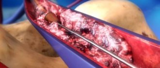
The great saphenous vein (GSV) runs under the skin from the ankle joint along the inner surface of the leg to the groin fold. In the groin it flows into the deep femoral vein. That is why thrombophlebitis of the GSV is dangerous due to the transition of thrombosis from the superficial (GSV) to the deep (femoral) vein - ascending thrombophlebitis of the GSV. But thrombosis of the femoral vein is dangerous due to the detachment of a blood clot and pulmonary embolism. Therefore, if there are signs of GSV thrombosis (redness, pain along the inner surface of the thigh), you should urgently consult a surgeon or call an ambulance. Such patients are hospitalized and, if there is a threat of thrombosis spreading to the femoral vein, the GSV is ligated closer to the groin - this is a simple operation under local anesthesia.
A similar situation, but much less frequently, occurs with thrombophlebitis of the small saphenous vein (SSV). It runs along the back of the leg and drains into the popliteal (deep) vein in the popliteal fossa.
Treatment of deep vein thrombosis
In almost all cases, deep vein thrombosis is treated in a hospital. An exception may be deep vein thrombosis of the leg, provided there is no threat of thromboembolism. The danger of thromboembolism can only be determined by ultrasound examination.
If deep vein thrombosis is suspected, the patient should be hospitalized immediately. In the hospital, an examination is carried out to clarify the prevalence of thrombosis, the degree of threat of pulmonary embolism, and treatment is started immediately.
Usually, drugs that reduce blood clotting (anticoagulants), antiplatelet agents, anti-inflammatory drugs, and phlebotropic agents are prescribed.
In case of massive thrombosis, in the early stages it is possible to carry out thrombolysis - the introduction of agents that “dissolve” thrombotic masses.
If there is a threat of thromboembolism, when the thrombus is attached to the vessel wall only in some part, and the tip of the thrombus “dangles” in the lumen of the vessel (floating thrombus), thromboembolism is prevented (installation of a vena cava filter, plication of the inferior vena cava, ligation of the femoral vein, etc. ).
The consequences of deep vein thrombosis vary. With a small thrombosis on the lower leg there may be no consequences. With massive thrombosis, especially “high” (at the level of the hip or above), swelling of the leg may persist for a long time, and in severe cases there may be trophic disorders, even the appearance of ulcers (post-thrombophlebitic disease (PTSD)). Therefore, after deep vein thrombosis, the patient is prescribed treatment for a long time in the form of elastic compression (special stockings), indirect anticoagulants (warfarin under the control of blood clotting), and other drugs.
You should see a phlebologist for at least six months after thrombosis.
In case of recurrent thrombosis, a genetic study is carried out; if the tests are positive, the issue of lifelong prescription of anticoagulants is decided.
Treatment of thrombophlebitis of superficial veins
The main therapeutic measures are reduced to elastic compression (elastic bandage or compression hosiery) and the prescription of medications.
Medicines used include phlebotropic drugs (detralex, phlebodia), antiplatelet agents (thrombo-ACC), and anti-inflammatory drugs (Voltaren). Lyoton-gel is applied topically.
All patients require venous ultrasound to exclude concomitant deep vein thrombosis and clarify the prevalence of superficial vein thrombophlebitis.