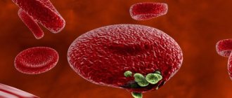
Toxocariasis
is a disease caused by helminths of the nematode group (roundworms). The symptoms are very extensive, as they depend on which organs the larvae have entered. In the acute period, the signs of the disease are similar to those of other helminthiases.
Toxocariasis is quite common in all countries. Most often, it affects children who become infected while playing in the sand and with animals, and adults in the course of their professional activities. The risk group includes veterinarians, dog handlers, agricultural workers, and utility workers.
tests
Definition of disease
Toxocariasis is the migration of animal helminth larvae into the human body. The source of the disease, toxocara, was discovered by the German scientist Werner in 1782. Only in 1950 was infection by these helminths identified as a separate disease. Toxocara eggs can be found in soil and contaminated water. The main carriers are dogs, cats, other domestic and wild animals.
Since almost all invasions are characterized by a long course in most cases with mild manifestations, it can be argued that almost 90% of the population are infected or have been infected at different times. According to WHO research, helminthiasis ranks 4th in the world ranking in terms of the degree of damage to human health.
Causes of toxocariasis
Infection with toxocariasis occurs when Toxocara eggs enter the human body with food and water, through household, contact, and fecal-oral routes. Infection is possible through contact with animals, especially homeless ones, and soil contaminated with eggs. The most common sources of infection are puppies, pregnant and lactating bitches. Cockroaches contribute to the spread of toxocariasis in everyday life; they absorb helminth eggs and subsequently release some of the living individuals.
There are currently 2 types of parasites known:
- T. canis - parasitic in animals of the canine family (dogs, wolves, arctic foxes, foxes);
- T. mystax are parasitic on animals of the cat family (cats, lynxes, panthers, lions).
Humans are not a natural host for the parasite. Therefore, most of the Toxocara die in the human body due to unsuitable conditions for survival. They do not reach sexual maturity. Humans are a dead-end branch; it is impossible to become infected from other people.
crbhotimsk.by
Ascariasis is a widespread helminthiasis, characterized by symptoms of general intoxication, most often affecting the intestines, lungs and liver. Found exclusively in humans. The disease is caused by parasitic worms - roundworms.
Development of roundworm
Adult roundworms parasitize the small intestine of humans and can also be localized in the appendix, large intestine, gall bladder and other organs. The female releases approximately 240 thousand eggs per day, which, when released into the external environment, under favorable conditions (ambient temperature +12-37 degrees, and humidity more than 8%) begin to intensively ripen to the stage of a mature larva.
Under unfavorable environmental conditions, roundworm eggs can survive in soil, water, fruits and vegetables for a long time, sometimes up to 7 years.
Mature larvae enter the human intestines with contaminated water, from the surface of unwashed vegetables and fruits, and from dirty hands. Penetrating through the intestinal walls, the larvae enter the liver through the bloodstream, then through the bloodstream into the heart cavity and lungs. From the lung tissue, the larvae penetrate into the small bronchi, irritate them, causing coughing, which facilitates the penetration of the larvae into the oral cavity and re-swallowing. This completes the development cycle of roundworms, and adult individuals colonize the human intestine.
Mature roundworms parasitize only humans, so the only source of infection is the people themselves. Once the feces enter a favorable environment for the development of eggs, there is a danger of their spreading to:
Fruits, vegetables, drinking water, food, as well as the transfer of mature helminth eggs through contaminated hands onto household items, clothing, and dishes.
Ordinary flies, whose presence is widespread during the warm season, can also participate in human infection.
By eating an unwashed apple or other fruits and vegetables, simply by putting dirty fingers in the mouth, a person introduces helminth eggs into his body, that is, into a favorable environment for further spread and development.
More often they become infected and get sick:
Preschool children who attend kindergarten, and where there is a high risk of spreading parasites from one child to another.
Work stations for sewage and wastewater treatment
Plumbers
Agricultural workers
There is a high probability of infection of gardeners
Symptoms of ascariasis
The manifestations of ascariasis disease depend on the stages that the parasite goes through before it finally settles in the intestines. At the early stage of the disease, general malaise, irritability, sometimes a slight increase in body temperature appear, there may be pain in the joints and muscles, and pinpoint skin rash. During this period, the course of the disease can be wave-like, the symptoms either subside or intensify. The listed signs characterize many diseases; careful questioning of the patient and special laboratory tests can help to suspect helminth infection.
If the disease is not detected at the initial stage and special treatment is not started, then when roundworms enter the liver, discomfort in the abdomen, pain in the right hypochondrium, sometimes enlarged liver, and nausea are noted.
When roundworm larvae pass through the pulmonary system, a dry cough, shortness of breath, and chest pain appear.
The stage of advanced clinical manifestations, when mature roundworms colonize the intestines, is characterized by aggravation of all of the above signs of damage to the gastrointestinal tract and damage to the central nervous system by helminth waste products. Patients lose their appetite, complain of abdominal pain, severe weakness, sleep disturbances, headaches, children become restless and shudder in their sleep. Both children and adults noticeably lose weight.
Features of ascariasis in children.
Infection with roundworms in children occurs much more often than in adults. Thus, in endemic areas, ascariasis is detected in 30% of preschool children. The high incidence rate is due to the fact that children do not sufficiently observe the rules of hygiene, more often eat unwashed vegetables and fruits, put their hands in their mouths, and bite their nails.
All parents should know the symptoms of ascariasis in children. The fact is that these helminths can significantly worsen health conditions, and they are also widespread among children from 1 to 14 years old. But children under one year old get sick relatively rarely, which is due to the peculiarities of their diet.
Symptoms of the early (migration) phase appear within 7-14 days.
An increase in body temperature to 38°C without a runny nose or sore throat.
A dry cough, which can be mistaken for allergic bronchitis, occurs when the larvae penetrate the lungs.
Dry and moist wheezing in the lungs.
Asthmatic-like attacks may be a reflex reaction to the penetration of larvae into the lungs.
Nausea, a sign of poisoning by toxins released when parasites molt.
Pain in joints and muscles is also associated with intoxication.
Nervousness, irritability, tearfulness and hysterical attacks due to poisoning of the nervous system.
Sleep disturbances: the child tosses and turns in his sleep, coughs, screams, complains of nightmares, and his night terrors worsen.
Pain in the right hypochondrium indicates migration of larvae to the liver.
A small rash on the body, sometimes itchy in the form of hives or blisters with transparent contents, which are localized on the hands and feet, is the result of an allergization of the body.
Symptoms of ascariasis in children worsen during the molting of the larvae. It occurs approximately on the 6th, 10th, 15th and 25-30th days of larval development. After adult worms enter the intestines, roundworm eggs appear in the stool.
Late (intestinal) phase symptoms are caused by damage to the lining of the small intestine and malabsorption of nutrients. Symptoms can last from 6 months to a year.
Nausea, belching and vomiting
Decreased or increased appetite
Unstable stool – constipation and diarrhea
Periodic abdominal pain in the navel area
Rumbling in the stomach and increased gas formation indicate dysbacteriosis caused by the activity of roundworms.
Itchy redness around the anus occurs due to skin irritation from roundworm waste products.
Weight loss, sometimes significant, is due to the fact that helminths absorb nutrients entering the intestines.
A decrease in blood pressure in some children is a symptom of intoxication.
Visual impairment is caused by impaired absorption of vitamin A.
Fatigue, developmental delays, and decreased academic performance are signs that the nervous system is suffering from vitamin deficiency and toxin poisoning.
Roundworms remain mobile. They may ascend into the stomach and pharynx or descend into the lower intestine. Therefore, long (up to 40 cm) roundworms can sometimes be released when coughing, vomiting or in feces.
An indirect sign indicating ascariasis in children is weakened immunity. Respiratory diseases often occur, which are severe and long-lasting; stomatitis, gingivitis, and purulent inflammation of the skin are not uncommon.
Complications of ascariasis
Complications of ascariasis are associated with massive proliferation of parasites in the intestines due to untreated disease.
The accumulation of a huge number of parasites in the intestine can lead to blockage of its lumen and the formation of obstruction or rupture of the intestine and the occurrence of inflammation of the peritoneal membranes (peritonitis). These conditions require immediate surgical treatment.
When roundworms penetrate the gallbladder or appendix, inflammation of these organs occurs. Rare cases of adult worms entering the heart cavity with a fatal outcome have been described.
Another dangerous helminthic disease is toxocariasis.
Toxocariasis is a widespread parasitic disease caused by Toxocara. The main carriers of these parasites are cats and dogs, and upon contact with them, humans are often infected.
Toxocara is a round worm that parasitizes members of the canine (dogs, foxes, wolves) and felid (domestic cats) families.
Life cycle of Toxocara.
The main life cycle of Toxocara, in which all stages of development of parasites are possible, is observed only in definitive animals - the canine or feline families. Dogs and cats can become infected with Toxocara eggs and larvae almost anywhere. For example, they can ingest worm eggs upon contact with the environment. After animals swallow the eggs, the latter enter the intestines and there they form larvae.
Penetrating through the intestines, the larvae enter the blood vessels and migrate with the blood throughout the body. Migration of larvae often leads to damage to various organs and systems of the animal.
After the larvae enter the lungs and penetrate the trachea, the animal’s cough reflex is triggered and the parasites are evacuated (probably some of you have noticed a cough in your pet. Be careful, this may be an infection with worms!). Some of the larvae leave the body, the other part is retained in the oral cavity and swallowed into the esophagus. The larvae travel through the esophagus to the intestines and settle there. Here they develop and form into adults, which can subsequently lay eggs.
A female helminth can lay up to two hundred thousand eggs in one day.
The eggs enter the external environment through the animal's excrement. Once in the soil, helminth eggs mature and then become infectious to humans and animals. Eggs usually mature within one month, but at optimal soil temperature and moisture, they may take as little as one week to fully mature. Due to the high resistance of parasites to environmental influences, eggs can remain viable and infective for years.
There is also an auxiliary life cycle of Toxocara, in which the final host of the helminth is a puppy or kitten infected from a sick mother.
Parasitism of Toxocara in the human body is possible only in the larval stage. After a person swallows helminth eggs, they enter the gastrointestinal tract, the eggs hatch into larvae and are carried through the bloodstream to all organs.
While in the human body, parasites can maintain their viability for years, but do not manifest themselves in any way. Activation of toxocara usually occurs when a person’s immunity decreases.
If toxocariasis is activated, depending on the location of the parasites, the patient develops symptoms of acute or chronic toxocariasis.
Symptoms of toxocariasis
Clinically, toxocariasis has no specific symptoms, and its symptoms are largely similar to the symptoms of other helminthiases in the acute period.
general malaise;
increased body temperature (can vary from 37 to 37.9 degrees);
decreased appetite and body weight;
change in blood composition
muscle pain;
allergic manifestations (skin rash, cough);
enlarged lymph nodes.
Chronic toxocariasis is characterized by alternating periods of exacerbation of these symptoms and rest. There may also be a hidden course of this disease, which can only be identified with the help of a special laboratory test.
The location of toxocara in the respiratory system, heart, brain, eyes is extremely dangerous, which without treatment leads to the formation of pulmonary and heart failure, epileptiform seizures, paralysis, decreased visual acuity up to its loss, strabismus, inflammation of eye tissue.
Treatment of ascariasis and toxocariasis.
Modern treatment methods make it possible to quickly and without any harmful consequences for the body get rid of the presence of parasitic helminths in the human body.
There are several types of anthelmintic drugs, the mechanisms of which include the inhibition of the vital activity of helminths, a paralyzing effect on the muscular apparatus of the worms, with the subsequent exit of the parasites from their usual habitat and their death.
We draw your attention to the fact that effective treatment of helminthiasis is possible only as prescribed and under the supervision of a specialist attending physician!
Is the treatment of helminthiasis effective with folk remedies?
This question comes up often. For a long time, herbal preparations have been known, a number of plants and their parts used for food that have anthelmintic properties. But it should be remembered that self-medication most often leads to prolongation of the disease and its aggravation, with a high risk of complications.
Specific treatment, auxiliary medications, diet should only be prescribed by a doctor! Cure from helminthiasis is confirmed only by repeated laboratory testing.
Prevention of ascariasis and toxocariasis.
Active identification of persons with possible carriage of parasites. These activities are carried out mainly in preschool institutions, through periodic medical examinations of children with the collection of smears from the perineal area for laboratory testing for the presence of helminth eggs.
Teaching children to observe basic hygiene rules:
Always wash your hands with soap after using the toilet and before eating.
Before eating a raw fruit or vegetable, be sure to wash the greens with hot boiled water.
Trim fingernails as they grow
Change underwear every day
Preventive administration of anthelmintic drugs to persons living in areas where parasites are spreading.
In order to prevent toxocariasis, it is necessary:
regular examination of pets, as well as their timely treatment;
implementation of planned deworming of animals;
taking various measures aimed at reducing homeless animals;
designating and equipping special areas for walking pets;
mandatory heat treatment of meat;
periodic replacement of sand in children's sandboxes;
protection of parks, squares and playgrounds from visiting animals;
combating mechanical carriers of helminth eggs (for example, cockroaches, flies);
Conducting educational work in organized children's groups and with parents.
Pathogenesis
The causative agent of toxocariasis is roundworms of the Nematoda class. Two species of Toxocara have been discovered; T. canis, parasites of the canine family, can be found in humans. The likelihood of infection with T. mystax has not been established. The pathogen enters the human body in the egg phase with a living larva (cyst), with an average size of 60 microns.
In the digestive tract, the shell of the eggs disintegrates. If a person’s immunity is weakened, the larvae (larvae) become active and begin their migration. Through the bloodstream they penetrate into various tissues, systems and organs. The next stage is the formation of lesions: inflammation, hemorrhage, necrosis. The antigenic effect of larvae leads to allergic reactions.
Granulomas form in the areas where the larvae invade. They are surrounded by fibrous tissue, as a result they cannot receive more nutrients and die. However, in such a short life, the larvae manage to have a significant negative impact on the liver, lymph nodes, lungs, myocardium, and brain. This can lead to the development of pneumonia, pancreatitis, respiratory failure, cause hepatitis or meningitis. Infestations can lead to partial or complete loss of vision and other diseases.
Development of the disease
Once swallowed, eggs enter the intestines. Here the larvae emerge from them and are carried through the bloodstream to the organs. The body's immune response suppresses the activity of the larvae. Capsules are formed around them, and Toxocara larvae are dormant in them. Such asymptomatic carriage can last for decades, while the parasite does not have any negative effect on the human body.
When the immune defense weakens, the larvae become activated and begin to migrate to different organs. At the same time, toxocara causes allergic reactions, as well as mechanical damage to tissues and an inflammatory process.
Classification
In the international classification, toxocariasis is assigned the code B83.0.
The disease is classified according to the following characteristics:
Type of disease development and severity of symptoms:
- Typical form.
- Atypical form (asymptomatic, with erased symptoms).
Form of defeat:
- visceral,
- ophthalmic,
- cutaneous,
- neurological,
- muscles affected
- the thyroid gland is affected,
- lymph nodes are affected.
Severity of illness:
- light;
- medium-heavy;
- severe form.
Nature of the disease:
- Smooth,
- Unsmooth (with complications, exacerbation of existing diseases).
Duration:
- Acute (up to 3 months from the moment of infection).
- Chronic, lasting more than 3 months.
Visceral form
The most common form of toxocariasis is visceral, diagnosed in 90% of cases. Characterized by low-grade fever, intoxication syndrome, and decreased appetite. More than half of patients have a nighttime, unproductive cough. The invasion is accompanied by disturbances in the gastrointestinal tract, including flatulence, diarrhea, and vomiting.
Ophthalmic toxocariasis
The ocular form is the second most frequently diagnosed, but still quite rare. Granulomas are found in the posterior and peripheral parts of the eyeball. This leads to inflammation of the choroid, vitreous abscesses, inflammation of the optic nerve and other abnormalities caused by the migration of larvae. As a rule, only one eye is affected; double damage is extremely rare. Ophthalmic toxocariasis leads to decreased vision, strabismus, and the formation of a dull (opaque) white lump (leukocoria).
Neurological form
Characterized by convulsions, paralysis, behavior disorder, muscle movement disorders, muscle pain. The neurological form is extremely rare and leads to meningitis or meningoencephalitis.
Damage to the thyroid gland
Symptoms of larvae penetration into the thyroid tissue are expressed in its enlargement. The lymph nodes in the submandibular and neck area also increase in volume. The liver and spleen are affected. Lymph nodes are painful when palpated, but no inflammatory changes occur.
Symptoms
The clinical picture of toxocariasis is varied, since the symptoms are directly related to the affected organs. The following symptoms are common to all forms of the disease:
- Temperature with chills from subfebrile in mild cases to +39 C, can last up to 3 weeks.
- Headaches of moderate intensity.
- Intoxication syndrome: loss of appetite, nausea, non-localized abdominal pain, diarrhea.
- Significant increase in the size of lymph nodes, liver, spleen.
- Respiratory syndrome: from mild night cough to bronchial obstruction, pneumonia, signs of suffocation, cyanosis.
- Skin syndrome, expressed in allergic reactions, urticaria, papular rashes. More often they appear on the skin of the lumbar region and extremities.
In the acute form of central nervous system damage, headaches, insomnia, and convulsions are observed. The pathological condition can lead to meningoencephalitis, myelitis, arachnoiditis, paresis and paralysis. In some cases, mental disorders are observed. Patients with ocular toxocariasis complain of deterioration in the quality of vision, the appearance of passing “dots” in front of the eyes and a blind spot that does not pass.
Symptoms are recurrent, on average they appear for 1 - 3 weeks, then go away, and after some time they make themselves felt again.
Diagnostics
Diagnosis of toxocariasis is carried out by an infectious disease specialist. It is necessary to interview the patient to identify the route of infection. Are there dogs in the house, have you had contact with stray animals? Is it possible to accept the possibility of consuming foods and water containing Toxocara eggs?
Laboratory diagnostics include the following methods:
- Biochemical - confirmation of nosology by detecting increased IgE antibodies, decreased albumin levels, hypergammaglobulinemia. A study can also be conducted to determine changes in the data of the hepatic and pancreatic complex.
- The serological ELISA method also allows you to confirm the diagnosis.
- Hematological - aimed at identifying the growth of eosinophils, an increase in leukocytes, a decrease in hemoglobin levels, and an increase in ESR. The method allows you to determine the severity of the disease.
- Sputum microscopy is used to identify the form of the disease.
To clarify the clinical form of toxocariasis and determine the severity of the disease, instrumental studies are prescribed:
- X-ray;
- Ultrasound of the peritoneum;
- external respiration testing.
Electrocardiography, computer and magnetic resonance imaging are performed. Also, if necessary, the reactivity of the bronchi can be studied, and ophthalmological, neurological and histomorphological studies of the affected organs and tissues can be performed.
Toxocariasis of the eye
If ophthalmic toxocariasis is suspected, an ophthalmological examination is performed. Its goal is to identify Toxocara larvae in the macula, optic nerve area, and vitreous body. Sometimes during examination you can record the movements of the parasite. The examination reveals not only the larvae themselves, but the degree of changes that have occurred: hemorrhages, fibrosis, retinal detachment.
Criteria for confirming the diagnosis
- Sort by diagnostic power score from highest to lowest:
- Eosinophilia in peripheral blood.
- Increases in total protein and IgE with a decrease in albumin volume.
- Detection of IgG antibodies in a titer of 1:800.
- An increase in ESR with a decrease in the level of hemoglobin and red blood cells.
- Detection of increased levels of liver transferases, alkaline phosphatase, and direct bilirubin amylase in the blood serum.
- Detection of increased amylase levels in urine.
- Detection of Toxocara larvae in sputum.
These data also help determine the severity of the disease. For example, high leukocytosis indicates the presence of complications along with severe eosinophilia and increased amylase in the urine.
The tasks of an infectious disease doctor also include differential diagnosis. First of all, it is necessary to exclude ascariasis, opisthorchiasis, and strongyloidiasis. If ophthalmic toxocariasis is suspected, it is necessary to exclude retinoblastoma, and allergic skin rashes from a reaction to allergens and insect bites.
Treatment
Therapy for toxocariasis can be carried out in a hospital or on an outpatient basis, depending on the nature and severity of the disease. Patients with manifest and atypical forms are subject to mandatory hospitalization, in the presence of complications. The activities are aimed at achieving the following tasks:
- stopping the development of pathology;
- prevention of complications;
- preventing the formation of residual and recurrent phenomena.
The treatment method depends on the clinical picture. First of all, anthelmintic drugs are prescribed to stop the migration of larvae.
For the purpose of detoxification, solutions consisting of potassium, magnesium, calcium, sodium, sodium acetate chlorides and containing dextrose are administered. Potassium and sodium chloride also helps restore electrolyte balance.
Healing is the complete disappearance of symptoms, a decrease in the level of antibodies and eosinophils in the blood, and the absence of signs of damage to other organs. Dispensary observation is recommended once every 2 months for a year. Children are seen by a pediatrician, adults by a general practitioner, family doctor, or infectious disease specialist. The list of laboratory tests used during the observation period includes a clinical blood test, coprological studies, and others, depending on the affected organs.
Clinical example
I have not yet encountered ocular toxocariasis; after all, the form is quite rare. But the visceral one had to be treated. A 56-year-old woman was sent to me for consultation. She was treated by a dermatologist for about six months for chronic urticaria. Periodically there was discomfort in the abdomen and occasional diarrhea. In a general blood test, the level of eosinophils was from 15 to 25 over six months. The dermatologist prescribed a test for helminthiases for the patient and an antibody titer to Toxocara was detected at 1:1600.
During the interview, it turned out that the patient has a dacha where she spends all the summer months and part of the autumn. She doesn't have dogs, but her neighbors in the country do. Considering all this data, it was possible to make a diagnosis of toxocariasis without a doubt. The patient completed the course of treatment, and two weeks after its completion, she donated blood for antibodies. At the follow-up appointment, she noted the disappearance of rashes and itching. The antibody titer decreased to 1:400. For control, I scheduled her to donate blood again in three months.
As a conclusion, let me remind you once again that we do not live in India, Africa or Thailand. The population of Russia is not completely infected with helminths. We do not need to take prophylactic anthelmintic drugs.
Prevention of toxocariasis is the treatment of pets and careful adherence to personal and food hygiene. Only an infectious disease specialist can diagnose and treat toxocariasis.
Be healthy.
Complications
Among the most common complications after suffering toxocariasis are:
- asthma of bronchitis type;
- Chronical bronchitis;
- epilepsy;
- retinal detachment;
- one-sided blindness.
In the acute phase of the disease, larvae located subcutaneously can cause the formation of infiltrates, abscesses, and phlegmons. Due to weakened immunity, a bacterial infection may occur. Damage to the lungs is dangerous, which causes the development of pneumonia, respiratory failure, which, if unfavorable, can cause death.
The degree of development of complications depends not only on which organs are affected, but also on the number of larvae. Extensive parasite infestations cause multiple organ failure. The patient requires urgent resuscitation care and the risk of death is high.
If infection occurs in a pregnant woman, there is a high probability of damage to the fetus. Toxocariasis leads to miscarriages, premature births and various intrauterine growth retardations.
Visceral migratory larval syndrome
Toxicosis can affect various organs:
- skin;
- lungs;
- liver;
- joints;
- heart;
- eyes;
- brain.
Thus, the syndrome manifests itself with various symptoms. However, the most common areas affected are the respiratory tract, making signs of upper respiratory tract inflammation one of the most common.
Unexpected manifestations of visceral larval migration syndrome are also possible after the sudden migration of a large number of larvae:
- acute pneumonia;
- eosinophilic meningoencephalitis.
This syndrome may increase the risk of death if the infection is accompanied by an abnormal immune response or when there is extensive migration of larvae into the myocardium or central nervous system.
toxocariasis
Prognosis and prevention
For mild and moderate forms of toxocariasis, the prognosis is favorable. Complete recovery without complications is observed in 90% of patients. In 5%, the development of iatrogenic complications is observed, in 5% new diseases caused by toxocariasis develop, and relapses are noted.
Prevention consists of carrying out general sanitary measures to protect the environment. First of all, it is necessary to create special walking areas for dogs. Feces must be disposed of and not left on the ground. Also, dog owners must undergo regular deworming, and street stray animals must be caught.
Particular attention should be paid to hygiene. It is necessary to wash your hands thoroughly after returning home and avoid contact with other people's animals. You cannot eat food on the street if you cannot wash or sanitize your hands. Vegetables and fruits should be well washed. Children need to be taught hygiene skills from childhood. While playing in the sandbox, it is important to prevent sand and soil from being eaten; afterward, it is equally important to wash the baby.
If there is a sick person in the house, all family members must be examined. The premises should be disinfected and individual, disposable care products should be provided to the patient.
Preventive actions
In recent years, every case of toxocariasis has been registered with the SES. Prevention of toxocariasis comes down to the following. One of the main activities is the examination of dogs and their timely deworming. Equally important is the limitation of special areas for their walking and maintenance of these areas in proper hygienic condition. It is still important to observe the rules of personal hygiene: washing hands after contact with soil or contact with animals, careful handling of greens, vegetables, and food products that may contain soil particles. And in the USA they even issued a postage stamp containing information about the need to immediately clean up dog feces in order to reduce the incidence of toxocariasis.
Infectious disease doctor-gastroenterologist Pugaeva S.V.
Advantages of JSC "SZTsDM"
You can take tests to identify Toxocara larvae in one of the laboratories of the SZTsDM. You can undergo a full examination in the comfortable conditions of the center:
with the latest diagnostic equipment;
with qualified, friendly employees;
without queues, with the possibility of a specialist visiting your home.
The laboratories are located in places with convenient transport accessibility in Pskov, Veliky Novgorod, Kaliningrad, St. Petersburg and other cities of the Leningrad region. Regardless of where the tests are taken, you are guaranteed quick availability of results, which can be obtained in several ways.
For more detailed information, please contact our managers by phone.


