Anomalies of the uterus and vagina are observed in 4.3-6.7% of all women of childbearing age. They cause infertility in every eighth case, and in 12.6-18.2% they cause recurrent miscarriage, placental abruption, abnormal fetal position and other complications.
One of the most common anomalies of the uterus is its infantility (also called hypoplasia) - a defect associated with the small size of the main reproductive organ. With a slight decrease in the size of the uterus, the anomaly does not manifest itself with any symptoms, but a significant degree of hypoplasia can be suspected even in adolescence - by the late appearance and extreme pain of menstruation.
Causes of uterine infantilism
A girl’s uterus begins to form during the prenatal period – at the 5th week of its development. By the time of birth, it is fully formed, but still very small. Until the age of 9-10, it grows slowly, spending the first 3 years in the abdominal cavity, and then descending into the pelvis. From 10 to 13-14 years, her growth accelerates greatly, and by the time of puberty she should reach her normal size: 48±1 mm in length, neck length 26±1 mm (that is, a total length of 72-76 mm), 33 mm in thickness, 41 mm in width. If this does not happen, then one of the situations has occurred:
- The uterus either initially failed to develop: it was prevented either by intoxication that affected the mother’s body during the prenatal period, or a disorder occurred at the level of genes or chromosomes, due to which the organ will no longer be able to grow;
- The uterus began to develop, but the girl’s body (mainly her endocrine system) was adversely affected. This could happen as a result of:
- suffered severe influenza: the virus “likes” to affect the main endocrine organs - the hypothalamus and pituitary gland;
- frequent respiratory tract infections: ARVI, chronic tonsillitis;
- nicotine or drug intoxication;
- constant stress (they also affect the hypothalamus), including those caused by significant mental and physical stress at school;
- hypovitaminosis;
- tumors of the pituitary gland or hypothalamus;
- damage to the ovaries by mumps, measles, rubella viruses;
- insufficient nutrition of the girl, including as a result of her following a diet for weight loss;
- surgery on a girl's ovaries.
Prevention of uterine hypoplasia
In order to avoid pathology, it is necessary to eliminate the impact of negative factors on the mother’s body so that problems do not arise in the intrauterine development of the embryo. Negative effects on the body should be excluded even in adolescence. Exhausting diets should be excluded, as they contribute to acute hypovitaminosis and dysfunction of many organs and systems. Good nutrition, absence of stressful situations, normal physical activity and timely treatment of infectious diseases are the best preventive measures to prevent uterine hyperplasia. Regular (twice a year) gynecological examinations and prevention of the development of various diseases are the key to the health of mother and child.
Attention!
This article is posted for informational purposes only and under no circumstances constitutes scientific material or medical advice and should not serve as a substitute for an in-person consultation with a professional physician.
For diagnostics, diagnosis and treatment, contact qualified doctors! Number of reads: Date of publication: 09/29/2018
Gynecologists - search service and appointment with gynecologists in Moscow
Signs of an infantile uterus
If the teenage uterus does not manifest itself in any way, and a woman can only find out about this anomaly during pregnancy or even after childbirth, then more severe degrees of hypoplasia have their own symptoms:
- late onset of menstruation (after 16 years);
- painful periods;
- irregular periods;
- decreased libido;
- difficulty achieving orgasm;
- infertility or recurrent miscarriage.
Uterine hypoplasia is often combined with other gynecological pathologies: mainly endometriosis, frequent inflammation of the vagina and cervix. They can mask the manifestations of uterine hypoplasia.
Often, girls with uterine infantility have a characteristic appearance: they are short, thin, with very small breasts and a narrow pelvis. Upon examination, the gynecologist sees a small amount of pubic hair, underdeveloped labia and a small vagina.
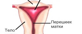
Classification of uterine hypoplasia
The uterus is considered underdeveloped if, by a certain age of the patient, its size is smaller than it should be, according to normal physiology.
Depending on when the organ stopped developing, 3 degrees of uterine hypoplasia are considered:
- Hypoplasia of the uterus 1 degree. The organ is called "embryo" or "vestigial". In this case, the size of the uterus is no more than three and a half centimeters. Moreover, most of the length with hypoplasia is the cervix. This anomaly is formed during the intrauterine development of the embryo.
- Hypoplasia of the uterus 2 degrees. The size of the organ can reach five and a half centimeters, and the proportions of the cervix and body are three to one. The organ is called infantile or childish.
- Hypoplasia of the uterus 3 degrees. The “teenage” uterus reaches seven centimeters, having a normal ratio of the cervix to the body of the organ.
Based on the degree of underdevelopment of the organ, the manifestations of the pathology also differ. Hypoplasia of the endometrium of the uterus is also found - in this case, the mucous layer of the organ becomes thinner (normally it is 7 mm). This type of underdevelopment is the easiest to treat.
Infantile uterus and pregnancy
With an embryonic type of uterus, pregnancy can occur only with the use of assisted reproductive technologies, but often it is necessary to resort to surrogacy with the woman’s egg.
The childhood type of hypoplasia makes pregnancy possible, but pregnancy is associated with the risks of premature birth, placental abruption, abnormal position of the fetus in the uterus, and premature rupture of amniotic fluid.
With the teenage type of anomaly in combination with preserved ovarian function, problems with conception and pregnancy usually do not arise. In the early postpartum period, uterine contractions need to be monitored.
Is it possible to warn?
Severe uterine hypoplasia is usually a congenital pathology, so it is difficult to prevent the development of the disease. However, thoughtful planning of pregnancy, maintaining a healthy lifestyle and eliminating the influence of harmful factors can reduce risks.
It is important to pay attention to the development of girls during puberty during the formation of the body. For the full formation of the reproductive system, you should avoid harmful factors, hypothermia, excessive stress, and bad habits. It is worth giving up diets in favor of a nutritious healthy diet.
Diagnostics and treatment in our clinic
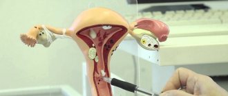
Having established a preliminary diagnosis of uterine hypoplasia and determined its degree by ultrasound, clinic specialists perform hysterosalpingography: this is how they find out the internal structure of the uterine cavity and determine the tortuosity of the fallopian tubes. After this, we need to create a hormonal profile of the woman.
Only on the basis of these studies will we be able to draw up a treatment plan for uterine hypoplasia. The main methods we use for this developmental anomaly are:
- physiotherapeutic methods: magnetic therapy, paraffin, ozokerite applications, endonasal galvanization, mud therapy and other methods;
- vitamin therapy;
- gynecological massage;
- Exercise therapy.
If the degree of uterine infantilism is high, and you want to get pregnant, we will select exactly the assisted reproductive technology that will solve your problem. This could be IVF or fertilization of your egg using the ICSI, PIXI, IMSI method, followed by the introduction of the embryo into a surrogate mother.
What is needed to confirm the diagnosis?
A gynecologist can assume the presence of uterine hypoplasia during examination when there are signs of general infantilism. During a vaginal examination, attention will be drawn to underdevelopment of the vulva, scant hair growth, a short and narrow vagina, and a disproportionate ratio of the uterus to the cervix. The leading criterion is a reduction in the size of the uterus.
Data from laboratory and instrumental studies help determine the diagnosis.
Ultrasound diagnostics provides the most information.
In addition to the reduced size, flattening of the uterine body and a pronounced anterior tilt - hyperanteflexia - will be characteristic. The fallopian tubes are usually long and twisted, which increases the likelihood of an ectopic pregnancy. Often, with general infantilism, ultrasound can detect ovarian hypoplasia. This is associated with a decrease in the formation of sex hormones, so determining the concentration of steroids plays a significant role in the diagnosis of uterine underdevelopment.
Another study that helps confirm uterine hypoplasia is hysterosalpingography (HSG). The uterine cavity and fallopian tubes are filled with a special contrast agent, after which a series of x-rays are taken. The resulting image allows you to see a small uterus with long fallopian tubes, which allows you to confirm the diagnosis.
To determine the hormonal status, the level of hormones in the blood is determined (androgens, estrogens, prolactin, follicle-stimulating and luteinizing hormones, T3, T4 - thyroid hormones).
In doubtful cases, the attending physician has the right to order an X-ray of the skull and an MRI of the brain.
Preventive measures and forecasts
When diagnosing the fertile type of uterine infantility, the possibility of conception is excluded. In this case, pregnancy is possible only with the use of assisted reproductive technologies (ART). If the generative function of the gonads is preserved, in vitro fertilization (IVF) is used using oocytes ready for insemination.
The course of pregnancy in patients with severe endometrial hypoplasia is associated with a high probability of spontaneous abortion and complicated delivery.
In women with miscarriage syndrome, intracytoplasmic sperm injection (ICSI) is performed as part of surrogacy. With minor changes in the structure of the reproductive organ and normal secretion of steroid hormones by the ovaries, the chances of conception and successful pregnancy increase by 45-50%.
Hormonal (endocrine) form
It develops as a result of a disruption in the hormonal regulation of menstrual cycles, which ensures the release of a mature egg from the follicle. A distinctive feature of hormonal infertility is anovulation (non-ripening of the egg or failure to leave the follicle of a mature and ready-to-fertilize egg). The cause may be injury or disease of the pituitary-hypothalamic zone, excessive hormonal secretion of prolactin and lack of progesterone, changes in the structure and function of the ovaries (Stein-Leventhal syndrome), inflammatory and tumor pathologies of the ovaries.
Immune form
The formation of immune infertility occurs against the background of the production in women of antibodies to sperm antigens - specific immunoglobulins that destroy the membrane structure of sperm.
In most cases, infertility is not caused by a single factor, but by a combination of two or more reasons.
Sometimes, even with a full examination of married couples, the cause of infertility cannot be determined. More than 15% of the examined partners are susceptible to a form of infertility of unknown origin.
Functional tests
When diagnosing female infertility, the most common ones are:
- Temperature test - analysis of the nature of changes in the lowest body temperature (basal) during night sleep or at the time of awakening. Allows you to assess the activity of ovarian hormones and the ovulation process.
- Cervical index test - assessment in points of the quality of mucus from the cervix to determine the degree of estrogen concentration in the body.
- Postcoitus test - is carried out to determine the degree of sperm motility in the cervical secretion and to detect the presence of antibodies to sperm antigens.
Determination of the hormonal profile using tests and samples:
- Urine examination for the level of dehydroepiandrosterone and ketosteroids - to assess the functional state of the adrenal glands.
- Assessing the influence of hormones on the phases of menstruation - studying plasma for the levels of cortisol, prolactin, thyroid hormones and testosterone. It is carried out on the 5th day of menstruation.
- Assessment of corpus luteum function and ovulation by determining plasma progesterone concentration. The study is carried out on the 20th day of the cycle.
- Detection of hormonal imbalances during menstrual dysfunction (amenorrhea, oligomenorrhea) - by determining the level of estradiol, prolactin, luteinizing and follicle-stimulating hormones in the blood.
When examining women with infertility, tests for reactions to various hormones are widely used, which makes it possible to more accurately assess the condition of a certain link in the reproductive system. The most common tests are progesterone, estrogen-gestagen, clomiphene, dexamethasone and metoclopramide.
The classic basis of the examination is:
- laboratory determination of the patient’s infectious status – detection of viral and bacterial infections that reduce the likelihood of conception;
- ultrasound examination of the OMT - to exclude pathological changes in the female genital organs;
- ultrasound and x-ray examination of the fallopian tubes - to exclude the causes of tubal obstruction.