Doctors Cost
Price list Doctors clinic
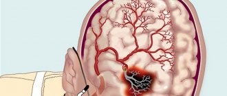
Cerebrovascular accident (CVA) is a reduction in blood access to the brain. This is a real vascular catastrophe. The brain weighs approximately 2% of a person's body weight. A relatively small organ consumes 20% of all incoming energy. The brain does not accumulate the substances necessary for its activity. For normal functioning, it needs a continuous flow of glucose, oxygen, and some other elements supplied with the blood.
Cerebral circulation is distinguished by a multi-level system of redundant vessels and several regulatory mechanisms. But sometimes this is not enough. Cutting off blood supply to the brain for 10 seconds is enough to cause irreparable harm.
Causes of circulatory disorders in the brain
- Systolic pressure is less than 60 or more than 180 millimeters of mercury.
- Insufficient metabolic control, accompanied by unreasonable dilation of cerebral vessels.
- Atherosclerosis of cerebral vessels.
- Decreased blood oxygen saturation.
The compensatory capabilities of the brain are great. Vascular anastomoses allow blood to be distributed throughout the entire volume of the organ, even if the inflow through the left or right carotid artery is disrupted. However, these mechanisms do not work for long; after some time, oxygen starvation develops, and the first signs of the disease appear. This is why it is important to seek medical help immediately.
Most often, NMC develops against the background of:
- history of traumatic brain injury;
- chronic venous insufficiency;
- hypertension,
- atherosclerosis, ischemic heart disease,
- osteochondrosis,
- diabetes mellitus,
- hormonal imbalance,
- excess weight,
- stress, stress
- chronic fatigue,
- lipid metabolism failure,
- drinking alcohol,
- smoking.
Why blood circulation may be impaired
Causes of fetoplacental insufficiency (impaired blood circulation between mother and child):
- Low placentation (attachment of the placenta to the wall of the lower parts of the uterus or “presentation”). The thin muscular layer of the lower uterus is not able to provide sufficient blood flow to the fetus. If there is no migration of the placenta (advancement in the upper part of the uterus), the situation threatens to worsen the pathology.
- Late toxicosis of pregnant women. It affects the small vessels of the uterus, which disrupts blood circulation.
- Drop in hemoglobin level or anemia. This condition causes an accelerated heartbeat in the mother, altering the normal blood circulation in the uteroplacental circle.
- Incompatibility of the Rh factors of the blood of mother and baby, causing anemia in the fetus and immune conflict.
- High blood pressure in the mother due to heart problems, swelling, stress.
- Pathology of the umbilical arteries , for example, the presence of only one umbilical artery.
- Multiple pregnancy , requiring more nutrients.
Some maternal diseases contribute to the spread of pathology, in particular:
- Acute infections, the pathogens of which are capable of penetrating the placenta;
- Uterine defect (“bicornuate” uterus, having a septum in the middle dividing it into two halves). Fetal development occurs in only one of them. The threat is posed by the compression factor of the growing fetus and the disruption of blood flow to it. In such situations, a disturbance of uteroplacental blood flow on the left, grade 1a or on the right, often occurs.
- Diabetes. It affects the walls of the uterine vessels.
- Deviations of the uterine epithelium (endometriosis).
- Uterine tumors. The size of a benign tumor (fibroid) determines how much the fetus will suffer from insufficient blood supply. The larger the fibroids, the higher the risk of failure. Hormonal changes caused by pregnancy stimulate the growth of tumors. The presence of this disease requires constant monitoring of the uterine blood supply.
Acute cerebral circulatory disorders. Symptoms
According to the World Health Organization (WHO), 17 million people are diagnosed with stroke every year. Five million of them die, another 5 million remain disabled.
Table 2. Types of NMC
| NMK | Kinds | Development mechanism |
| Ischemic. | Cerebral infarction (ischemic stroke). | Stopping the blood supply to a part of the brain. |
| Transient NMC. | Lasts up to a day. | |
| Transient cerebrovascular ischemic attack. | Occurs periodically. | |
| Hemorrhagic. | Intracerebral hemorrhage. | Rupture of a vessel with subsequent effusion of blood into the brain tissue. |
| Subarachnoid hemorrhage. | Hemorrhage into the subarachnoid space. | |
| Non-purulent thromboembolism. | Thrombosis. | Blockage of a vessel by a thrombus. |
| Embolism. | Blocking of a vessel with an embolus. |
Features of blood circulation between mother and child during pregnancy
Disturbance in the blood flow of the placenta causes a lack of nutrition and oxygen in the child and causes his death.
The state of placental-uterine blood flow requires close attention during pregnancy. To assess his condition, routine diagnostics are carried out, and preventive and therapeutic measures are taken. The work of blood circulation between mother and baby is based on the functioning of the umbilical artery, veins, and placenta. The uterine arteries are able to contract, blocking the flow of blood due to the thickness of the muscle layer they have. This structure of the uterine arteries is designed to reduce blood loss during menstruation.
During pregnancy at 4-5 weeks, during the gestation of the egg, the muscle layer in the arteries disappears under the influence of hormones. At 16 weeks, another transformation of the arteries occurs, during which they open to constant filling with blood.
What happens in the arteries:
- connection of two flows of different directions;
- diffusion of substances necessary for a growing baby;
- enrichment of the fetal bloodstream with oxygen and beneficial substances brought by the maternal circulation.
Part of the work of blood circulation falls on the arteries and veins of the umbilical cord. Blood flows through the arteries to the baby, and through the veins it returns to the placenta. Violation of the fetal-placental blood flow leads to inhibition of the growth of the child’s organs and poses a threat to his health.
Signs and symptoms of acute cerebrovascular accidents depending on the area of brain damage
- The basin of the internal carotid artery – paresthesia, paresis, paralysis on the opposite side of the face and body. Pathology of speech and vision in one eye.
- Vertebrobasilar basin - systemic dizziness, pain in the back of the head, unsteady gait, loss of visual fields, double vision, inability to swallow and speak.
- Brain stem - eye paralysis, hearing loss, swallowing and speech disorders, paresthesia of the facial skin.
- Medulla oblongata – bilateral paralysis.
- Temporal lobe – loss of spatial and temporal orientation, memory impairment.
How is placental circulation disrupted?
Poor blood flow associated with the placenta is called placental insufficiency. It can occur at any stage of pregnancy in two forms.
Acute appears suddenly, even during childbirth, and does not depend on the duration of pregnancy. The fetus falls into a state of hypoxia (oxygen deficiency), which threatens its death.
The main pathological mechanisms of this condition:
- premature placental abruption;
- heart attack due to thrombosis.
Chronic often complicates the course of pregnancy after 13 weeks. Symptoms appear in the third trimester. The mechanism of formation is early aging of the placenta due to the deposition of fibrin on the villi.
As a result of changes in the structure of chorionic villi (placental tissue), the functioning of the hematoplacental barrier ceases, metabolic processes between the maternal body and the fetus are disrupted
Negative consequences in such conditions, depending on the degree of violation, can lead to inevitable death of the fetus.
Diagnostics of NMC
- Duplex ultrasound examination of blood vessels.
- Contrast venography, angiography.
- CT, MRI.
- Transcranial Doppler.
- Laboratory tests - clinical, biochemical blood tests with determination of lipid profile, hematocrit.
- Fundus examination.
- Detection of hearing loss, smell, taste, and pathology of the vestibular apparatus.
Treatment
In case of acute cerebrovascular accident, treatment should be started immediately. The minutes count down.
For chronic pathology, the course of therapy is drawn up individually after diagnosis. The doctor takes into account age, concomitant diseases, and stage of the process.
Basic treatment includes, among other things, 3 mandatory recommendations.
- Nutrition correction. It should help normalize lipid metabolism and reduce blood cholesterol. Usually the consumption of meat, deli meats, and animal fats is limited. It is recommended to eat more vegetables, herbs, and dairy products.
- Increased physical activity. Helps normalize blood flow and increase vascular tone.
- Weight loss. Correcting your diet and increasing physical activity usually helps you lose extra pounds and be in good physical shape. If this is not enough, seek the help of an endocrinologist.
In case of cerebrovascular accident, with various symptoms, treatment is prescribed:
- physiotherapy;
- physical therapy;
- medicines;
- surgical treatment.
Fetoplacental insufficiency - symptoms and treatment
For timely diagnosis of FPN, it is necessary to establish the exact duration of pregnancy. This is done based on data about the woman’s menstrual cycle.
Methods for detecting fetoplacental insufficiency:
- bimanual examination of the uterus;
- assessment of fetal motor activity;
- auscultation of fetal heart rate;
- control of a woman’s body weight;
- ultrasound examination (ultrasound);
- vascular Doppler ultrasound (USDG);
- cardiotocography (CTG).
A bimanual examination of the uterus to determine its size is carried out upon registration at the antenatal clinic. At each examination, the uterine fundus height (UFH) is measured. VSDM is 2 cm less than normal and the absence of dynamics within 2-3 weeks in 80% of cases indicates a violation of fetal development.
Assessment of fetal motor activity . To do this, use the “Count to 10” test. Every day, from nine in the morning, the woman counts the episodes of fetal movements, and on the tenth time she makes a note in a special table. Normally, the fetus should make the tenth movement no later than 21 hours. If the fetus makes fewer movements, this indicates hypoxia - lack of oxygen. The study is carried out no earlier than 28 weeks of pregnancy [10].
Auscultation (“listening”) of the fetal heart rate. Normally, the fetal heart rate is 120-160 beats per minute.
Controlling a pregnant woman's body weight - normal weight gain averages 250-400 g per week.
The most informative method for diagnosing fetoplacental insufficiency is vascular Doppler ultrasound (USDG) . With ultrasound, the ratio of two indicators is analyzed:
- maximum systolic blood flow velocity, depending on the pumping function of the fetal heart and the capacity of its arterial vessels;
- the speed of diastolic blood flow, which is caused by the resistance of the peripheral part of the vascular bed.
A planned ultrasound examination is carried out during the second and third ultrasound screenings, that is, at 22 and 30-34 weeks. Additional examinations are prescribed according to indications.
Degrees of blood flow disturbance according to Doppler ultrasound:
I degree:
- I A degree - blood flow in the uterine arteries is impaired, while in the umbilical cord it remains normal. A similar picture is often observed with preeclampsia and extragenital diseases, accompanied by a prolonged increase in blood pressure. This degree of blood flow disturbance is not dangerous for the fetus.
- I B degree - blood flow is impaired in the arteries of the umbilical cord, but in the uterine vessels remains normal. The condition is typical for primary FPN, associated with a violation of the location and attachment of the placenta, as well as the formation of the main systems and organs of the fetus.
II degree - blood flow is impaired in the uterine arteries and in the umbilical cord, but the deviation does not reach critical values. The condition occurs during the development of a pathological process and indicates decompensation of adaptive capabilities, which leads to severe FPN and delayed fetal development.
III degree - critical disturbances of blood flow in the umbilical cord with normal or deteriorated blood circulation in the uterine arteries. At this degree, reverse (reverse) diastolic blood flow or its absence may be detected. Violations indicate decompensated FPN, in which the fetus may die [7].
Fetal growth retardation, as a complication of FPN, is diagnosed by ultrasound. The examination reveals a discrepancy between the size of the fetus and the gestational age and delayed growth of the uterus.
Fetal hypotrophy occurs:
- Symmetrical - lack of body weight is proportional to the lag in length.
- Asymmetrical - lag in body weight with normal fetal length. Asymmetrical malnutrition threatens the birth of a child with defective development of the central nervous system, less capable of rehabilitation due to prolonged brain hypoxia.
The asymmetric type of malnutrition is the most common. In this case, early and late malnutrition can be detected, as well as a temporary slowdown in fetal growth, which levels off as the mother’s condition improves.
Ultrasound diagnosis of intrauterine growth restriction (IUGR) includes measuring several indicators of the fetal body: biparietal size (distance between the temples), thigh length and abdominal circumference. Based on these indicators, the weight of the fetus is calculated.
There are three degrees of severity of IUGR:
- 1st degree - a delay of two weeks;
- 2nd degree - from two to four weeks;
- Grade 3 – more than four weeks [5].
The amount of amniotic fluid is also determined by ultrasound. The combination of IUGR and oligohydramnios is an unfavorable prognostic sign requiring early delivery.
On ultrasound, FPN can be suspected based on the maturity of the placenta, which does not correspond to the gestational age. An “overripe” placenta is determined by the presence of characteristic areas: calcifications, multiple cysts and depressions.
There are four degrees of placental maturity:
- Grade 0 is typical for up to 30 weeks and means that the placenta is normal and is able to fully perform its functions;
- Grade 1 is acceptable from 27 to 34 weeks and indicates active growth and minor deviations from the norm, which does not interfere with the placenta performing its functions;
- Grade 2 is considered normal for 34-38 weeks, deviations already exist, and they are dangerous for the child if this degree is diagnosed at an earlier stage;
- Grade 3 is allowed from 37 weeks. At this stage, the uterus begins to lose its functions, so if this degree is diagnosed at 32-34 weeks, the condition is considered dangerous to the baby’s health [1][2].
Cardiotocography (CTG) - measurement of heart rate and motor activity of the fetus.
A CTG study is prescribed from 32 weeks. During the examination, the integral indicator of fetal condition (ISP) is assessed:
- less than 1.0 indicates the normal condition of the fetus;
- from 1.01 to 2.0 - initial signs of a disorder requiring observation and control CTG;
- from 2.01 to 3.0 - severe condition of the fetus, hospitalization in the parental home is required for observation and determination of the timing and method of delivery;
- more than 3.0 – critical condition of the fetus, requiring emergency delivery [8].
Prevention
To avoid the sudden appearance of cerebrovascular accidents, you need to lead an active lifestyle. Do as much physical activity as possible, morning exercises. Walk more, swim, take a contrast shower. Be sure to monitor blood pressure.
You should stop smoking and drinking alcoholic beverages.
It is recommended to consume more foods rich in vitamins C, D, E, and fiber. Minimize the intake of fried, fatty, spicy, salty foods. If you have chronic diseases, they must be treated. If you have hypertension, monitor your blood pressure.
Symptoms of uteroplacental blood flow disorder 1a degree
If this pathology is in the stage of compensation, the expectant mother will not feel any pronounced deviations. In this case, you can find out about the disease only after examination. Obvious signs of the disease accompany the acute form and chronic decompensation. This pathology is accompanied by the following symptoms:
- a sharp increase or cessation of motor activity of the embryo;
- too slow growth of the abdomen (the diameter of its circumference does not correspond to the normative indicators corresponding to the specific gestational age);
- gestosis;
- arterial hypertension;
- strong weight gain of the expectant mother;
- swelling of the legs below the knees;
- proteinuria.
In some cases, bleeding may occur. This symptom most likely indicates placental abruption. If bleeding occurs, you should immediately see a gynecologist.
- Disturbance of uteroplacental blood flow 1b degree