E.P.
Tikhonova, S.V. Lipnyagova, T.Yu. Kuzmina, S.V. Levitsky Department of Infectious Diseases and Epidemiology Krasnoyarsk State Medical University Municipal Clinical Hospital No. 6, Krasnoyarsk
The problem of opisthorchiasis in our region continues to be very relevant for both infectious disease specialists and general practitioners. In the structure of parasitic morbidity, opisthorchiasis in the adult population is 84.5%. Early diagnosis at the first examination of the patient presents great difficulties due to the peculiarities of the clinical picture of the disease, often the lack of data from laboratory and instrumental studies. The purpose of our message was to remind about this helminthiasis and present an interesting case from practice.
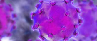
Opisthorchiasis
(code according to ICD10-B66.0) – natural focal biohelminthiasis, characterized by damage to the hepatobiliary system and pancreas. Humans are parasitized by the cat (Siberian) fluke Opisthorchis felineus and Opisthorchi viverrini (squirrel fluke). The definitive hosts are humans, cats, dogs, and civets (family Viverridae of the order of carnivorous mammals). Intermediate hosts are various species of mollusks of the genus Codiella and others; additional hosts are fish of the carp family - ide, dace, chebak, European roach, roach, tench, rudd, carp, bream, silver bream, podust, asp, bleak.
Source of infection
are people infected with opisthorchiasis, as well as domestic and wild carnivores whose diet includes fish. Human infection occurs through consumption of raw or undisinfected fish containing viable metacercariae by heating, freezing, or salting.
The world's largest natural focus of opisthorchiasis is located in the Ob-Irtysh basin; the highest incidence rates of the population are recorded here (up to 500 per 100 thousand). In the basins of the Northern Dvina, Kama, Volga, Ural, Don, Dnieper, Desna, Southern Bug, Northern Donets and Neman rivers there are also natural foci of opisthorchiasis, but they are less intense.
The main foci of opisthorchiasis in the Krasnoyarsk region are the five territories of the Chulym region, where up to 70% of all cases of opisthorchiasis in the region are registered annually. An increase in the incidence of opisthorchiasis was noted in 19 territories. The incidence has increased not only in the endemic areas of the Chulym basin, but also in a number of territories of the region due to population migration and an increase in imported cases.
Every year, from 20,050 to 26,100 cases of parasitic diseases are registered in the Krasnoyarsk Territory. In the general structure of infectious and parasitic diseases, they occupy second place and account for 3.6%. Opisthorchiasis is also detected in a number of European countries - France, Bulgaria, Holland, Italy, Switzerland, Sweden, Poland and Romania.
The main role in the pathogenesis of opisthorchiasis is played by:
– allergic reactions
(especially pronounced in the early phase of the disease), which arise as a result of the release of metabolic products by helminths;
– mechanical impact of helminths
, which consists of damage to the walls of the bile and pancreatic ducts and gallbladder by suckers and spines covering the surface of the helminth’s body. The accumulation of parasites causes a slowdown in the flow of bile and pancreatic secretions;
– neuro-reflex influences
through helminth irritation of the nerve elements of the ducts, resulting in pathological nerve impulses transmitted primarily to the stomach and duodenum;
– occurrence of conditions ( biliary dyskinesia
, accumulation of parasites, eggs, desquamated epithelial cells in them, temporary and complete cessation of bile flow), favorable for the addition of a secondary infection of the biliary tract;
– glandular proliferation
epithelium of the bile and pancreatic ducts, which should be considered as a precancerous condition.
Why is opisthorchiasis dangerous?
The causative agent of the disease, the cat fluke, parasitizes the gallbladder and bile ducts and devours the mucous membrane of organs. The normal digestive process gradually deteriorates. Dysbacteriosis occurs, an allergic reaction of unknown origin appears, the internal organs closest to the gallbladder become inflamed, and stones form.
If the pathology appears during pregnancy, the parasite has a depressing effect on the development of the fetus, physical and mental development. One of the most terrible consequences is cancer of the liver, gall bladder, and pancreas.
In addition to the gastrointestinal tract, the patient’s heart and blood vessels, metabolism, and nervous system suffer. As a result, vegetative-vascular dystonia, insomnia, nervousness may occur, tremors of the limbs appear, appetite decreases, and the menstrual cycle changes.
Chronic opisthorchiasis from the perspective of a systems approach. Clinic, diagnosis, pathomorphosis, treatment
Paltsev A.I.
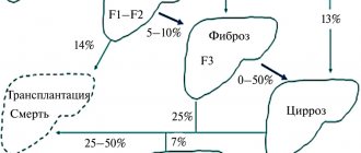
Despite a certain improvement in the sanitary state of the environment and an increase in the sanitary literacy of the population, chronic opisthorchiasis remains a very common disease. This problem is of particular medical and social importance for the West Siberian region, since the Ob-Irtysh basin is the largest endemic focus of opisthorchiasis in the world. In the Middle Ob basin, the incidence of helminthiasis among residents reaches 51-82%, and in some areas over 95%. In connection with the ongoing migration processes of the country's population, it should be noted that already a year after arriving in the center of opisthorchiasis, from 11.5% to 17.9% become infected, after 1.5 years - 42%, after 5 years - 46.7%, subsequently, the level of invasion increases and reaches 73.7% [26,29].
But it would be wrong to consider opisthorchiasis only a regional pathology of the Ob-Irtysh basin, covering more than 10 territories and regions of Russia and Kazakhstan. D.D. Yablokov (1979) believed that diseases associated with this helminthiasis are common in the basins of the Dnieper and its tributaries, the Volga-Kama, the Neman River, and therefore it is diagnosed in residents of Ukraine, Central Russia, the Perm region, Tatarstan and European countries
Clinical characteristics of patients and research methods
We observed 750 patients infected with Opistorchis felineus, aged from 18 to 67 years (among them were 502 men and 248 women). Opisthorchiasis affected mainly young and middle-aged people, and only 35 people (or 4.7%) were elderly patients.
The diagnosis of chronic opisthorchiasis was based on the detection of Opistorchis felineus eggs during microscopy of bile obtained during duodenal intubation or during coprooscopic examination.
The state of the secretory and motor functions of the stomach was assessed using generally accepted methods. To resolve the issue of contamination of the mucous membrane of the stomach and duodenum with Helicobacter pylori, a morphological method and a urease test were used. In the diagnosis of pathology of the liver, biliary system, and pancreas, general clinical, biochemical, immunological, bacteriological methods, ultrasound, X-ray, and magnetic resonance imaging were used (the latter as a method of differential diagnosis). We attached significant importance in determining the type of biliary dyskinesia and the state of the exocrine function of the liver to chromatic duodenal sounding [13].
Statistical processing of the material was carried out on a personal computer. Differences in the compared parameters were considered significant if the probability of error p was less than 0.05.
Research results and discussion
Analysis of the study results allowed us to identify a number of syndromes that occurred with varying frequency: with high frequency - such as cholangiocholecystitis, cholestasis, biliary dyskinesia, allergic, gastrointestinal manifestations, disorders of intestinal microbiocenosis, vegetative-vascular dystonia, pancreatopathy, and less often - cholangiohepatitis. The degree of their severity, as a rule, depended on the duration and intensity of the invasion.
The syndromic approach, consideration of the main links of pathogenesis showed that chronic opisthorchiasis is a systemic disease that is not limited only to the pathology of the parasite’s organs. It shows that the influence of the main pathogenetic factors described by Academician of the USSR Academy of Medical Sciences D.D. Yablokov back in 1979, applies both to organs located on migration routes and to intact organs and systems. All this allowed us to revise the previously given definitions of opisthorchiasis as a predominantly local disease of the liver, gallbladder and pancreas [14,21,23,29,31].
In the light of the presented data, chronic opisthorchiasis should be considered as a systemic human disease caused by the trematode Opistorchis felineus (seu viverrini), parasitizing the ducts of the liver, gall bladder and pancreas, which has an allergic, mechanical, neurogenic effect with the possible addition of a secondary infection and affecting the organs of permanent habitats of the helminth located on its migration routes, as well as intact organs and systems.
As noted earlier by us and other researchers, the manifestations of clinical syndromes depend on the duration and intensity of invasion [1,3,8,9,12]. Thus, an analysis of the clinical course of dyskinetic syndrome using modern diagnostic methods showed that in the first three years after infection, 86% of patients had a hyperkinetic type of biliary dyskinesia, 11% had a normokinetic type, and only 3% had a hypokinetic type. After 5-7 years, and especially after 10 years, the hypokinetic type of dyskinesia was determined in 87% of cases, normokinetic in 6%, and hyperkinetic in 7%.
As our research in recent years has shown, allergic syndrome is much more common than is commonly believed. In 90% or more cases of chronic opisthorchiasis, patients experience changes in the conjunctiva of the eyelids, which manifests itself in hyperemia of varying intensity, edema, vascular injection, proliferative changes in the form of small white or yellow rashes. Based on the severity of these phenomena, one can judge the duration and intensity of the invasion: bright hyperemia, vascular injection, distinct swelling indicate a high intensity of invasion, often superinvasion. Small rashes reflect a long-term process, often with reinvasion. Our observations show the high sensitivity of this diagnostic test. We gave a detailed description of changes in the conjunctiva of the eyelids in chronic opisthorchiasis back in 1999. Later they were called Paltsev’s symptom.
The stomach and intestines are not the permanent habitat of opisthorchises, however, they are involved in the pathological process in more than 90% of cases. Thus, pain syndrome was observed in 93% of patients, and dyspeptic syndrome - in 78%. 5% had a peculiar symptom complex reminiscent of dumping syndrome, which is probably due to the special insulin-competitive relationship between the parasite and the host, described by N.N. Ozeretskovskaya [16].
Chronic opisthorchiasis in local and especially in the indigenous population of Siberia occurs subclinically, or with minor manifestations. At the same time, a thorough history taking and a close, targeted examination make it possible to identify a number of characteristic signs for the pathology of the biliary system and pancreas.
Invasion of Opistorchis felineus in 83% of cases was accompanied by disruption of intestinal microbiocenosis. The microorganisms that determined it were the following: in 16.1% of cases Proteus, in 5.2% - staphylococci, in 4.5% - yeast-like fungi, in 1.1% - lactose-negative Escherichia and in 73.1% various associations were found . Disorders of microbiocenosis are characterized by changes in the tongue in the form of an increase in volume, often a crimson color and “cracks” on it, larger or smaller, which depends on the degree of dysbiosis. We diagnosed the last symptom in 83% of cases.
Various manifestations of vegetative-vascular dystonia were observed in 78% of patients, while its characteristics depended on the intensity and especially the duration of the invasion. Thus, up to three years of invasion, normotonus of the autonomic nervous system was determined in 32% of cases, sympathetic - in 49% and parasympathetic - in 19% of patients. 5-7 years after the invasion, the percentage of people with normotonus of the ANS was 17%, sympathetic tone of the ANS was recorded in 7% and parasympathetic in 76%. All this was reflected in changes in hemodynamic parameters, dermographism, and such characteristics of the skin as color, humidity, and temperature. Of the observed patients, 3-5% developed vagoinsular, and 7-9% sympathoadrenal crises. More than 90% of patients had tongue tremor.
Domestic morphologist N.A. Zubov (1973) at autopsy found opisthorchises in the pancreatic ducts in 30-35% of cases. It seems to us that lifetime this percentage is much higher. In this regard, it should be noted that in endemic foci of opisthorchiasis, the incidence of pancreatitis and pancreatic cancer exceeds the Russian average. We diagnosed pancreatopathy syndrome in 74% of observed patients.
Back in 1985, Academician of the USSR Academy of Medical Sciences A.S. Loginov wrote that chronic hepatitis is not a single disease, but is a clinical and pathomorphological syndrome that has several causes. One of these reasons, in particular, may be opisthorchiasis invasion. We diagnosed cholangiohepatitis syndrome in 48 people (13% of cases). Its manifestations can be different: from those that need to be actively identified to bright manifest ones. The leading symptoms are asthenic, dyspeptic, cholestatic, anemia, fever, hypovitaminosis, and weight loss are possible.
The results of our studies showed that at present, for various reasons, opisthorchiasis has significantly changed the clinical course, which allows us to talk about its pathomorphosis [6,15,17,21]. The term “pathomorphosis”, in its modern understanding, was used by Doerr in 1956. It was introduced into Russian literature by Ya.L. Rapoport (1962, 1982), giving it the following definition: “Pathomorphosis is persistent changes in quantitative and qualitative shifts in nosology, as well as clinical and anatomical forms of diseases under the influence of various influences.” Doerr also emphasized the need to distinguish in pathomorphosis changes in the picture of the disease or natural pathomorphosis (it is also considered spontaneous). Its next type is called induced or therapeutic, since it is caused by the use of chemotherapy drugs and antibiotics. There is also a false or imaginary pathomorphosis associated with the pathology of therapy.
At present, biologists have not fully resolved the question of the homogeneity or heterogeneity of the cat fluke. It has been suggested that helminths interact with microbes. It is believed that the latter are capable of using parasites as habitat. So, E.N. Ilyinskikh (2002) found that opisthorchiasis, as the author puts it, is literally “stuffed” with microorganisms, including salmonella, Helicobacter pylori and, possibly, herpes viruses.
We have analyzed the comparative characteristics of the course of chronic opisthorchiasis according to data from various authors since 1953 [2,18,23].
The clinical course of chronic opisthorchiasis has undergone significant changes over the past half century, which requires clinicians to new approaches to diagnosis. However, as before, general practitioners, parasitologists, gastroenterologists, and infectious disease specialists in making a diagnosis largely depend on whether the laboratory assistant finds opisthorchis eggs during microscopy or not. The proposed immunological diagnostic methods can today be used primarily as screening methods, since they give both false-positive and false-negative results. This is why clinicians continue to face the important challenge of recognizing signs that are a “thing in themselves” so that they become a “thing for us.”
The analysis allowed us to identify a triad of symptoms characterized by high specificity and sensitivity, namely: 1) changes in the conjunctiva (Paltsev’s symptom); 2) tremor of the tongue and 3) “cracked tongue.” The presence of this triad allows the clinician to set specific tasks for the laboratory assistant conducting the study, to judge the duration and intensity of invasion, the severity of syndromes such as allergic, microbiocenosis disorders, asthenovegetative, cholangiohepatitis. Monitoring the severity of the manifestations of the triad of symptoms allows us to further judge the effectiveness of the therapy.
Principles of staged therapy of patients with chronic opisthorchiasis
Treatment of patients with chronic opisthorchiasis is carried out in three stages, including preparatory therapy, specific chemotherapy and rehabilitation [15,17].
Preparatory therapy requires taking into account the influence of pathogenetic factors on the clinical manifestations of the disease and the structure of the main clinical syndromes. The main tasks of this stage are:
- Relief of allergic syndrome, inflammatory (caused by both the addition of a bacterial infection and those arising on an immune basis).
- Ensuring proper outflow from the bile ducts and pancreatic ducts.
- Restoration of the motor-kinetic function of the biliary system.
- Conducting detoxification therapy, using anticholestatic drugs, as well as pathogenetic therapy drugs aimed at regulating leading syndromes.
We would like to especially emphasize the importance of carrying out this stage, since the effectiveness of the next one largely depends on how well the first one is carried out. Its duration is on average 10-14 days. It is necessary to prescribe modern antiallergic drugs. H1-histamine receptor blockers have proven themselves to be effective and are prescribed during the course of preparatory therapy, during specific therapy and, if indicated, during the rehabilitation period. And today the words of N.N. remain relevant. Plotnikov, who wrote that “there is no opisthorchiasis without angiocholitis” [21], therefore, in the indicated cases, broad-spectrum antibiotics are used for a short course (usually a 5-day course). Selective antispasmodic and choleretic therapy should be carried out differentially, taking into account the type of biliary dyskinesia: selective antispasmodics, cholekinetics, choleretics or mixed-action drugs are used. They are prescribed for at least three months, so we consider it appropriate to provide a classification of drugs [17].
They are divided into:
- Drugs that stimulate the bile-forming function of the liver - true choleretics (choleretics):
Preparations containing bile acids.
- Synthetic drugs (hydroxymethylnicotinamide, oxafenamide, holonerton, cyclone).
- Combined choleric patients (allochol, digestal, festal, cholenzim).
- Herbal preparations (galstena, kurenar, flamin, fumeterre, cholagogum, cholagol, holosas, cholaflux).
- Drugs that increase the secretion of bile due to its water component (hydrocholeretics).
- Cholekinetics that increase the tone of the gallbladder and reduce the tone of the bile ducts (berberine biosulfite, xylitol, magnesium sulfate, sorbitol, cyclone, olimethine, cholagol).
The complexity of pathogenetic mechanisms forced us to find antispasmodics that are most effective for this symptom complex. These drugs most likely include mebeverine hydrochloride (Duspatalin). The advantages of the drug are selectivity for the gastrointestinal tract and the absence of side effects; dual mechanism of action (it reduces the tone and reduces the contractile activity of smooth muscles, without affecting normal peristalsis), high affinity for the sphincter of Oddi (20-40 times higher than papaverine); a modern dosage form containing microspheres with gradual release of the active substance, which allows the drug to be used 2 times a day. It seems appropriate to use Duspatalin at the stage of preparation for specific chemotherapy and at the stage of rehabilitation of patients with chronic opisthorchiasis.
For symptoms of cholestasis, ursodeoxycholic acid drugs are prescribed. According to indications, prokinetics, enzyme preparations, pre- and probiotics, nootropics, and eradication therapy agents are used.
Specific chemotherapy is currently carried out with praziquantel. Praziquantel and its analogues affect the ionic regulation of the helminth. By opening the pores of the parasite's cell membranes, they promote increased release of calcium ions and, as a result, disruption of the cell membranes themselves. Its direct effect on glycogen catabolism has been revealed. Drugs in this group are considered inducers of spastic paralysis in opisthorchises. It is prescribed at the rate of 60 mg per 1 kg of patient body weight. We have developed a gentle method of using praziquantel, when the specified daily dose is divided into two days. The anthelmintic effectiveness remains the same. A day after taking the chemotherapy, tubage and techniques to increase the passage of bile are prescribed - electrical stimulation of the right phrenic nerve, pulsed magnetic field, intestinal irrigation. This is where early rehabilitation of patients with chronic opisthorchiasis begins. If the parasite dies, an exacerbation of the allergic syndrome and an increase in intoxication phenomena are possible, and therefore desensitizing therapy is intensified and detoxification therapy and sorbents are prescribed.
A week after taking praziquantel, in the last two years we have been using Ecorsol, consisting of aspen bark extract - 25%, Solyanka holmovaya extract - 1% and glucose - 74% and populin, which also contains a complex of biologically active substances of aspen bark and mineral concentrate waters of Lake Shira. Studies conducted in clinics in Tomsk and Novosibirsk [20,22] showed that their effectiveness is 70-85%. The mechanism of action of these parapharmaceuticals remains unclear. The authors of the drug are the head of the Department of Pharmacology of the Siberian Medical University, Professor A.S. Saratikov and colleagues.
In the future (at least three months), the patient receives differentiated therapy, depending on the severity of certain syndromes, but without fail, as we have already noted, antispasmodic, cholekinetic or choleretic. After 3 months, duodenal intubation is carried out three times (with a weekly interval). After which a conclusion is made about helminthological recovery. The patient may be recommended to undergo treatment in a gastroenterological sanatorium or drink mineral waters at home.
Thus, opisthorchiasis, being a systemic disease, remains an important medical problem, but it needs to be solved only in a complex of government, sanitary and anti-epidemic, veterinary and some other measures [5,18,26,30].
Literature 1. Alekseeva A.S. Features of autonomic regulation of the cardiovascular system and clinical and functional parallels in secondary neurocirculatory dystonia in patients with chronic opisthorchiasis: Abstract of thesis. dis…. Candidate of Medical Sciences - Tomsk, 1998. - 21 p. 2. Akhrem-Akhremovich R.M. Human opisthorchiasis. - M.: "Medicine", 1963. - 146 p. 3. Beloborodova E.I., Kalyuzhina M.I., Buzhak N.S. The influence of specific treatment on the functional state of the small intestine in opisthorchiasis // Med. Parasitol. and parasite. diseases. - 1990. - No. 3. - pp. 31-33. 4. Vinogradov K.N. About a new species of fluke (Distomum sibiricum) in the human liver. Separate reprint // Proceedings of the Tomsk Society of Naturalists. - Tomsk, 1881 - p.15. 5. Gichev Yu.P. Environmental causes of major diseases and reduced life expectancy. - Novosibirsk, 2000. - 89 p. 6. Zubov N.A. On pathomorphosis in opisthorchiasis // Medical parasitol and parasitic diseases. —1968. - No. 4. - pp. 409-410. 7. Ilyinskikh E.N. Current issues in studying the problem of opisthorchiasis in Siberia // Bulletin of Siberian Medicine, 2002, No. 1 - p. 63-69. 8. Kalyuzhina E.V. Clinical and functional state of the small intestine in patients with diabetes mellitus in combination with chronic opisthorchiasis: Abstract of thesis…. Candidate of Medical Sciences - Tomsk, 1999. - 22 p. 9. Kalyuzhina M.I. The state of the digestive organs in patients during the residual period of chronic opisthorchiasis: Abstract of thesis.... Doctor of Medical Sciences - Tomsk, 2000. - 53 p. 10. Clinical use of the drug enterosgel in patients with pathology of the digestive system. New approaches to therapy. Guidelines for doctors edited by prof. I.A.Maeva, prof. Yu.N. Shevchenko, associate professor A.B. Petukhova. - Moscow, 2000. - 89 p. 11. Kuznetsova V.G. Pathogenetic mechanisms and clinical features of the consequences of opisthorchiasis: Abstract of thesis.... Doctor of Medical Sciences - Novosibirsk, 2000. - 31 p. 12. Leutskaya Z.K. Some aspects of immunity in helminth infections. - M.: "Science", 1990. - 205 p. 13. Maksimov V.A., Chernyshev A.A., Tarasov K.M. Duodenal sounding. - Moscow, 1998 - 191 p. 14. Nogoller A.M. Diseases of the gallbladder and biliary tract. - M.: "Medicine", 1969. - 375 p. 15. Loginov A.S. Advanced frontiers of hepatology // Ter.arch. - 1999. - No. 2. - pp. 3-6. 16. Ozeretskovskaya N.N., Sergiev V.P. Mass treatment of opisthorchiasis with praziquantel from the position of a clinician and epidemiologist // Medical parasitol. and parasite. diseases. - 1993. - No. 5. — p.6-13. 17. Okorokov A.N. Treatment of diseases of internal organs. — 2nd ed., revised. and additional — M.: Med. lit. - 1999 18. Paltsev A.I. Diseases of the digestive system in chronic opisthorchiasis. - Novosibirsk, 1996. - 170 p. 19. Paltsev A.I., Miguskina E.I. Diseases of the digestive system in chronic opisthorchiasis // Practitioner. - 1999. - No. 36 (3). - p.23-26. 20. Paltsev A.I. Chronic opisthorchiasis // Medical newspaper, N 14 (6242) - 02.22.2002 - p. 8 -10. 21. Plotnikov N.N. Opisthorchiasis. - M.: "Medicine", 1953. - 128 p. 22. Poddubnaya O.A. New treatment and rehabilitation complex for chronic opisthorchiasis // Siberian Journal of Gastroenterology and Hepatology. - 1997. - p.239. 23. Podymova S.D. Liver diseases. - M.: "Medicine", 1998. - 703 p. 24. Use of the drug galstena in the complex treatment of patients with acute and chronic viral hepatitis. // Method. recommendations No. 43. Compiled by: Doctor of Medical Sciences ON THE. Malyshev, Ph.D. E.P. Selkova - Moscow, 2000 -11 p. 25. Rapoport Ya.L. Problems of pathomorphosis // Arch.pathol. - 1962. - No. 2. — p.3 26. Sergiev V.P. Registered and true prevalence of parasitic diseases // Medical parasitol. and parasitic diseases. - 1991. - No. 2. — pp. 3-5. 27. Tarasov K.M. Biliary insufficiency in diseases of the liver and biliary tract. Author's abstract. diss…. dr. honey. Sciences - Moscow, 2001 - 32 p. 28. Yakovenko E.P. Impaired bile formation and methods for their correction // Consilium medicum. Emergency issue - Media Medica - Moscow, 2002 - p. 3-5. 29. Yablokov D.D. Human opisthorchiasis. - Tomsk, 1979. - 237 p. 30. Opistorchiasis and the control program in Thailand // Symposium on Medical Parasitology, Abstracts. — Shanghai. - 1990. - p.40-41. 31. Renger FG Erkraukungen dar Leber und Gallenwede. - Jena, 1989. - 418 P.
The first symptoms of opisthorchiasis
The first signs appear when the protective function of the immune system decreases. In the early stages, the patient experiences the appearance of general weakness and sweating increases. The acute form of opisthorchiasis manifests itself as follows:
- the appearance of insomnia and nervousness;
- pain in the right hypochondrium;
- pain in the back and abdomen;
- presence of asthmatic bronchitis;
- feeling of body aches and fever;
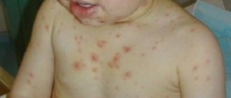
- the appearance of allergic rashes on the skin;
- the presence of flatulence, nausea, vomiting, abdominal pain, general discomfort;
- a sharp decrease in appetite, too liquid stool consistency.
When examining the body, the doctor may detect an enlarged liver and an increased number of eosinophils in a general blood test.
Symptoms of opisthorchiasis in adults are somewhat different from the first signs of children. In little fidgets, the disease is expressed in the manifestation of persistent allergosis, intoxication of the whole body and an immunosuppressive state. The temperature may also rise and last up to 2-3 weeks.
Symptoms in women are most pronounced during the menstrual cycle. If a woman has a parasite in her liver, her periods become very painful and her cycle becomes unstable.
Prevention of helminthic diseases
- cook the fish for 15-25 minutes;
- cooking cutlets, meatballs, etc. - 15-25 minutes;
- baking pies with fish for at least 45-60 minutes;
- cold smoking of fish must be carried out either after pre-salting for 2-3 days, or after freezing;
- fry large pieces of fish spread out and always in fat for at least 20 minutes, fry small fish whole for 15-20 minutes;
- salt the caviar (at a temperature of 5-6°C), the ratio of the amount of salt is 6% to the weight of the caviar for 12 hours, for example: 60 g of salt per 1 kg of caviar, or salting the caviar in a 5% solution (50 g of salt per 1 kg of caviar ) at least 2 days with periodic mixing of caviar;
- salt: small fish for 14 days, large fish (over 25 cm) for 40 days with the addition of 2 kg of salt per 10 kg of fish;
- drying of fish for at least 3 weeks with preliminary salting for 2-3 days; d freezing: at 40°C-7 hours, at 35°C-14 hours, -28°C-32 hours.
The danger of contracting opisthorchiasis and diphyllobothriasis does not disappear all year round.
How quickly does pathology manifest itself?
When parasites enter the human body, the protective functions of the immune system begin to decline. After this, the first signs may appear within 20 days. A person experiences general weakness, malaise, rapid heartbeat, increased body temperature, and severe sweating.
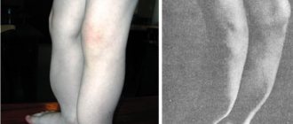
After about 30 days, the disease enters the chronic stage and its symptoms become similar to chronic cholecystitis, gastroduodenitis, pancreatitis, and hepatitis. I am worried about attacks of pain in the right hypochondrium, similar to biliary colic. Nervousness, sleep disturbances, chronic fatigue, frequent allergic skin rashes, trembling hands and eyelids appear.
The period of the acute stage of the disease ranges from several days to 3 months, sometimes longer. Symptoms are accompanied by signs of pulmonary diseases, the liver is enlarged, erosions and ulcers of the mucous membranes and tissues of the stomach and duodenum may be revealed during the study.
Diphyllobothriasis
Diphyllobothriasis is a helminthic disease affecting the digestive organs. The causative agent is the broad tapeworm. This is the largest of the human helminths, its length can reach 10 and sometimes 20 meters. The parasite consists of a head, neck and body. The head is oblong oval in shape, flattened on the sides and has on its narrow sides two longitudinal suction slits (bothria), with which the tapeworm is attached to the intestinal wall. The body consists of many segments, and the width is much greater than the length, which is due to the name of the parasite (wide tapeworm). The number of segments can reach 3000-4000 pieces. The tapeworm lives in the upper parts of the small intestines, feeds on the entire surface of the body, while absorbing various nutrients, including vitamins Bi2 and folic acid. The wide tapeworm is hermaphrodite. During the day, up to 2 million eggs are released into the external environment with feces. The number of parasites can reach up to 100 copies. The lifespan of parasites in the human body reaches 28 years.
For the development of a wide tapeworm, like opisthorchiasis, the presence of three hosts is necessary.
The definitive host is humans, domestic and wild animals. All of them secrete eggs, which end up in water bodies with meltwater. Diphyllobothriasis cannot be contracted through water.
The intermediate host is Cyclops (crustaceans). The eggs are swallowed by crustaceans (cyclops) and larvae develop in their bodies. Cyclops are swallowed as food by freshwater predatory fish.
Additional hosts are fish of predatory species: pike, burbot, perch, ruff; pike caviar is especially dangerous.
Having attached themselves to the wall of the intestines, the parasites injure the intestinal mucosa with bothria and can be one of the reasons for its necrosis. Sometimes intestinal blockage occurs.
Diphyllobothriasis occurs in mild or severe form, which is associated with the intensity of invasion, the presence of concomitant diseases and the general condition of the body. Sometimes the disease is asymptomatic.
In mild cases, patients complain of general weakness, poor appetite, nausea, pain and rumbling in the abdomen, intestinal disorders, and decreased ability to work.
In severe cases, intestinal obstruction occurs. In 2-3% of patients, a severe form of anemia (anemia) occurs. Patients complain of weakness, drowsiness, dizziness. Bright red spots and cracks appear on the tongue. The skin becomes pale with a yellowish tint; The liver and spleen may enlarge. Body temperature reaches 36-38 degrees.
The diagnosis of these diseases is established based on the detection of tapeworm and opisthorchid eggs in the feces.
Stages of acute opisthorchiasis
When it comes to acute opisthorchiasis, it is divided into 4 classes.
- Typhoid-like course - there are symptoms of pancreatitis, jaundice and hepatitis, an enlarged liver, gastritis, eosinophils in the blood up to 90%.
- Hepatocholangetic type - pancreatitis, aching abdominal pain, the liver is affected, the function of the pancreas is impaired.
- Gastroenterological – the presence of enterocolitis, stomach ulcers, erosive gastritis, stool disorders are observed.
- Damage to the respiratory tract - the presence of asthmatic-type bronchitis, pleurisy and pneumonia.
How is the disease diagnosed?
The first thing you need to do if you find signs of opisthorchiasis is not to self-medicate, but to seek the advice of a gastroenterologist. The doctor will conduct a visual examination, collect anamnesis and refer for tests and duodenal intubation. This is a method of examining a patient, which is carried out for diseases of the biliary tract and gallbladder to collect bile for laboratory testing.
Other types of diagnostics:
- general blood analysis;
- examination of internal organs using ultrasound;
- stool analysis for worm eggs;
- polymerase chain reaction (PCR).
After the diagnosis, the doctor examines the results of all tests and studies and gives the patient an accurate diagnosis. After an accurate diagnosis is made, the patient is prescribed treatment. It is carried out under strict supervision.
Treatment of opisthorchiasis
Treatment of the pathology should be carried out regardless of the stage and type of opisthorchiasis. The sooner treatment is started, the less likely it is that the parasite will cause severe damage to your body.
In order to start treatment, it is necessary to have the latest results of a general blood and urine test, a biochemical blood test, FGDS, ultrasound of the abdominal organs, and for patients over 40 years old, also an ECG.
The use of medications relieves inflammation. If there are allergic reactions or intoxication, then this problem is eliminated through the use of antihistamines. Choleretic drugs are prescribed, which are taken at the time prescribed by the doctor, and antibiotics are prescribed. The drugs are most often taken for up to 14 days. A special diet is established for the patient, which he must follow.
After the course of treatment, a control duodenal intubation will be done, you will receive a full medical report and doctor’s recommendations, which you must adhere to throughout your life.
After antiparasitic therapy, a rehabilitation period begins. It can last for 3-4 months, its main tasks are:
- restoration of normal functioning of the digestive system;
- complete disposal of dead helminths that remain in the body;
- elimination of the consequences of helminth activity in the human body.
Treatment of opisthorchiasis in Krasnoyarsk
Diagnosis and treatment of parasites can be done at the Medunion private medical clinic. During the pandemic, we provide all medical services in accordance with the mask regime. A distance is maintained between patients and clinic staff, and all work surfaces are sanitized every hour. Our patients can use masks and hand sanitizers.
The cost of diagnosis and treatment will depend on the procedures performed. To find out more information, make an appointment with a gastroenterologist who will conduct an examination, prescribe tests and treatment. To sign up, call or fill out the online form. Indicate the procedure you need and leave your details - our specialist will call you back and discuss a convenient time for the procedure.
In addition, the clinic operates a comprehensive diagnostic program “Gold Standard”. It includes all types of tests, including tests for parasites.
Spread of opisthorchiasis
There are several species of flukes, but opisthorchiasis is caused by three species - O.felineus, O.viverini, O.sinensis.
Our country is characterized by O.felineus - the cat or Siberian fluke. The center of its distribution area is Western Siberia, but the European part of the country is also inhabited by this fluke.
The risk of infection with the parasite is high: cats, dogs, animals of prey, and humans are the final hosts of the cat fluke.
Through an intermediate host:
- 1st - freshwater mollusk,
- 2nd - fish of the carp family
– human infection occurs through consumption of raw contaminated fish.
The incubation period is about 2 weeks.